
Abdominal Injury Mohammed Aref Malabarey MD FRCPC DABEM
Abdominal Injury Mohammed Aref Malabarey MD, FRCPC, DABEM Consultant Physician Assistant Professor of Emergency Medicine

Relevant Anatomy Anterior Abdomen: Between the anterior axillary line from the anterior costal margins to the groin creases Low chest: Begins at the nipple line or 4 th intercostal space anteriorly and the inferior scapular tip or 7 th intercostal space posteriorly Flank: Between the anterior and posterior axillary lines bilaterally and from the inferior scapular tip to the iliac crest Back: Between the posterior axillary lines, beginning at the inferior scapular tip and extending to the iliac crest
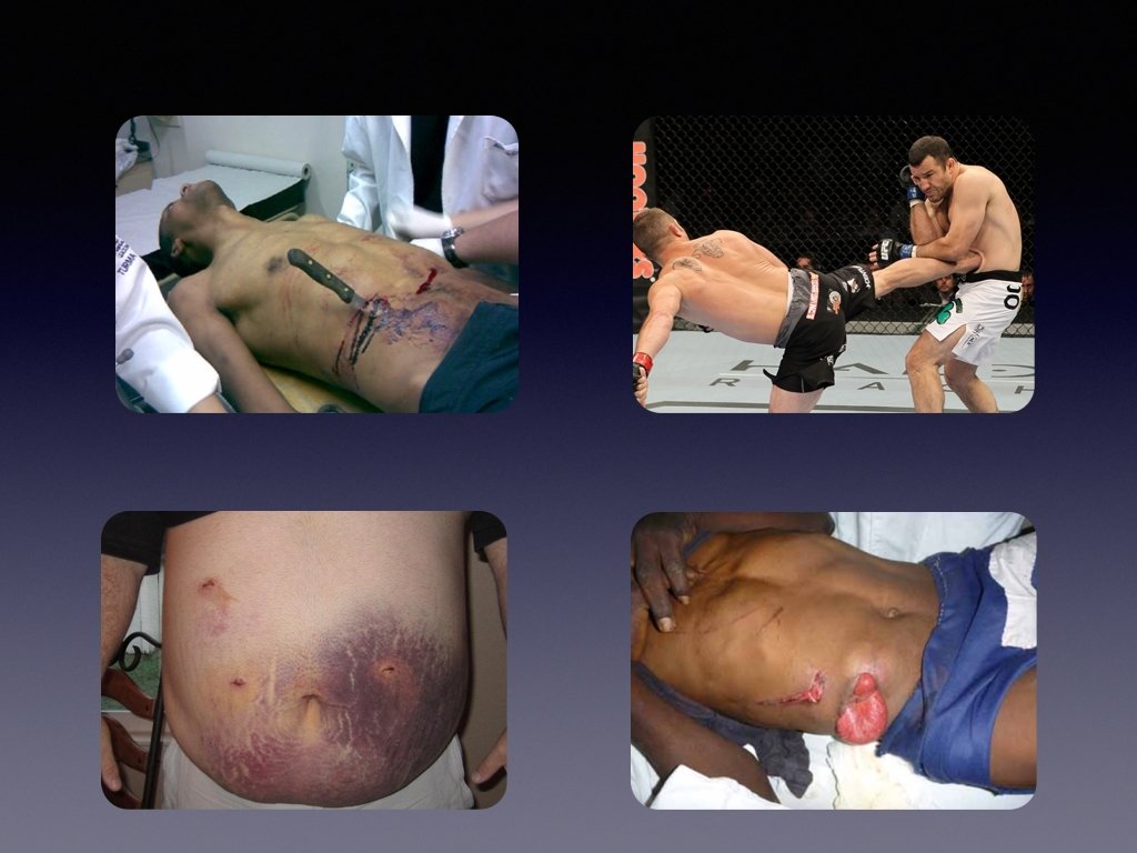
• Penetrating Abdominal Trauma – Stabbing 3 x more common than firearm wounds – GSW cause 90% of the deaths – Most commonly injured organs: small intestine > colon > liver • Blunt Abdominal Trauma – Greater mortality than PAT (more difficult to diagnose, commonly associated with trauma to multiple organs/systems) – Most commonly injured organs: spleen > liver, intestine is the most likely hollow viscus. – Most common causes: MVC (50 - 75% of cases) > blows to abdomen (15%) > falls (6 - 9%)

Mechanism of blunt injury ? • Rupture or burst injury of a hollow organ by sudden rises in intra-abdominal pressures • Crushing effect • Acceleration and deceleration forces → shear injury • Seat belt injuries – “seat belt sign” = highly correlated with intraperitoneal injury
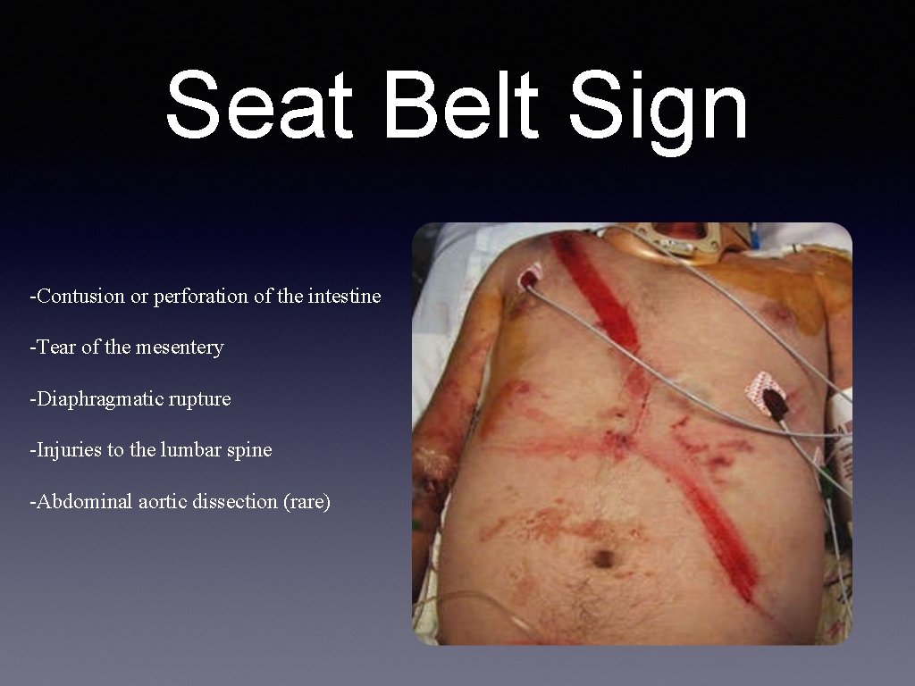
Seat Belt Sign -Contusion or perforation of the intestine -Tear of the mesentery -Diaphragmatic rupture -Injuries to the lumbar spine -Abdominal aortic dissection (rare)
Physical Exam is generally unreliable • Distracting Injuries • Altered Mental Status • Spinal cord injuries
Signs of Intraperitoneal Injuries? – Abdominal tenderness/ peritoneal signs, gastrointestinal hemorrhage – Entrance and exit wounds to determine path of injury – Abdominal distention —> Pneumoperitoneum, gastric dilatation, or Ileus – Ecchymosis of flanks (Gray-Turner sign) or umbilicus (Cullen's sign) - retroperitoneal hemorrhage – Abdominal contusions – eg lap belts
Investigations • Blood Tests —> Not very helpful! • Imaging: X-Rays, CT-Scan, Ultrasound
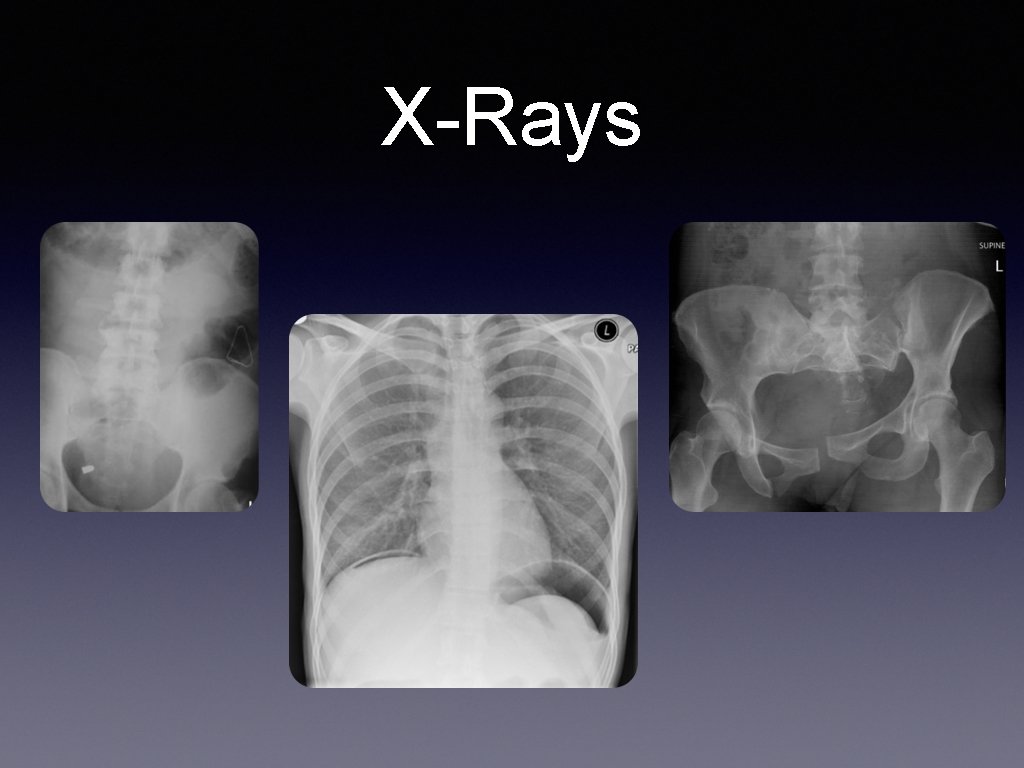
X-Rays
CT Scan NEVER send an unstable patient to CT – Accurate for solid visceral lesions and intraperitoneal hemorrhage – Guide nonoperative management of solid organ damage – IV not oral contrast Disadvantages : insensitive for injury of the pancreas, diaphragm, hollow viscus and mesentery
ULTRASOUND The FAST Exam • Perihepatic & hepato-renal space (Morrison’s pouch) • Perisplenic • Pelvis (Pouch of Douglas/rectovesical pouch) • Pericardium (subxiphoid) – sensitivity 60 to 95% for detecting 100 m. L - 500 m. L of fluid • Extended FAST (E-FAST): – Add thoracic windows to look for pneumothorax. – Sensitivity 59%, specificity up to 99% for PTX (c/w CXR 20%)
• Advantages: – Portable and rapid – No radiation or contrast – Less expensive • Disadvantages – Not as good for solid parenchymal damage, retroperitoneum, or diaphragmatic defects. – Limited by obesity, substantial bowel gas & subcutanous air – Can’t distinguish blood from ascites.
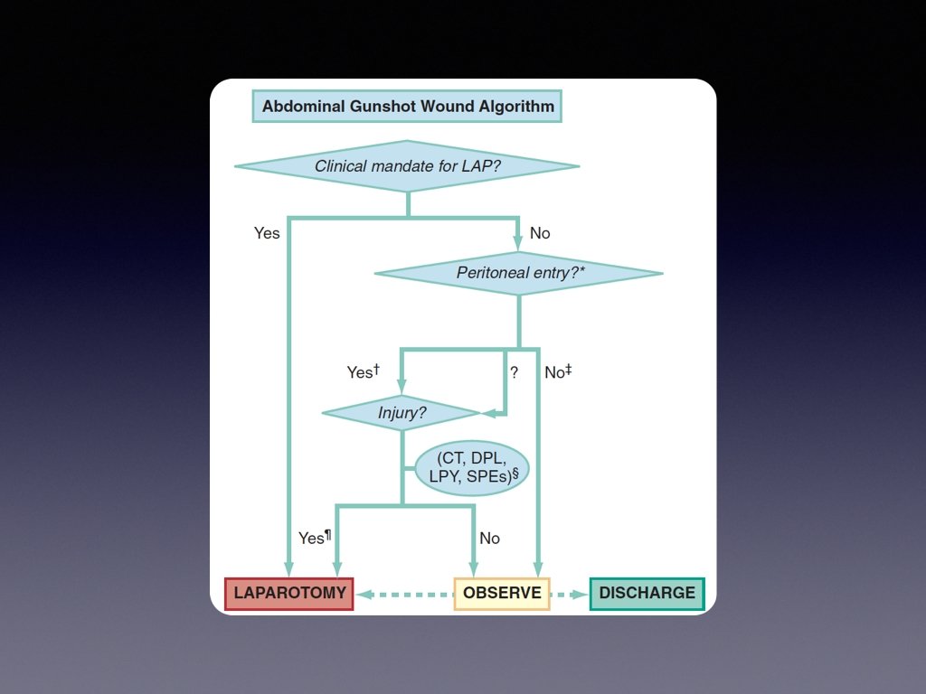
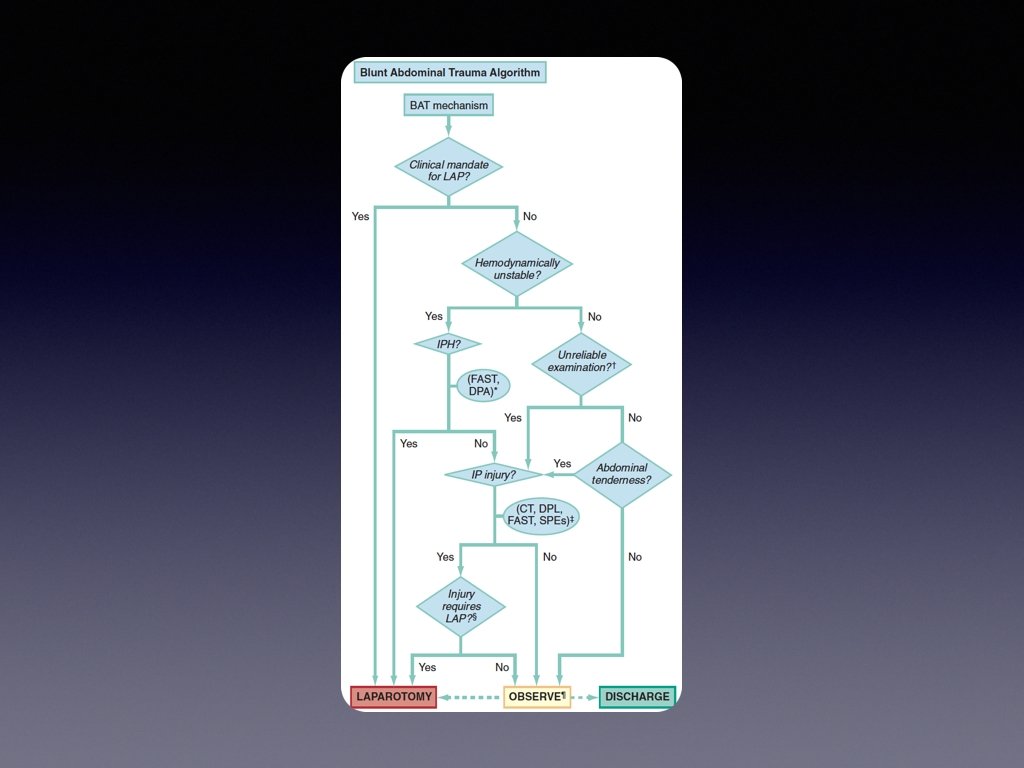
Management? Refer to approach to trauma lecture BUT WHEN TO TAKE THE PATIENT TO OR?
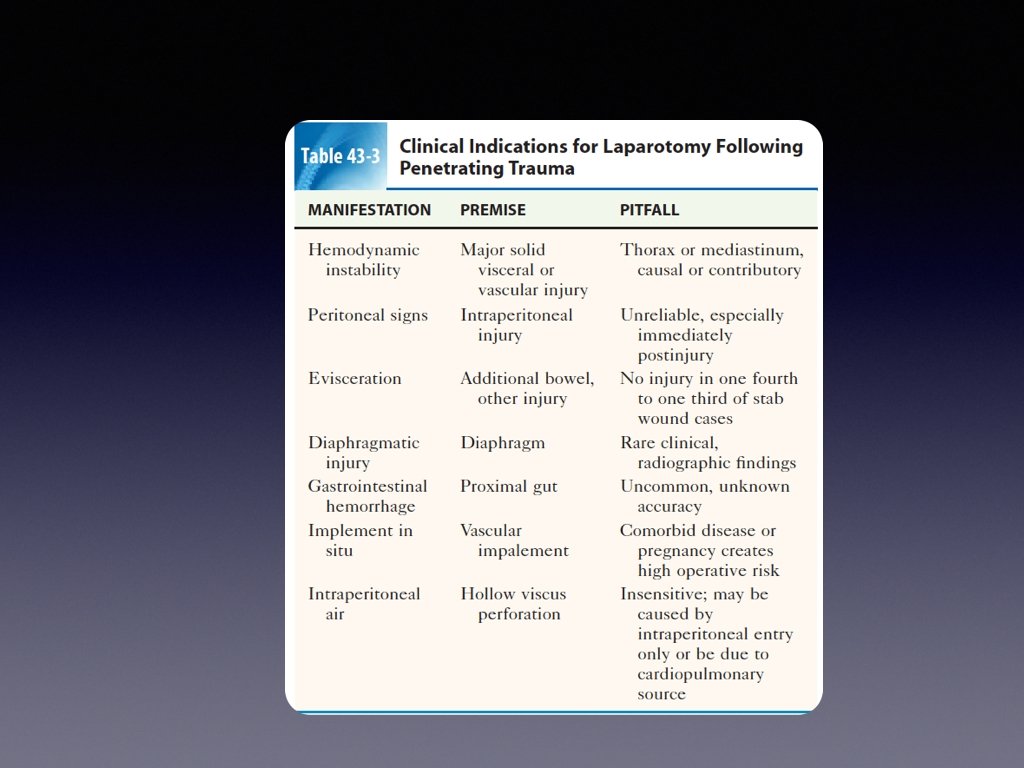
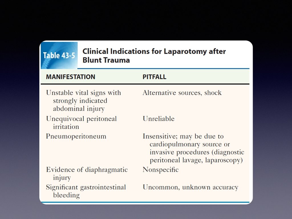
APPROACH
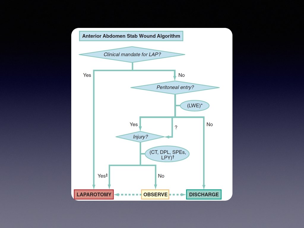
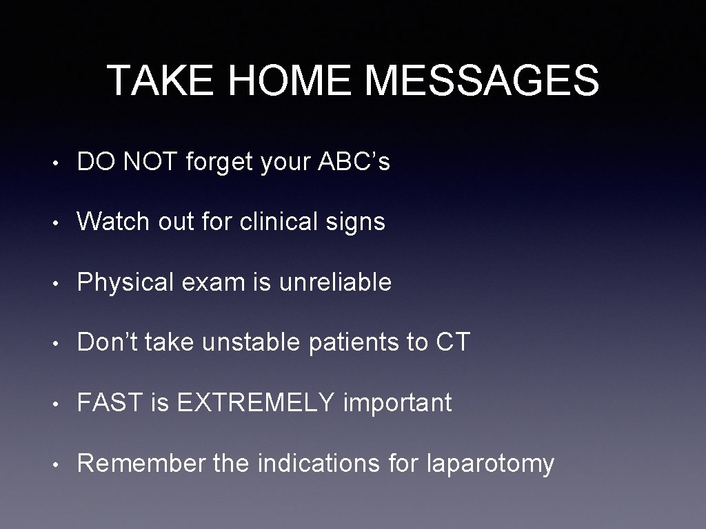
TAKE HOME MESSAGES • DO NOT forget your ABC’s • Watch out for clinical signs • Physical exam is unreliable • Don’t take unstable patients to CT • FAST is EXTREMELY important • Remember the indications for laparotomy

dr_aref@hotmail. com
- Slides: 22