
ABDOMEN The abdomen is the region of the
ABDOMEN
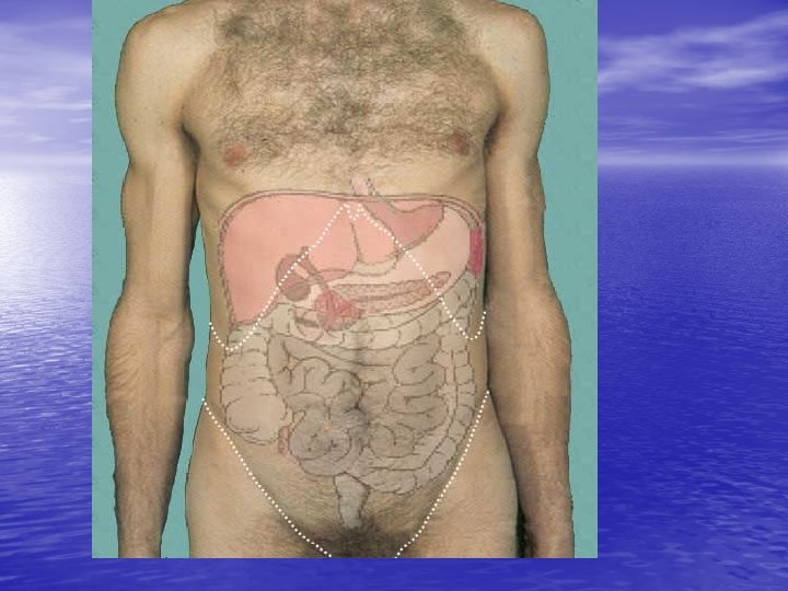
• The abdomen is the region of the trunk between the diaphragm above and the pelvic inlet below also known as superior pelvic aperture. So abdomen (proper) lies between thorax and pelvis • When a person is in supine position the upper limit of the abdomen is marked by an imaginary line that passes at the fifth intercostal space. This plane will also touch the upper surface of the diaphragm. The fifth intercostal space lies at the level of the 10 th or 11 th thoracic vertebrae
• abdomen has the abdominal wall, and the abdominal cavity. • The abdominal wall is constructed in such a way that it provides protection to the delicate visceral organs, blood vessels and nerves that lies in the abdominal cavity.
• Abdominal cavity is separated from thoracic cavity above by the diaphragm. And from pelvic cavity below by bony pelvis. • Behind is separated by lumbar vertebrae and intervertebral discs
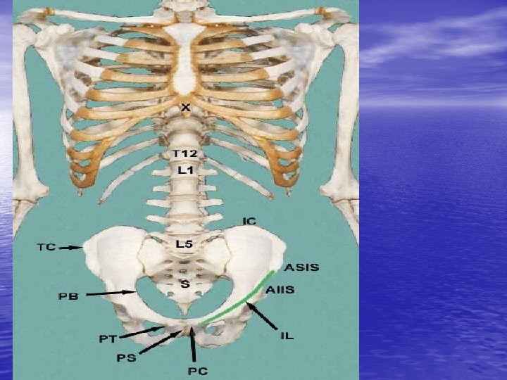
Skeleton of the Abdomen • The skeleton of the abdomen is represented in the adjacent figure. • Where we see the – Diaphragm – Five lumbar vertebral and their intervertebral discs – Bony pelvic
Lumbar vertebrae • There are 5 lumbar vertebrae that lie between the • • thoracic and sacral vertebrae. They are generally stronger, bodies and the vertebral foramina are generally kidney shaped and with a long transverse diameter. The spinous processes are short and pointing backward. Transverse processes are short, broad and thin. Lumbar vertebrae are frequently involved in back pain because they carry the more body weight and are subject to the largest forces and stresses along the spine.
• The inguinal ligament extends from the anterior superior iliac spine (ASIS) to the pubic tubercle (PT) and is used as one of the lower borders of the abdomen. • The thoracic diaphragm separates the abdominal cavity from the thoracic cavity

Abdominal Wall • The abdominal wall is split into anterior wall, • • right and left lateral walls and the posterior wall. For the purpose of description the anterior and lateral walls are described together using a combined term anterolateral wall. This is because the boundary between the two is not well defined and they contain nerves and muscles that are present in both areas.
A. The anterior abdominal wall • The anterior abdominal wall when viewed from the front is characterized by the presence of umbilicus (also called navel or belly button). This is a plucked scar that represents the former attachment site of the umbilical cord during fetal life
• i) Boundaries of the abdomen • Above: – The anterior abdominal wall is bounded above by the xiphoid process, the diverging left and right costal margins consisting of the costal cartilages of 7 th, 8 th, 9 th, and 10 th ribs and the 11 th and 12 th ribs. • Below: – on each side it is bounded by the iliac crest that ends in front at the anterior superior iliac spine, inguinal ligaments, pubic tubercle, pubic crest and pubic symphysis. The inguinal ligament (also called Poupart’s ligament) extends from the anterior superior iliac spine to the pubic tubercle.
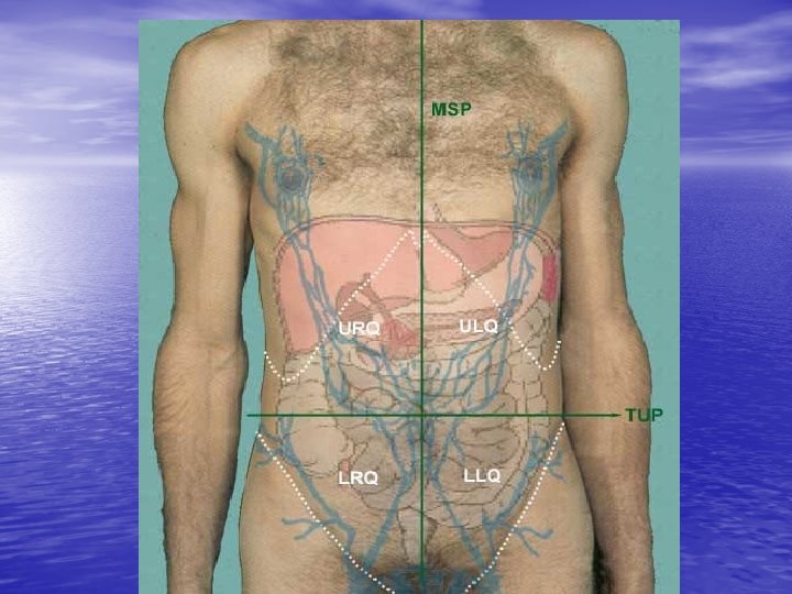
• To be able to visualize the relative positions of • abdominal organs as they lie within the abdomen, several different ways are available of subdividing the surface of the anterior abdominal wall. The easiest is to separate the surface into 4 quadrants: – – upper left quadrant ULQ lower left quadrant LLQ upper right quadrant URQ lower right quadrant LRQ • These quadrants are developed by dropping a vertical line down the middle of the sternum MSP and a horizontal line across and through the umbilicus TUP.
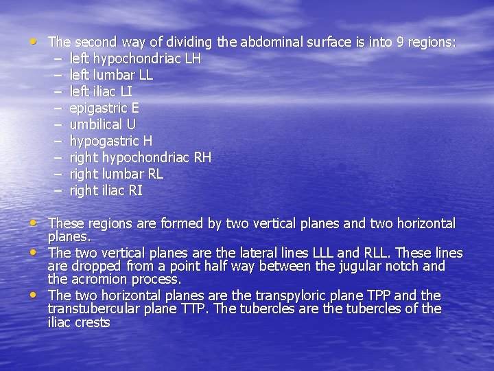
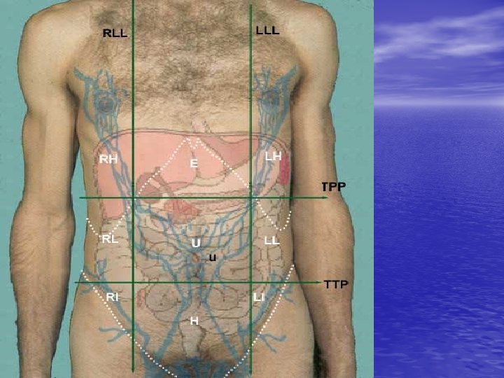
• The second way of dividing the abdominal surface is into 9 regions: – – – – – left hypochondriac LH left lumbar LL left iliac LI epigastric E umbilical U hypogastric H right hypochondriac RH right lumbar RL right iliac RI • These regions are formed by two vertical planes and two horizontal • • planes. The two vertical planes are the lateral lines LLL and RLL. These lines are dropped from a point half way between the jugular notch and the acromion process. The two horizontal planes are the transpyloric plane TPP and the transtubercular plane TTP. The tubercles are the tubercles of the iliac crests
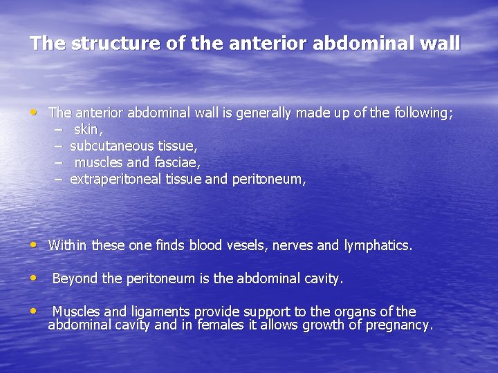
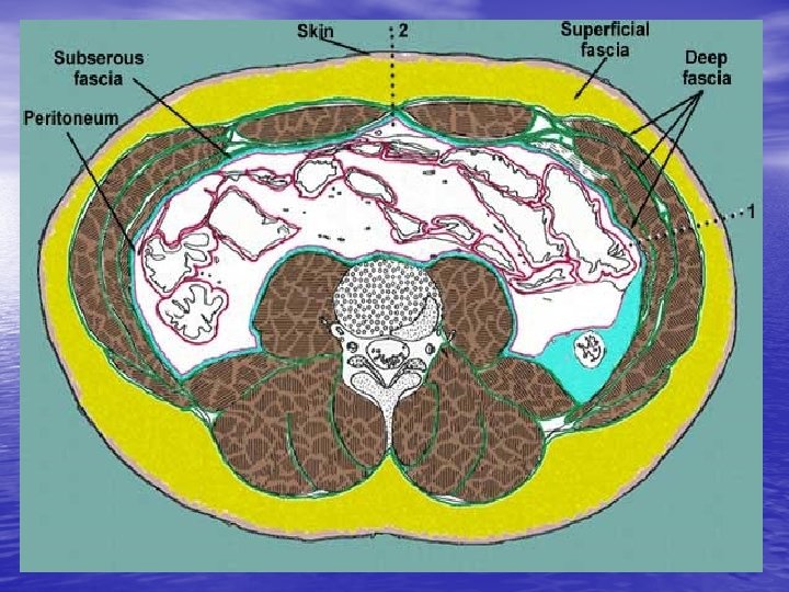
The structure of the anterior abdominal wall • The anterior abdominal wall is generally made up of the following; – – skin, subcutaneous tissue, muscles and fasciae, extraperitoneal tissue and peritoneum, • Within these one finds blood vesels, nerves and lymphatics. • Beyond the peritoneum is the abdominal cavity. • Muscles and ligaments provide support to the organs of the abdominal cavity and in females it allows growth of pregnancy.
• The muscles include the – external oblique and pyramidalis – internal oblique, – transversus abdominis muscles. – rectus abdominis • The sides of the abdomen are formed by the aponeurosis of three muscles, part of iliacus muscle and iliac bone.

Functions of anterolateral abdominal muscles Functions of the three flat muscles: • These muscles are the chief lateral flexors of the vertebral column; they also flex and extend the vertebral column. This is important in the maintenance of posture. • They support and protect the abdominal viscera • When they contract together with the diaphragm, they result into increase intrabdominal pressure, and this action provides the force required for defecation, micturation and parturition. • When contracting alternately with the diaphragm as antagonist help to bring about the expiratory act during forced breathing and are necessary to the abdominal form of respiration. • When one is lying down and head or leg are raised from the horizontal. They make trunk rigid, a requirement for pushing or lifting heavy objects

• Function of strap muscles: • The rectus abdominis muscle flexes the trunk against resistance • when one lies down it helps lifting of the chest and indirectly head. • It also depresses ribs and stabilizes the pelvis during walking.
Vessels and nerves of the anterior abdominal wall • i) The anterolateral abdominal wall is supplied by the spinal nerves. • The lower six thoracic nerves and the first lumbar nerves. • ii) The arteries of the anterior abdominal wall include the – superior and inferior epigastric arteries, intercostals and lumbar arteries, the deep circumflex iliac artery and three superficial inguinal arteries, which are the superficial eppigastric, external pudendal artery and superficial circumflex iliac artery
. • iii) The cutaneous veins drain in two directions as from the level of the umbilicus, upward to the thoracoepigastric and lateral thoracic veins and downwad to the great saphenous vein. – Subcutaneous veins around the umbilicus anastomose with the portal vein by way of branches along the ligament teres of the liver. • The lymph vessels drain upward into the axillary nodes and below to the superficial inguinal nodes.
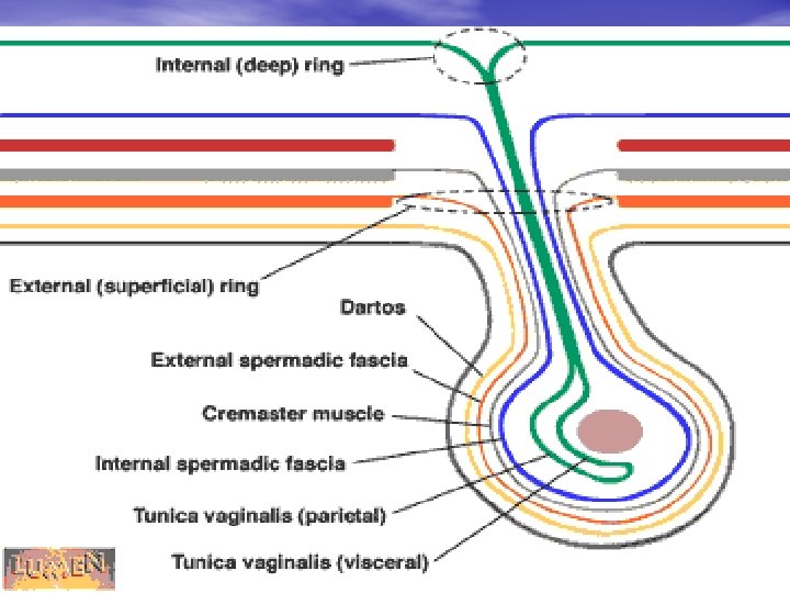
STRUCTURES IN THE ANTERIOR ABDOMINAL WALL • The abdomen must allow structures to pass from the abdominal cavity to enter the perineum and the lower limb. • This is possible because the lower part of the abdomen contain structures that allow communication between the abdomen and the perineum. • If the anterior abdominal wall was a closed region then it would have been difficult for the testes, lower limb and other structures in the pelvis and perineum to get blood and nerve supply. • During development structures develop in the lower abdomen to allow such communication. These structures include the inguinal ligament, deep and superficial inguinal rings, the inguinal canal and the spermatic cord.

• i) The Inguinal ligament: – The inguinal ligament is a thick folded lower free edge of the aponeurosis of the external oblique muscle. – It extends from the anterior superior iliac spine to the pubic tubercle and the skin crease at the junction of the thigh and abdomen lies just below and parallel to the inguinal ligament. – In adult it is about 12 -14 cm long and is inclined 4035 degrees to the horizontal. – The inguinal ligament bridges the muscles, vessels and nerves passing from the abdomen to the front of the thigh. Its medial end supports the spermatic cord.

• ii) The deep inguinal ring: – In order for structures to pass to and from the abdomen to the perineum there must be an opening at the innermost layer of the abdominal wall. – This opening is called the deep inguinal ring and is in the fascia transversalis. – It lies 1. 25 cm above the midinguinal (halfway between anterior superior iliac spine and symphysis pubis) point lateral to the origin of the inferior epigastric artery. – It is larger in male than in female and it allows passage of the spermatic cord in male and round ligament in female to enter the inguinal canal.

• iii) The inguinal canal: – The inguinal canal is an oblique passage at the anterior abdominal wall that extends from the deep inguinal ring to the superficial inguinal ring. – It is about 3 -5 cm and it lies above and parallel to the medial half of the inguinal ligament. – Its anterior wall is formed by the skin, subcutaneous tissue and the external oblique aponeurosis, – posterior wall is formed by fascia transversalis and between these two are the internal oblique and the transversus abdominis muscle. – Above is the internal oblique and transversus, below union of transversalis fascia and inguinal ligament. Knowledge into the structures in the wall is important in understanding the strength i. e. contents are kept from herniating.
• ➫The contraction of external oblique • • • approximate the anterior wall to the posterior wall. ➫During standing there is continuous contraction of internal oblique and transversus in the inguinal region. ➫During straining (coughing etc) there is contraction of internal oblique and transversus abdominis muscle, this is sphincteric action. The inguinal canal contains the spermatic cord, ilioinguinal nerve as it travels to the perineum through the superficial inguinal ring.
• iv) The superificial inguinal ring: – The superficial inguinal ring is a triangular opening at the aponeurosis of the external oblique that allow passage of the spermatic cord in male or round ligament of the uterus in female from the inguinal canal to the perineum (i. e. scrotum and labia majora in female) and ilio-inguinal nerve. – It lies 1 cm above and lateral to the pubic tubercle and it is generally smaller in the female.
• v) The spermatic cord: – The spermatic cord consists of structures running to and from the testes, – These structures assemble at the deep inguinal ring, passing through the inguinal canal and then descends in the scrotum to the testes. – The contents of the spermatic cord include the ductus deferens, three arteries (testicular, artery to the ductus deferens, and cremasteric artery), testicular vein, nerves (sympathetic fibers to the arteries, sympathetic and parasympathetic fibers on ductus deferens, the genital branch of genitofemoral nerve) lymph vessels and the remnants of procesus vaginalis. – In female the equivalent is the round ligament, it also passes through the inguinal canal and merges with subcutaneous tissue of the labia.
Posterior abdominal wall • It is bounded above by the 12 th rib and below • by the pelvic brim and at the midline it is supported by the bodies, intervetebral discs and transverse processes of the five lumbar vertebrae. The muscles of the posterior abdominal wall include – the psoas major, psoas minor, quadratus lumborum, iliacus, part of the diaphragm and also part of the transversus abdominis muscle. Psoas and iliacus are commonly referred to as iliopsoas. • All muscles are covered by the fascia.
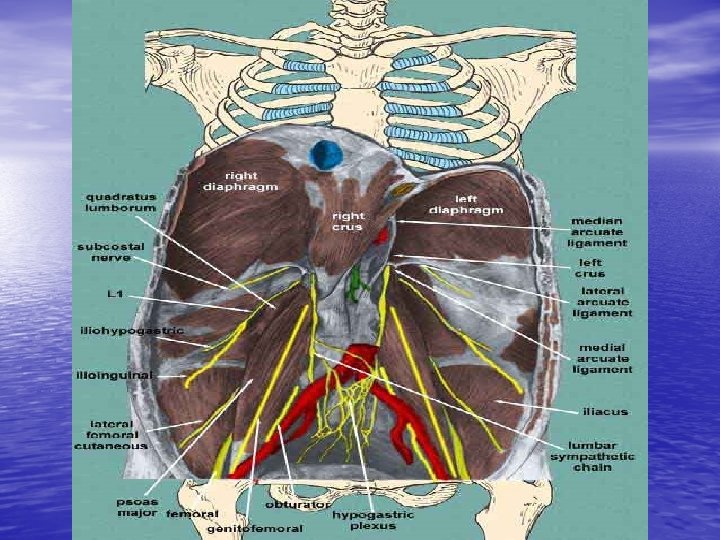

Nerves of the posterior abdominal wall: • The posterior abdominal wall contains two types of nerves; – the somatic nerves of lumbar plexus and visceral or splanchnic nerves of the autonomic nervous system, lumbosacral trunk.
Blood vessels of the posterior abdominal wall • The major vein of the posterior abdominal wall is the inferior vena cava • The arteries of the posterior abdominal wall except the subcostal • • artery arise from the abdominal aorta. . Arteries of the posterior abdominal wall: a) The subcostal artery: The subcostal arteries arise from the thoracic aorta and ech is situated below the 12 th rib. • b) The abdominal aorta: The abdominal aorta is a continuation of • • the thoracic aorta that enters the abdomen by pasing behind the diaphragm at the level of thoracic vetebra 12. In the abdomen it lies opposite the vertebral L 1 -L 4 and it gives out branches and at the level of L 4 birfucates into right and left common iliac artery that supplies the pelvis and lower limb. The area of birfucation is approximately to the left and just below the umbilicus.

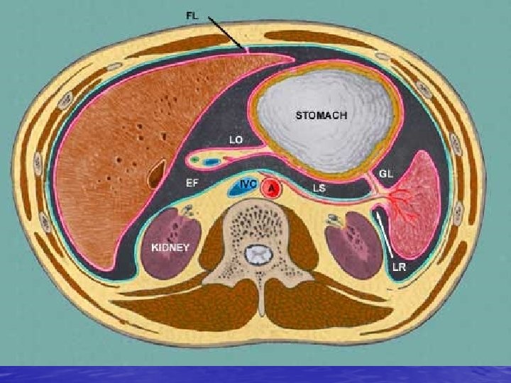
The abdominal cavity • The abdominal cavity is a space enclosed by the • • anterolateral and posterolateral abdominal walls. Above it is separated from the thoracic cavity by the diaphragm and below it is continuous with the pelvic cavity at the pelvic inlet or pelvic brim. The abdominal cavity is lined by the peritoneum and it lodges visceral organs, blood vessels and nerves that are bathed by serous fluid secreted by the peritoneum. .
– In man the peritoneal cavity is a closed sac but in female it communicates with the external environment via the uterine tubes, uterine cavity and vaginal orifice • Peritoneal folds or general arrangement of the peritoneum – The peritoneum forms certain folds and double layers, these are omentum, mesentery and peritoneal ligaments that perfom the main role of holding the abdominal viscera in certain position/location within the peritoneal cavity
• Functions of the peritoneum: – The peritoneum minimizes function by providing slippery surface that permits free movement of the abdominal viscera. – It acts against infection because it has phagocytic cells and it also localize infection and prevent spread of the infection. – The peritoneum also stores fat, greater omentum – Drug absorption, anaesthesize animals.
Contents of the abdominal cavity • i) The abdominal cavity contains the viscera, blood vessels, lymphatics, nerves and lymphnodes. The abdominal viscera: The abdominal viscera include the esophagus, stomach, small intestines, large intestines, liver, gall bladder, spleen, pancreas, kidneys ureters and suprarenal glands
DIAPHRAGM • Is a muscular and tendinous septum that separates the abdominal and thoracic cavity. • It pierced by structures that pass between thorax and abdomen • It’s a primary muscle of respiration • Is dome shaped

• Openings of the Diaphragm • The diaphragm has several structures passing through it and these openings are found at different vertebral levels as follows: • opening for inferior vena cava - T 8 • esophageal hiatus - T 10 • aortic hiatus - T 12
• Aortic opening transmits aorta, thoracic duct and azygos vein • Esophageal opening transmits esophagus, right and left vagus nerves, esophageal branches of left gastric vessels and lymphatics • Caval opening transmits inferior vena cava and terminal branches of right phrenic nerve
Functions • Muscle of inspiration • Muscle of abdominal straining e. g. In micturition, defecation, parturition • Weight lifting muscle
- Slides: 45