
AB balance Acidosis Alkalosis log H p H
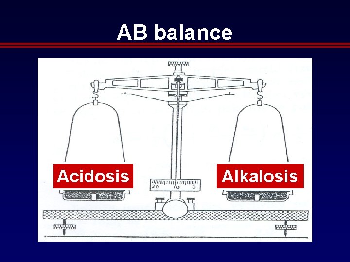
AB balance Acidosis Alkalosis
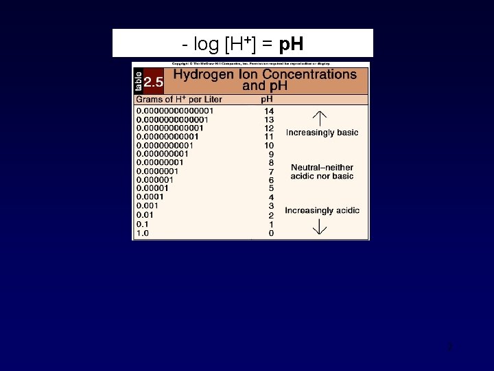
log [H+] = p. H 2
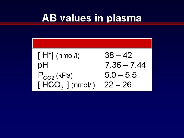
AB values in plasma [ H+] (nmol/l) 38 – 42 p. H 7. 36 – 7. 44 PCO 2 (k. Pa) 5. 0 – 5. 5 [ HCO 3 ] (nmol/l) 22 – 26
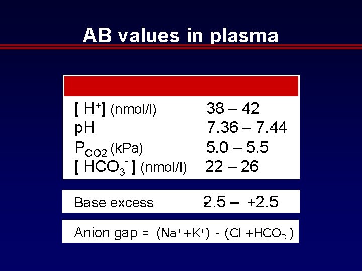
AB values in plasma [ H+] (nmol/l) 38 – 42 p. H 7. 36 – 7. 44 PCO 2 (k. Pa) 5. 0 – 5. 5 [ HCO 3 ] (nmol/l) 22 – 26 Base excess 2. 5 – +2. 5 Anion gap = (Na++K+) - (Cl +HCO 3 )
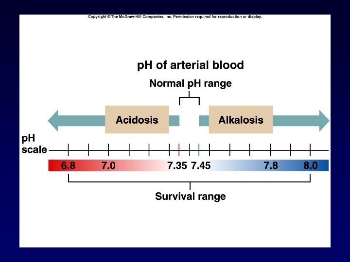
5
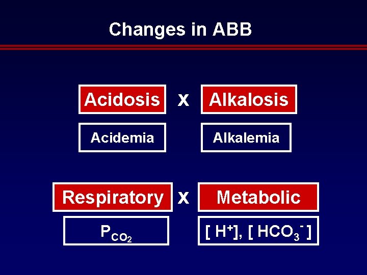
Changes in ABB Acidosis x Alkalosis Acidemia Respiratory PCO 2 Alkalemia x Metabolic [ H+], - [ HCO 3 ]
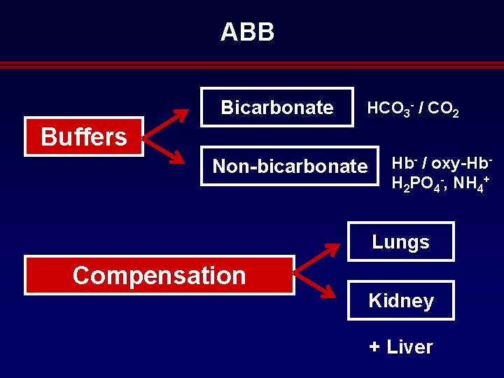
ABB Bicarbonate HCO 3 - / CO 2 Buffers Non-bicarbonate Hb- / oxy-Hb. H 2 PO 4 -, NH 4+ Lungs Compensation Kidney + Liver
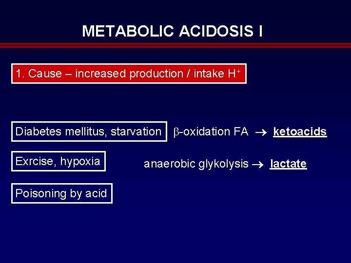
METABOLIC ACIDOSIS I 1. Cause – increased production / intake H+ Diabetes mellitus, starvation oxidation FA ketoacids Exrcise, hypoxia Poisoning by acid anaerobic glykolysis lactate
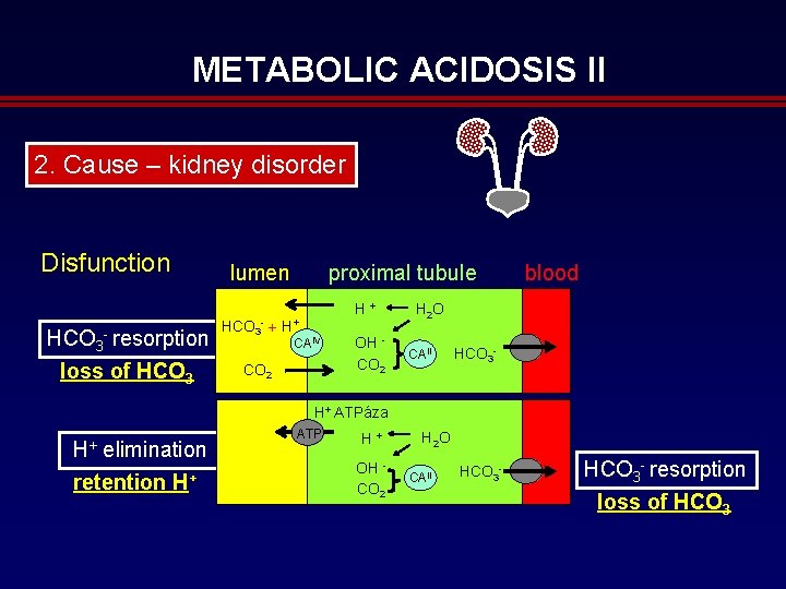
METABOLIC ACIDOSIS II 2. Cause – kidney disorder Disfunction lumen HCO 3 resorption loss of HCO 3 + proximal tubule H+ CAIV CO 2 H + H 2 O OH CO 2 CAII blood HCO 3 H+ ATPáza H+ elimination retention H+ ATP H + OH CO 2 H 2 O CAII HCO 3 resorption loss of HCO 3
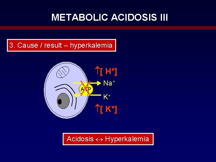
METABOLIC ACIDOSIS III 3. Cause / result – hyperkalemia [ H+] ATP Na+ K+ [ K+] Acidosis Hyperkalemia
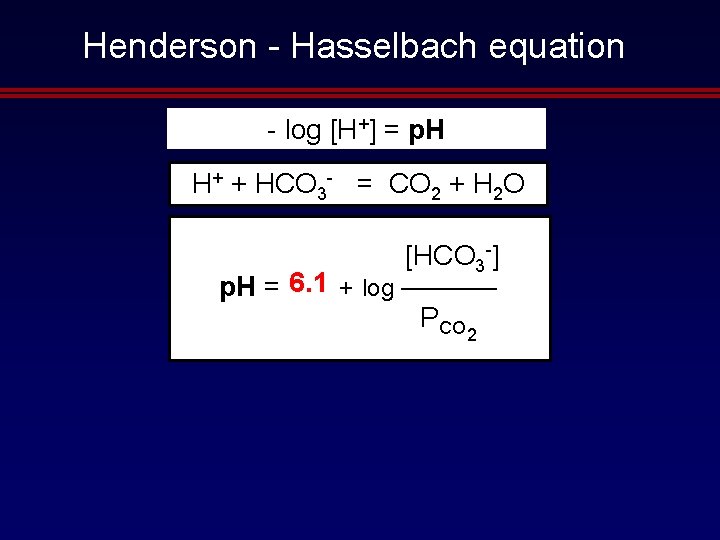
Henderson Hasselbach equation log [H+] = p. H H+ + HCO 3 = CO 2 + H 2 O [HCO 3 ] 6. 1 a + log ———— p. H = p. K PCO 2
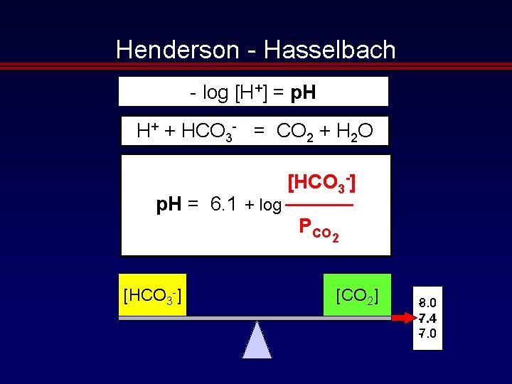
Henderson Hasselbach log [H+] = p. H H+ + HCO 3 = CO 2 + H 2 O [HCO 3 -] p. H = 6. 1 + log ———— PCO 2 [HCO 3 ] [CO 2] . 0 8 7. 4 7. 0
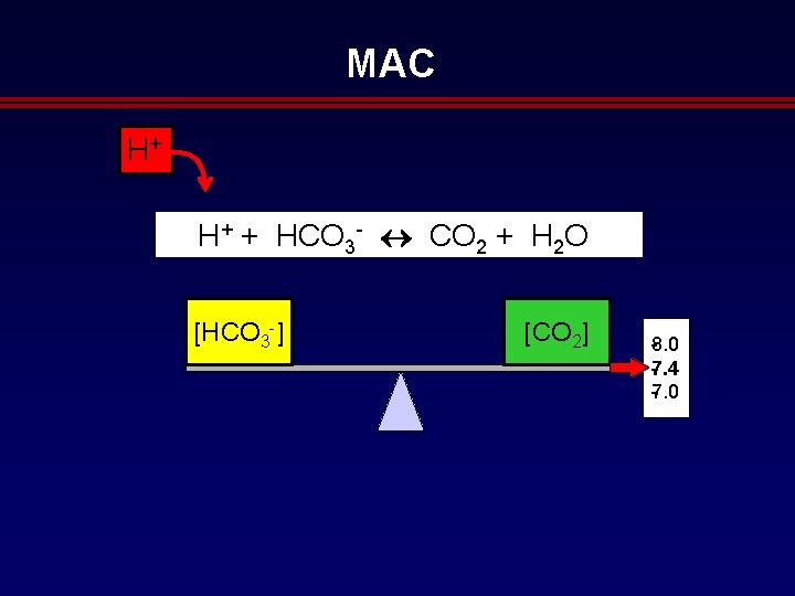
MAC H+ H+ + HCO 3 CO 2 + H 2 O [HCO 3 ] [CO 2] . 0 8 7. 4 7. 0
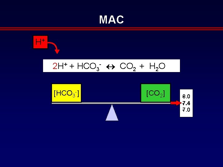
MAC H+ 2 H+ + HCO 3 CO 2 + H 2 O [HCO 3 ] [CO 2] . 0 8 7. 4 7. 0
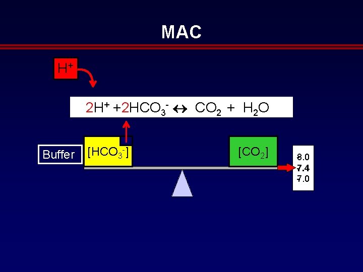
MAC H+ 2 H+ +2 HCO 3 CO 2 + H 2 O Buffer [HCO 3 ] [CO 2] . 0 8 7. 4 7. 0
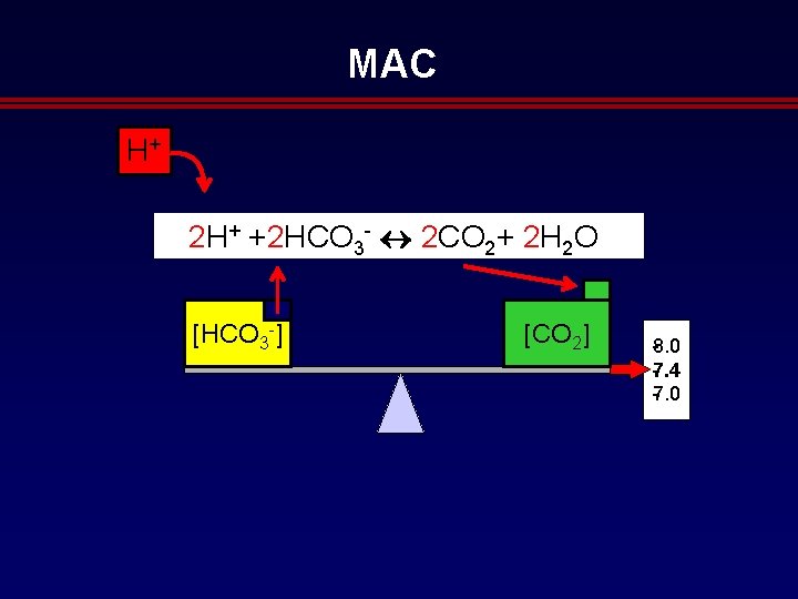
MAC H+ 2 H+ +2 HCO 3 2 CO 2+ 2 H 2 O [HCO 3 ] [CO 2] . 0 8 7. 4 7. 0
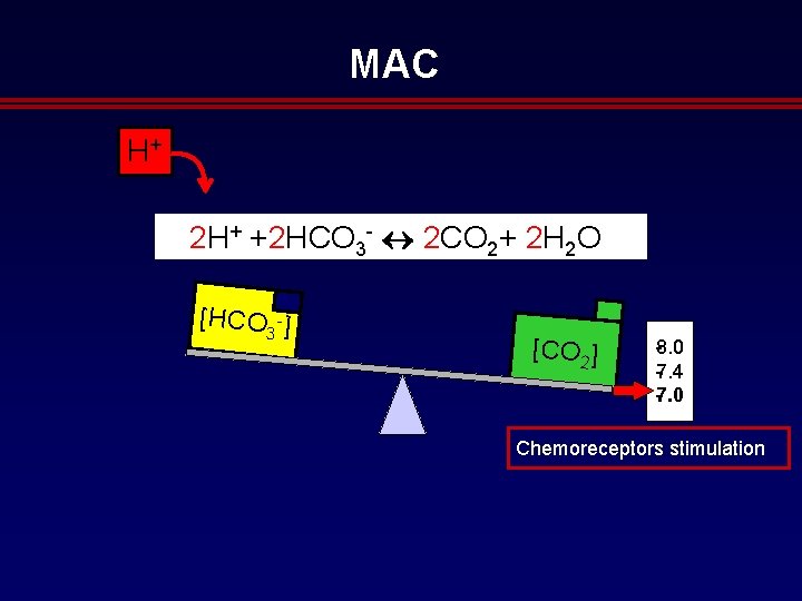
MAC H+ 2 H+ +2 HCO 3 2 CO 2+ 2 H 2 O [HCO ] 3 [CO 2] . 0 8 7. 4 7. 0 Chemoreceptors stimulation
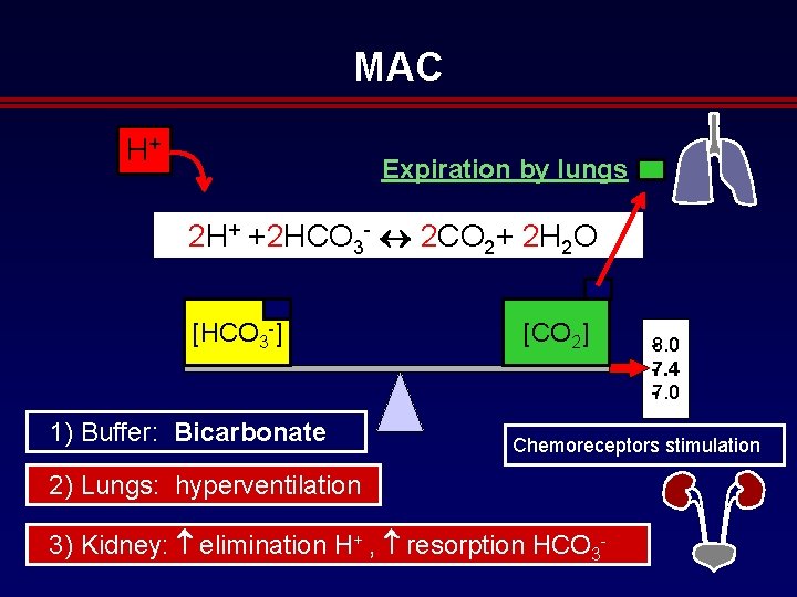
MAC H+ Expiration by lungs 2 H+ +2 HCO 3 2 CO 2+ 2 H 2 O [HCO 3 ] 1) Buffer: Bicarbonate [CO 2] . 0 8 7. 4 7. 0 Chemoreceptors stimulation 2) Lungs: hyperventilation 3) Kidney: elimination H+ , resorption HCO 3
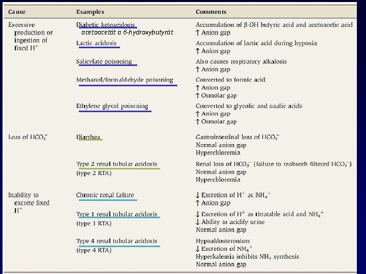
acetoacetát a β-hydroxybutyrát Metabolic acidosis - summary
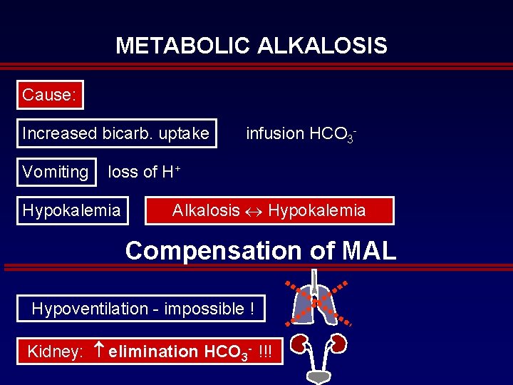
METABOLIC ALKALOSIS Cause: Increased bicarb. uptake Vomiting infusion HCO 3 loss of H+ Hypokalemia Alkalosis Hypokalemia Compensation of MAL Hypoventilation impossible ! Kidney: elimination HCO 3 - !!!
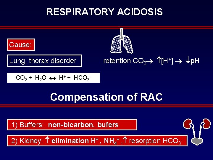
RESPIRATORY ACIDOSIS Cause: Lung, thorax disorder retention CO 2 [H+] p. H CO 2 + H 2 O H+ + HCO 3 Compensation of RAC 1) Buffers: non-bicarbon. bufers 2) Kidney: elimination H+ , NH 4+ , resorption HCO 3
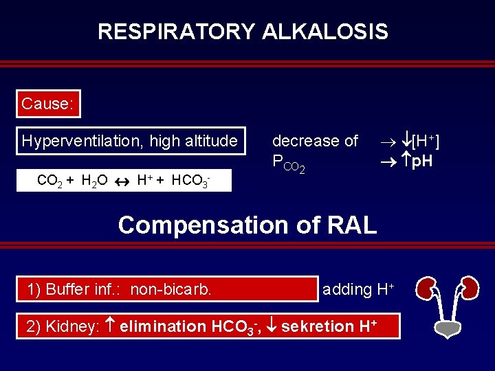
RESPIRATORY ALKALOSIS Cause: Hyperventilation, high altitude CO 2 + H 2 O H+ + HCO 3 decrease of PCO 2 ® [H+] p. H Compensation of RAL 1) Buffer inf. : non bicarb. adding H+ 2) Kidney: elimination HCO 3 -, sekretion H+

Questions
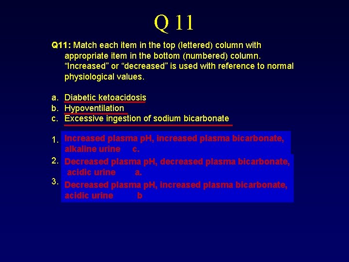
Q 11: Match each item in the top (lettered) column with appropriate item in the bottom (numbered) column. “Increased” or “decreased” is used with reference to normal physiological values. a. b. c. 1. Diabetic ketoacidosis Hypoventilation Excessive ingestion of sodium bicarbonate Increased plasma p. H, increased plasma bicarbonate, alkaline urine c. alkaline urine 2. Decreased plasma p. H, decreased plasma bicarbonate, acidic urine a. 3. Decreased plasma p. H, increased plasma bicarbonate, acidic urine b
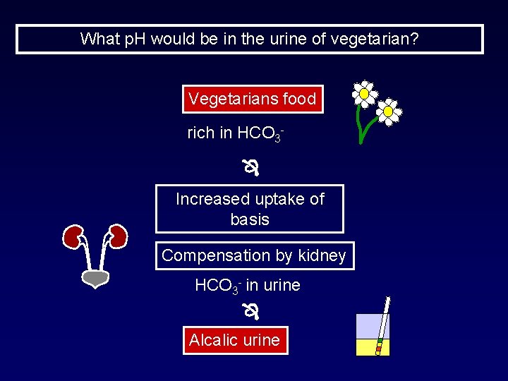
What p. H would be in the urine of vegetarian? Vegetarians food rich in HCO 3 Increased uptake of basis Compensation by kidney HCO 3 in urine Alcalic urine
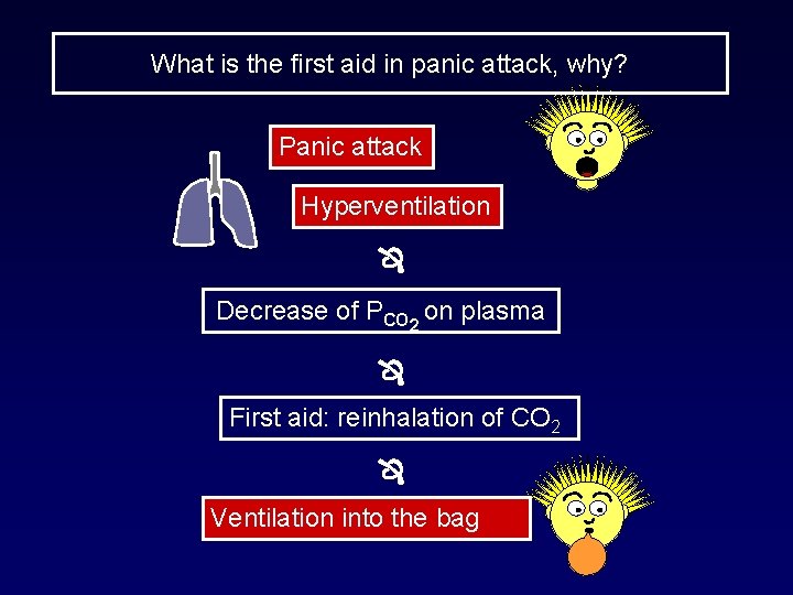
What is the first aid in panic attack, why? Panic attack Hyperventilation Decrease of PCO 2 on plasma First aid: reinhalation of CO 2 Ventilation into the bag
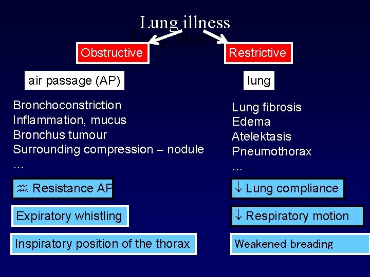
Lung illness Obstructive air passage (AP) Restrictive lung Bronchoconstriction Inflammation, mucus Bronchus tumour Surrounding compression – nodule … Lung fibrosis Edema Atelektasis Pneumothorax … Resistance AP Lung compliance Expiratory whistling Respiratory motion Inspiratory position of the thorax Weakened breading
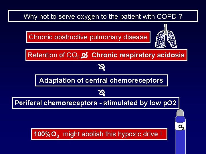
Why not to serve oxygen to the patient with COPD ? Chronic obstructive pulmonary disease Retention of CO 2 Chronic respiratory acidosis Adaptation of central chemoreceptors Periferal chemoreceptors - stimulated by low p. O 2 100%O 2 might abolish this hypoxic drive ! O 2
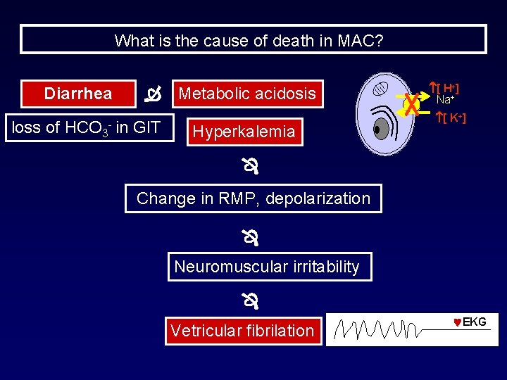
What is the cause of death in MAC? Diarrhea loss of HCO 3 Metabolic acidosis in GIT Hyperkalemia [ H+] Na+ [ K+] Change in RMP, depolarization Neuromuscular irritability Vetricular fibrilation EKG
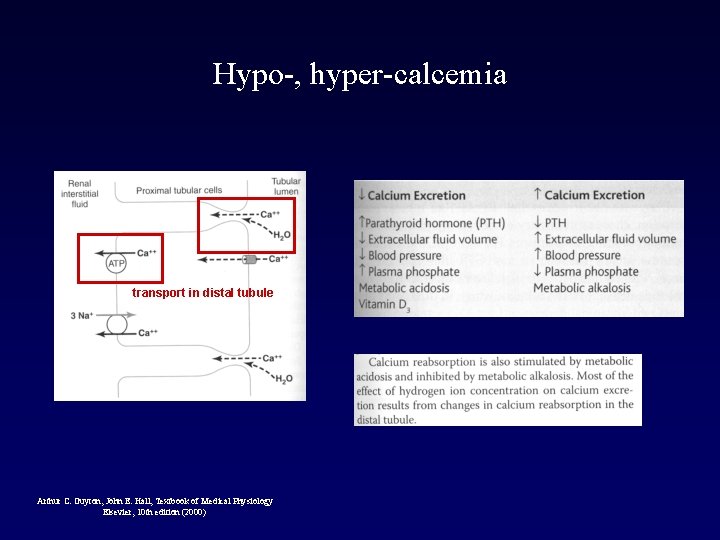
Hypo-, hyper-calcemia transport in distal tubule Arthur C. Guyton, John E. Hall, Textbook of Medical Physiology Elsevier, 10 th edition (2000)
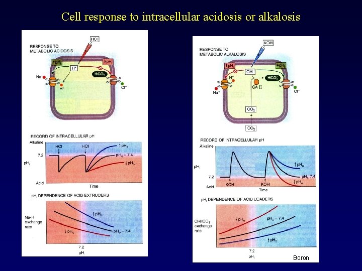
Cell response to intracellular acidosis or alkalosis Boron
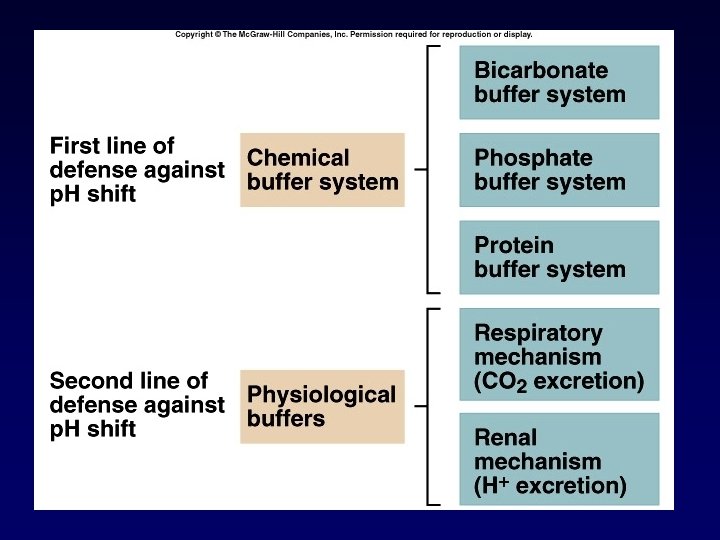
Buffer systems
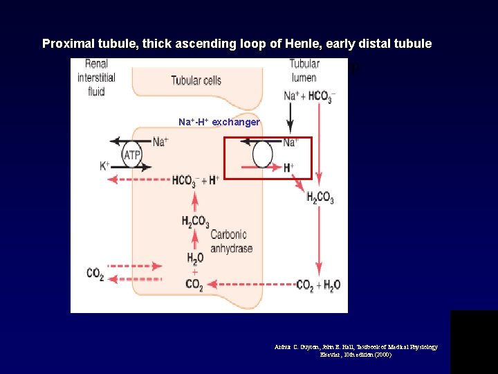
Proximal tubule, thick ascending loop of Henle, early distal tubule HCO 3 - proximal, ascending loop Na+-H+ exchanger Arthur C. Guyton, John E. Hall, Textbook of Medical Physiology Elsevier, 10 th edition (2000)
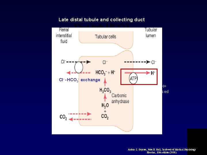
Late distal tubule and collecting duct Dist tub HCO 3 - Cl- - HCO 3 - exchange Urine H+ incerased 900 x Arthur C. Guyton, John E. Hall, Textbook of Medical Physiology Elsevier, 10 th edition (2000)
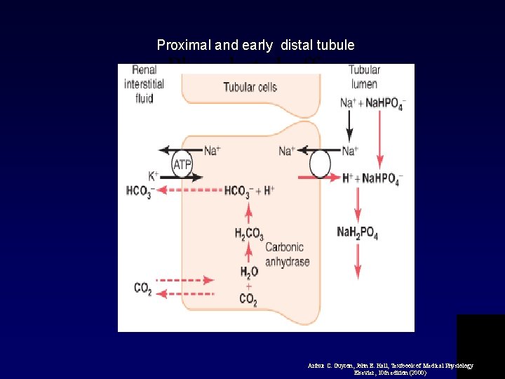
Proximal and early distal tubule Phosphate buffer Arthur C. Guyton, John E. Hall, Textbook of Medical Physiology Elsevier, 10 th edition (2000)
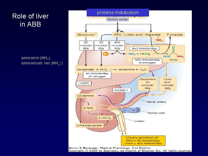
Role of liver in ABB ammonia (NH 3) ammonium ion (NH 4+) proteine metabolism
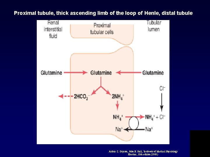
Proximal tubule, thick ascending limb of the loop of Henle, distal tubule NH 4+ proxi Arthur C. Guyton, John E. Hall, Textbook of Medical Physiology Elsevier, 10 th edition (2000)
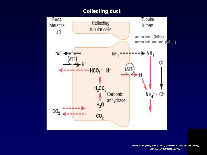
Collecting duct NH 4+ collect ammonia (NH 3) ammonium ion (NH 4+) Arthur C. Guyton, John E. Hall, Textbook of Medical Physiology Elsevier, 10 th edition (2000)
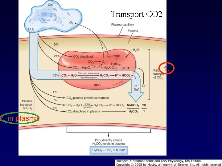
2. in plasma Transport CO 2 1.
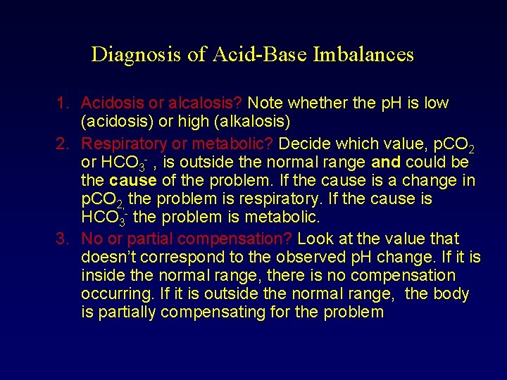
Diagnosis of Acid-Base Imbalances 1. Acidosis or alcalosis? Note whether the p. H is low (acidosis) or high (alkalosis) 2. Respiratory or metabolic? Decide which value, p. CO 2 or HCO 3 , is outside the normal range and could be the cause of the problem. If the cause is a change in p. CO 2, the problem is respiratory. If the cause is HCO 3 the problem is metabolic. 3. No or partial compensation? Look at the value that doesn’t correspond to the observed p. H change. If it is inside the normal range, there is no compensation occurring. If it is outside the normal range, the body is partially compensating for the problem
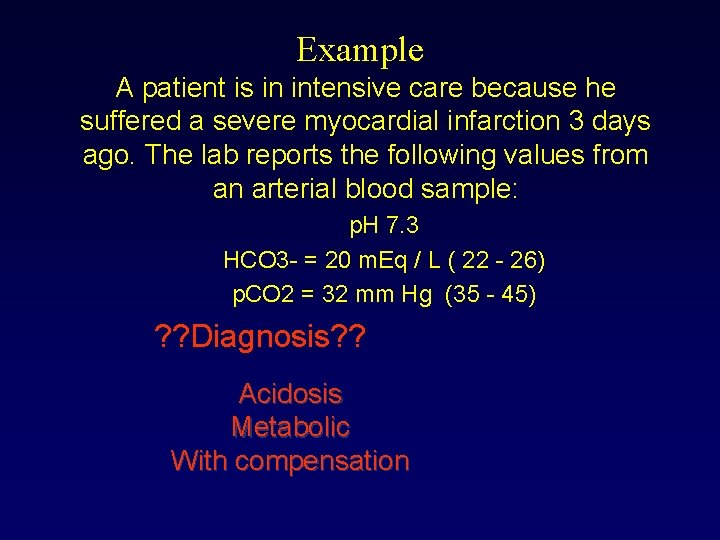
Example A patient is in intensive care because he suffered a severe myocardial infarction 3 days ago. The lab reports the following values from an arterial blood sample: p. H 7. 3 HCO 3 = 20 m. Eq / L ( 22 26) p. CO 2 = 32 mm Hg (35 45) ? ? Diagnosis? ? Acidosis Metabolic With compensation
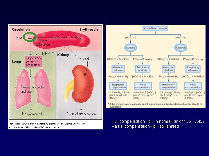
summary Full compensation p. H in normal rank (7. 35 7. 45) Partial compensation p. H still shifted
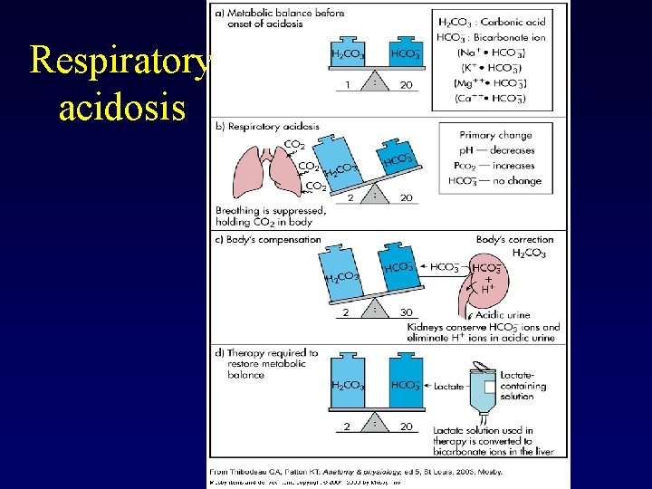
Respiratory acidosis
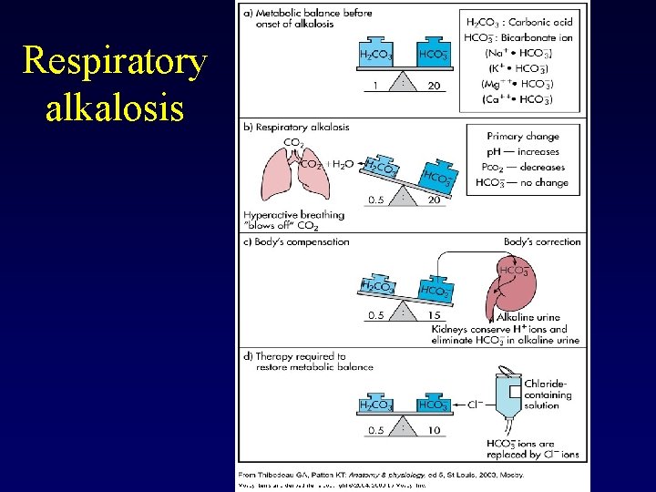
Respiratory alkalosis
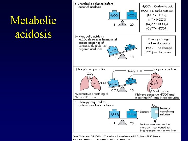
Metabolic acidosis
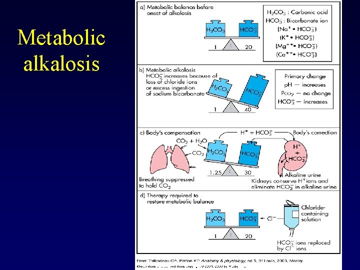
Metabolic alkalosis

- Slides: 47