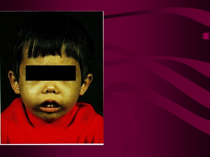
A one year old boy was brought by

• A one year old boy was brought by his mother in Child OPD MH with h/o Pallor, inability to feed and lethargy • O/E boy is markedly pallor TLC : 7. 8 x 10⁹/l Hb : 5 g/dl Plt : 290 x 109/l • Numerous circulating normoblasts seen. 1. What is your diagnosis 2. What investigations you will carry to confirm diagnosis 3. Outline management.
, MRCPath (London), FRCPath")
The Thalassemias Maj Gen Dr Muhammad Ayyub MBBS, Ph. D (London), MRCPath (London), FRCPath (UK) Prof & HOD AM College

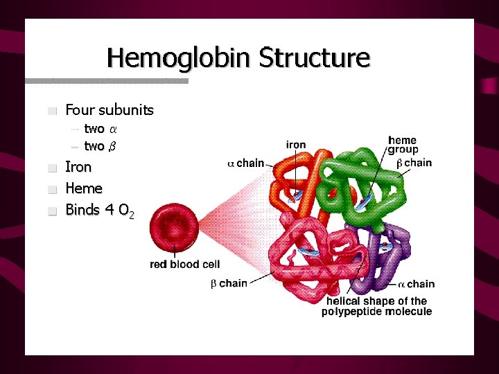
Hemoglobin structure heme α β β α

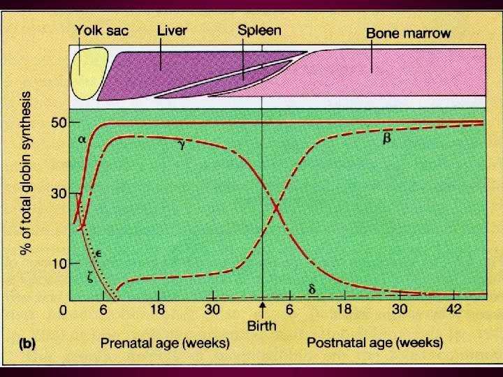
Hemoglobins in normal adults α β α γ α δ α Hb. A Hb. F Hb. A 2 98% ~1% <3. 5%

Introduction • Heritable, hypochromic anemias-varying degrees of severity • Genetic defects result in decreased or absent production of m. RNA and globin chain synthesis • At least 100 distinct mutations • High incidence in Asia, Africa, Mideast, and Mediterrenean countries

Hemoglobin Review • Each complex consists of : – Four polypeptide chains, non-covalently bound – Four heme complexes with iron bound – Four O 2 binding sites

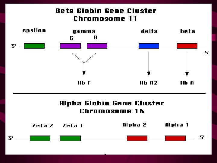
Globin Chains • Alpha Globin – 141 amino acids – Coded for on Chromosome 16 – Found in normal adult hemoglobin, A 1 and A 2 • Beta Globin – 146 amino acids – Coded for on Chromosome 11, found in Hgb A 1 • Delta Globin – Found in Hemoglobin A 2 --small amounts in all adults • Gamma Globin – Found in Fetal Hemoglobin • Zeta Globin – Found in embryonic hemoglobin

Hemoglobin Types • • Hemoglobin Type Globin Chains Hgb A 1— 92%----- a 2 b 2 Hgb A 2— 2. 5%---- a 2 d 2 Hgb F — <1%----- a 2 g 2 Hgb H --------- b 4 Bart’s Hgb------- g 4 glu val Hgb S---------- a 2 b 26 glu lys Hgb C---------- a 2 b 26

Genetics • Alpha globins are coded on chromosome 16 – Two genes on each chromosome – Four genes in each diploid cell – Gene deletions result in Alpha-Thalassemias • Also on chromosome 16 are Zeta globin genes— Gower’s hemoglobin (embryonic) • Beta globins are coded on chromosome 11 – One gene on each chromosome – Two genes in each diploid cell – Point mutations result in Beta-Thalassemias • Also on chromosome 11 are Delta (Hgb A 2) and Gamma (Hgb F) and Epsilon (Embryonic)

Beta Thalassemias • Result from Point Mutations on genes • Severity depends on where the hit(s) lie – b 0 -no b-globin synthesis; – b+ reduced synthesis • Disease results in an overproduction of aglobin chains, which precipitate in the cells and cause splenic sequestration of RBCs • Erythropoiesis increases, sometimes becomes extramedullary

b-Thal--Clinical • b-Thalassemia Minor – Minor point mutation – Minimal anemia; no treatment indicated • b-Thalassemia Intermedia – Homozygous minor point mutation or more severe heterozygote – Can be a spectrum; most often do not require chronic transfusions • b-Thalassemia Major-Cooley’s Anemia – Severe gene mutations – Need careful observation and intensive treatment

Beta Thalassemia Major • Reduced or nonexistent production of b-globin – Poor oxygen-carrying capacity of RBCs • Failure to thrive, poor brain development – Increased alpha globin production and precipitation • RBC precursors are destroyed within the marrow • Increased splenic destruction of dysfunctional RBCs – Anemia, jaundice, splenomegaly • Hyperplastic Bone Marrow – Ineffective erythropoiesis—RBC precursors destroyed • Poor bone growth, frontal bossing, bone pain – Increase in extramedullary erythropoiesis • Iron overload—increased absorption and transfusions – Endocrine disorders, Cardiomyopathy, Liver failure

b-Thalassemia Major—Lab findings • Hypochromic, microcytic anemia – Target Cells, nucleated RBCs, anisocytosis • Reticulocytosis • Hemoglobin electrophoresis shows – Increased Hgb A 2—delta globin production – Increased Hgb F—gamma globin production • Hyperbilirubinemia • LFT abnormalities (late finding) • TFT abnormalities, hyperglycemia (late endocrine findings)

b-Thalassemia Major--Treatment • Chronic Transfusion Therapy – Maximizes growth and development – Suppresses the patient’s own ineffective erythropoiesis and excessive dietary iron absorption – PRBC transfusions often monthly to maintain Hgb 10 -12 • Chelation Therapy – Binds free iron and reduces hemosiderin deposits – 8 -hour subcutaneous infusion of deferoxamine, 5 nights/week – Start after 1 year of chronic transfusions or ferritin>1000 ng/dl • Splenectomy--indications – Trasfusion requirements increase 50% in 6 mo – PRBCs per year >250 cc/kg – Severe leukopenia or thrombocytopenia

b-Thalassemia Major Complications and Emergencies • Sepsis—Encapsulated organisms – Strep Pneumo • Cardiomyopathy—presentation in CHF – Use diuretics, digoxin, and deferoxamine • Endocrinopathies—presentation in DKA – Take care during hydration so as not to precipitate CHF from fluid overload

Anticipatory Guidance and Follow Up • Immunizations—Hepatitis B, Pneumovax • Follow for signs of diabetes, hypothyroid, gonadotropin deficiency • Follow for signs of cardiomyopathy or CHF • Follow for signs of hepatic dysfunction • Osteoporosis prevention – Diet, exercise – Hormone supplementation – Osteoclast-inhibiting medications • Follow ferritin levels

On The Horizon • Oral Chelation Agents • Pharmacologically upregulating gamma globin synthesis, increasing Hgb F – Carries O 2 better than Hgb A 2 – Will help bind a globins and decrease precipitate • Bone Marrow transplant • Gene Therapy – Inserting healthy b genes into stem cells and transplanting

Alpha Thalassemias • Result from gene deletions • One deletion—Silent carrier; no clinical significance • Two deletions—a Thal trait; mild hypochromic microcytic anemia • Three deletions—Hgb H; variable severity, but less severe than Beta Thal Major • Four deletions—Bart’s Hgb; Hydrops Fetalis; In Utero or early neonatal death

Alpha Thalassemias • Usually no treatment indicated • 4 deletions incompatible with life • 3 or fewer deletions have only mild anemia
- Slides: 23