A nucleic acid is a macromolecule composed of

")

 programs cells Bacteriophages Martha Chase")

![Chargaff’s Rules: [A] = [T] [G] = [C] [A] + [G] = [T] +](https://slidetodoc.com/presentation_image_h2/5cfdf3a4493d3131cfeb43056095ad45/image-6.jpg "Chargaff’s Rules: [A] = [T] [G] = [C] [A] + [G] = [T] +")


 • Made up of nucleotides (DNA molecule) in a DNA double")


 2. Guanine (G) • PYRIMIDINES 3. Thymine")
 Thymine (T) A=T Guanine (G) Cytosine (C)")


 = Thymine (30%) Guanine (20%)")


 2. m. RNA 3. r.")


- number of small RNA molecules found")
- Slides: 42
• A nucleic acid is a macromolecule composed of nucleotide chains. • These molecules carry genetic information or form structures within cells. • The most common nucleic acids are • deoxyribonucleic acid (DNA) and ribonucleic acid (RNA). . • Artificial nucleic acids include 1. peptide nucleic acid (PNA), 2. Morpholino 3. locked nucleic acid (LNA), 4. Glycol nucleic acid (GNA) and threose nucleic acid (TNA).
DNA as Genetic Material • established by several critical experiments – Fred Griffith (1928) – Oswald T. Avery, C. M. Mac. Leod, and M. J. Mc. Carty (1944) – Alfred D. Hershey and Martha Chase (1952)
1944 - Avery, Mac. Leod & Mc. Carty Purified DNA as transforming factor • Work not well-received • Protein more complex & better able to store information Oswald Avery Colin Mac. Leod Maclyn Mc. Carty
1952 - Hershey & Chase Viral DNA (not protein) programs cells Bacteriophages Martha Chase & Alfred Hershey
DNA • Discovery of the DNA double helix A. 1950’s Chargaff’s Rule B. Rosalind Franklin - X-ray photo of DNA. C. Watson and Crick - described the DNA molecule from Franklin’s X-ray.
Chargaff’s Rules: [A] = [T] [G] = [C] [A] + [G] = [T] + [C] purines pyrimidines
Chargaff’s Rule • Adenine must pair with Thymine • Guanine must pair with Cytosine • Their amounts in a given DNA molecule will be about the same T A G C
X-ray Photograph of DNA The periodicity of the arcs on this photograph indicated that: 1) DNA had a regular helical structure. 2) There are regular repeats every 3. 4 and 34 Angstroms
DNA structure and Replication Chapter 16
The Structure of DNA • Four features summarize the molecular architecture of DNA: – The DNA molecule is a doublestranded helix. – The diameter of the DNA molecule is uniform. – The twist in DNA is right-handed. – The two strands run in different directions (they are antiparallel).
The Structure of DNA • The sugar–phosphate backbones of each strand coil around the outside of the helix. • The nitrogenous bases point toward the center of the helix. • Hydrogen bonds between complementary bases hold the two strands together. • A always pairs with T (two hydrogen bonds). • G always pairs with C (three hydrogen bonds).
WATSON AND CRICK MODEL 1. A double helix with regular dimensions. Two antiparallel chains. 2. The sugar phosphate backbone is out; the bases are inside the helix. 3. The helix is right handed. 4. MAJOR POINT: The two chains are held together by hydrogen bonds.
The Double Helix
Space Filling Model
II. The sugar phosphate backbone is outside; III. the bases are inside.
Major forces holding the helix together: 1. base stacking forces 2. hydrophobic interactions among the bases 3. hydrogen bonds
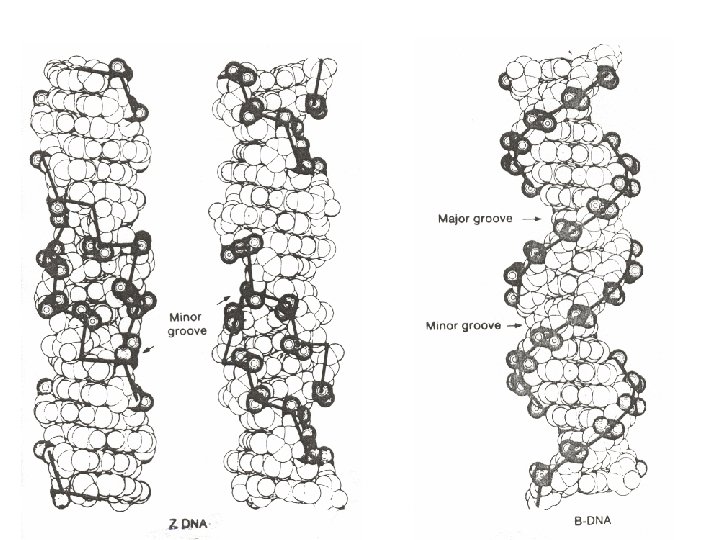
III. The helix is generally right handed. However left handed Z-DNA exists.
Different forms of DNA Parameters Direction of helical rotation A Form Right B Form Right Z-Form Left Residues per turn of helix Rotation of helix per residue (in degrees) 11 10 33 36 12 base pairs -30 Base tilt relative to helix axis (in degrees) 20 6 7
Different forms of DNA Parameters A Form B Form Z-Form Major groove narrow and deep wide and deep Flat Minor groove wide and shallow narrow and deep Anti for Py, Syn for Pu most prevalent within cells occurs in stretches of alternating purinepyrimidine base pairs Orientation of Nglycosidic Bond Comments
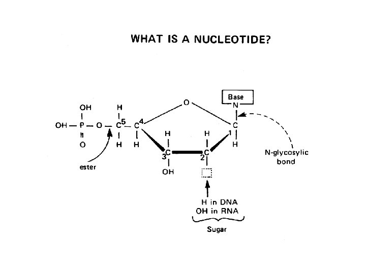
Deoxyribonucleic Acid (DNA) • Made up of nucleotides (DNA molecule) in a DNA double helix. • Nucleotide: 1. Phosphate group 2. 5 -carbon sugar 3. Nitrogenous base • ~2 nm wide
Phosphate Group O O=P-O O DNA Nucleotide 5 CH 2 O N C 1 C 4 Sugar (deoxyribose) C 3 C 2 Nitrogenous base (A, G, C, or T)
DNA Double Helix 5 O 3 3 P 5 O O C G 1 P 5 3 2 4 4 2 3 P 1 T 5 A P 3 O O P 5 O 3 5 P
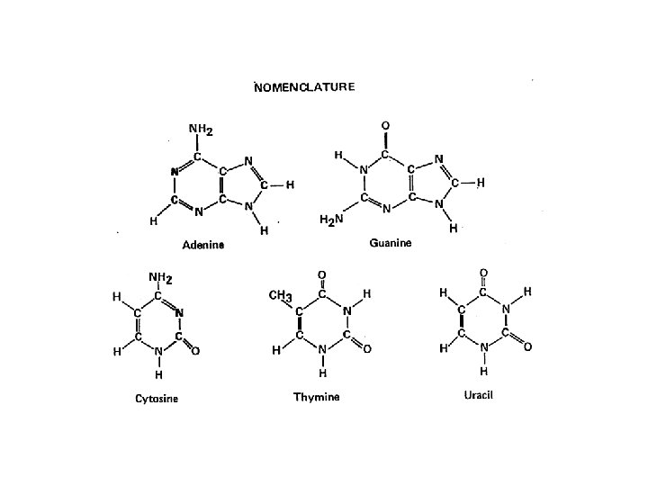
Nitrogenous Bases • PURINES 1. Adenine (A) 2. Guanine (G) • PYRIMIDINES 3. Thymine (T) 4. Cytosine (C) A or G T or C
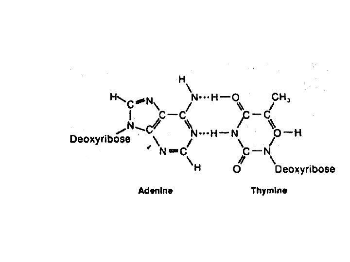
BASE-PAIRINGS Purines Pyrimidines Base Pairs Adenine (A) Thymine (T) A=T Guanine (G) Cytosine (C) C G # of H-Bonds 2 3
H-bonds G C T A
Question: • If there is 30% Adenine, Adenine how much Cytosine is present?
• There would be 20% Cytosine Adenine (30%) = Thymine (30%) Guanine (20%) = Cytosine (20%) (50%) = (50%)
Figure 11. 7 Base Pairing in DNA Is Complementary
• The genetic material performs four important functions: – It stores all of an organism’s genetic information. – It must be precisely replicated in the cell division cycle. – It is expressed as the phenotype. – It is susceptible to mutation.
Watson & Crick predicted that each DNA strand could serve as a template for the replication of a new strand Q: What is the mode of replication?
RNA
4 types of RNA 1. t. RNA (transfer RNA) 2. m. RNA 3. r. RNA 4. sn. RNA
t. RNA There are 4 arms and 3 loops. The acceptor, D, T pseudouridine C and anticodon arms, D, T pseudouridine C and anticodon loops. Sometimes t. RNA molecules have an extra or variable loop (this is shown in yellow in the adjacent figure).
m. RNA
r RNA
ribosomal RNA is found in the ribosomes. Prokaryotic ribosomes have 3 r. RNA molecules: 23 S, 16 S and 5 S. Eukaryotic ribosomes have 4 r. RNA molecules: 28 S, 18 S, 5. 8 S and 5 S.
sn. RNA • Small nuclear RNA (sn. RNA)- number of small RNA molecules found in the nucleus. • These RNA molecules are important RNA splicing (removal of the introns from hn. RNA) and maintenance of the telomeres, or chromosome ends. • They are always found associated with specific proteins and the complexes are referred to as small nuclear ribonucleoproteins (SNRNP) or sometimes as snurps. • Antibodies against snurps are found in a number of autoimmune diseases.