
A new approach to the diagnosis of cervical
- Slides: 20
“A new approach to the diagnosis of cervical, oesophageal and prostate cancer based on a combination of infrared and terahertz techniques. ” Peter Weightman Physics Department, University of Liverpool.
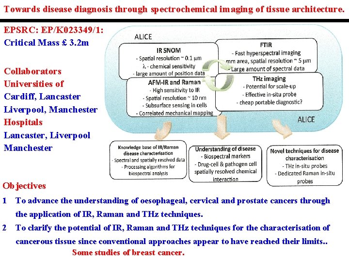
Towards disease diagnosis through spectrochemical imaging of tissue architecture. EPSRC: EP/K 023349/1: Critical Mass £ 3. 2 m Collaborators Universities of Cardiff, Lancaster Liverpool, Manchester Hospitals Lancaster, Liverpool Manchester Objectives 1 To advance the understanding of oesophageal, cervical and prostate cancers through the application of IR, Raman and THz techniques. 2 To clarify the potential of IR, Raman and THz techniques for the characterisation of cancerous tissue since conventional approaches appear to have reached their limits. . Some studies of breast cancer.
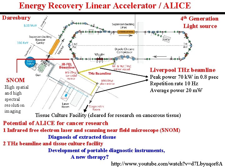
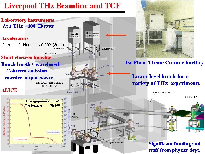
Energy Recovery Linear Accelerator / ALICE Daresbury 4 th Generation Light source Liverpool THz beamline Peak power 70 k. W in 0. 8 psec Repetition rate 10 Hz Average power 20 m. W SNOM High spatial and high spectral resolution imaging Tissue Culture Facility (cleared for research on cancerous tissue) Potential of ALICE for cancer research 1 Infrared free electron laser and scanning near field microscope (SNOM) Diagnosis of extracted tissue 2 THz beamline and tissue culture facility Development of portable diagnostic instruments, A new therapy? http: //www. youtube. com/watch? v=d 7 Lbyuqor 8 A
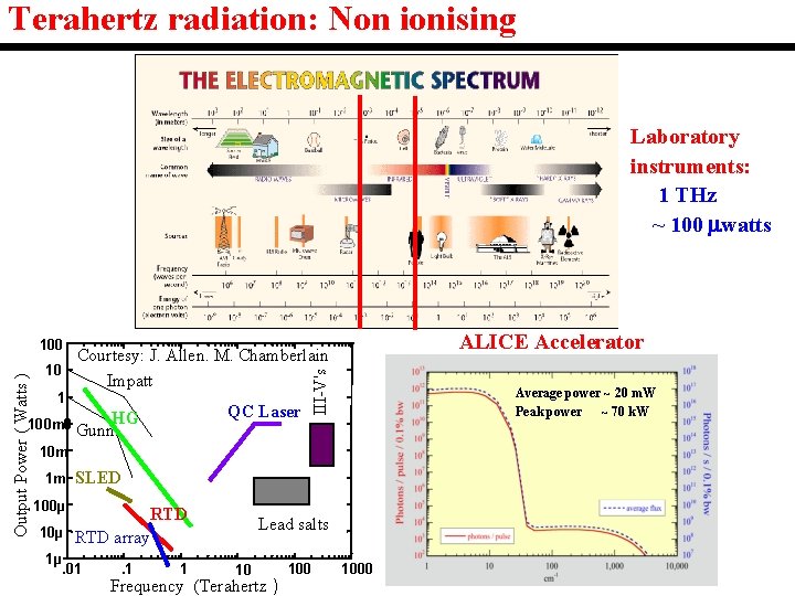
Terahertz radiation: Non ionising Laboratory instruments: 1 THz ~ 100 watts ALICE Accelerator Courtesy: J. Allen. M. Chamberlain 10 Impatt 1 QC Laser 100 m 0 Gunn. HG III-V's Output Power ( Watts ) 100 Average power ~ 20 m. W Peak power ~ 70 k. W 10 m 1 m SLED 100µ RTD 10µ RTD array 1µ . 01 . 1 1 Lead salts 10 Frequency (Terahertz ) 1000
. . Liverpool THz Beamline and TCF Laboratory instruments At 1 THz ~ 100 �watts Accelerators Carr et. al. Nature 420 153 (2002) Short electron bunches Bunch length < wavelength Coherent emission massive output power 1 st Floor Tissue Culture Facility Lower level hutch for a variety of THz experiments ALICE Average power ~ 20 m. W Peak power ~ 70 k. W Significant funding and staff from physics dept.
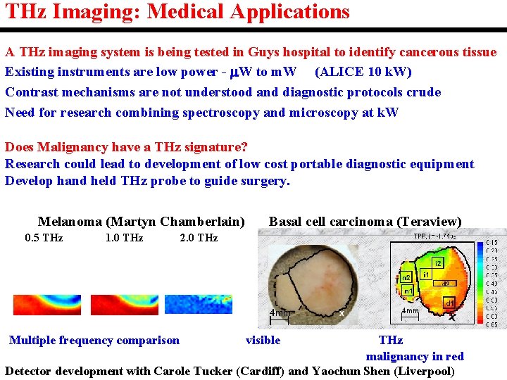
THz Imaging: Medical Applications A THz imaging system is being tested in Guys hospital to identify cancerous tissue Existing instruments are low power - W to m. W (ALICE 10 k. W) Contrast mechanisms are not understood and diagnostic protocols crude Need for research combining spectroscopy and microscopy at k. W Does Malignancy have a THz signature? Research could lead to development of low cost portable diagnostic equipment Develop hand held THz probe to guide surgery. Melanoma (Martyn Chamberlain) 0. 5 THz 1. 0 THz Multiple frequency comparison Basal cell carcinoma (Teraview) 2. 0 THz visible THz malignancy in red Detector development with Carole Tucker (Cardiff) and Yaochun Shen (Liverpool)
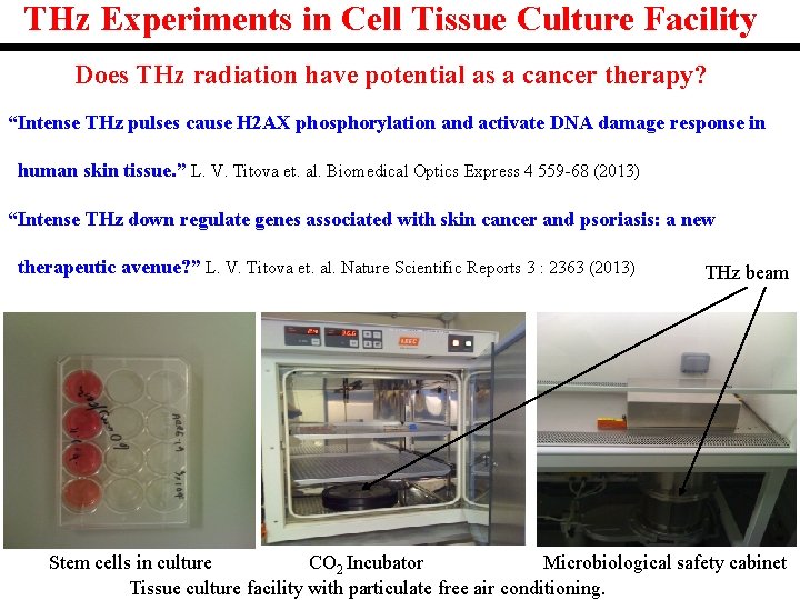
THz Experiments in Cell Tissue Culture Facility Does THz radiation have potential as a cancer therapy? “Intense THz pulses cause H 2 AX phosphorylation and activate DNA damage response in human skin tissue. ” L. V. Titova et. al. Biomedical Optics Express 4 559 -68 (2013) “Intense THz down regulate genes associated with skin cancer and psoriasis: a new therapeutic avenue? ” L. V. Titova et. al. Nature Scientific Reports 3 : 2363 (2013) THz beam Stem cells in culture CO 2 Incubator Microbiological safety cabinet Tissue culture facility with particulate free air conditioning.
Application of the SNOM on the IR FEL to the study of oesophageal cancer.
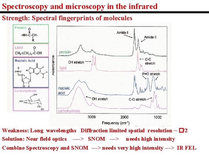
Spectroscopy and microscopy in the infrared Strength: Spectral fingerprints of molecules Weakness: Long wavelengths Diffraction limited spatial resolution ~ �/2 Solution: Near field optics ----> SNOM ---> needs high intensity Combine Spectroscopy and SNOM ---> needs very high intensity ---> IR FEL
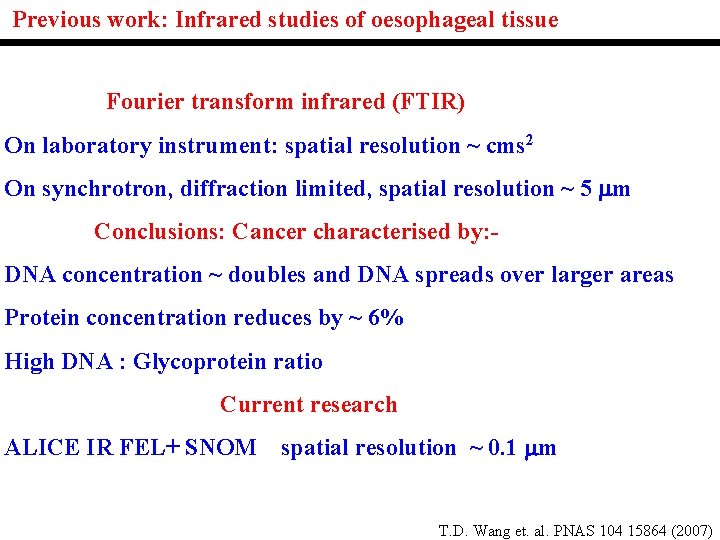
Previous work: Infrared studies of oesophageal tissue Fourier transform infrared (FTIR) On laboratory instrument: spatial resolution ~ cms 2 On synchrotron, diffraction limited, spatial resolution ~ 5 m Conclusions: Cancer characterised by: DNA concentration ~ doubles and DNA spreads over larger areas Protein concentration reduces by ~ 6% High DNA : Glycoprotein ratio Current research ALICE IR FEL+ SNOM spatial resolution ~ 0. 1 m T. D. Wang et. al. PNAS 104 15864 (2007)
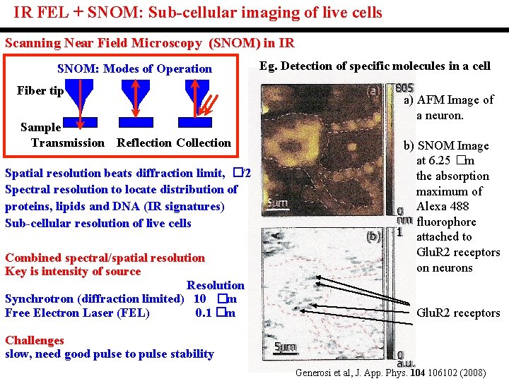
IR FEL + SNOM: Sub-cellular imaging of live cells Scanning Near Field Microscopy (SNOM) in IR SNOM: Modes of Operation Fiber tip Sample Transmission Reflection Collection Spatial resolution beats diffraction limit, �/2 Spectral resolution to locate distribution of proteins, lipids and DNA (IR signatures) Sub-cellular resolution of live cells Combined spectral/spatial resolution Key is intensity of source Resolution Synchrotron (diffraction limited) 10 �m Free Electron Laser (FEL) 0. 1 �m Eg. Detection of specific molecules in a cell a) AFM Image of a neuron. b) SNOM Image at 6. 25 �m the absorption maximum of Alexa 488 fluorophore attached to Glu. R 2 receptors on neurons Glu. R 2 receptors Challenges slow, need good pulse to pulse stability Generosi et al, J. App. Phys. 104 106102 (2008)
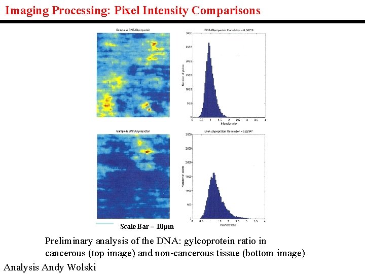
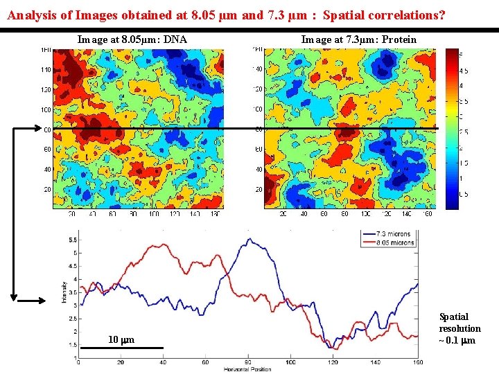
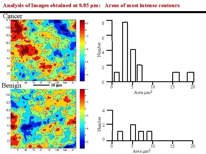
Imaging Processing: Pixel Intensity Comparisons Scale Bar = 10µm Preliminary analysis of the DNA: gylcoprotein ratio in cancerous (top image) and non-cancerous tissue (bottom image) Analysis Andy Wolski
Analysis of Images obtained at 8. 05 µm and 7. 3 µm : Spatial correlations? Image at 8. 05µm: DNA 10 m Image at 7. 3µm: Protein Spatial resolution ~ 0. 1 m
Analysis of Images obtained at 8. 05 µm: Areas of most intense contours Cancer 8 Number 6 4 2 0 10 m 0 5 10 15 20 0 5 10 2 Area mm 15 20 Area mm 2 4 Number Benign 2 0
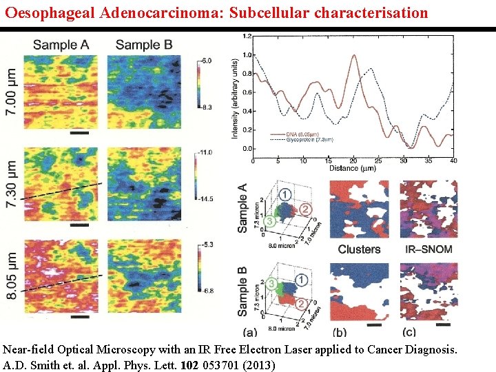
Oesophageal Adenocarcinoma: Subcellular characterisation Near-field Optical Microscopy with an IR Free Electron Laser applied to Cancer Diagnosis. A. D. Smith et. al. Appl. Phys. Lett. 102 053701 (2013)
Analysis of FTIR Imaging of oesophageal cancer.
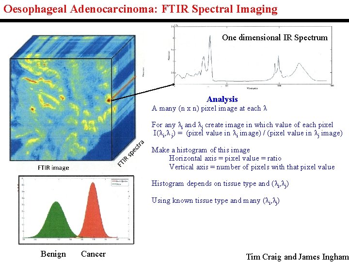
Oesophageal Adenocarcinoma: FTIR Spectral Imaging One dimensional IR Spectrum Analysis A many (n x n) pixel image at each λ For any λi and λj create image in which value of each pixel I(λi, λ j) = (pixel value in λi image) / (pixel value in λj image) Make a histogram of this image Horizontal axis = pixel value = ratio Vertical axis = number of pixels with that pixel value Histogram depends on tissue type and (λi, λj) Using known tissue type and many (λi, λj) Benign Cancer Tim Craig and James Ingham
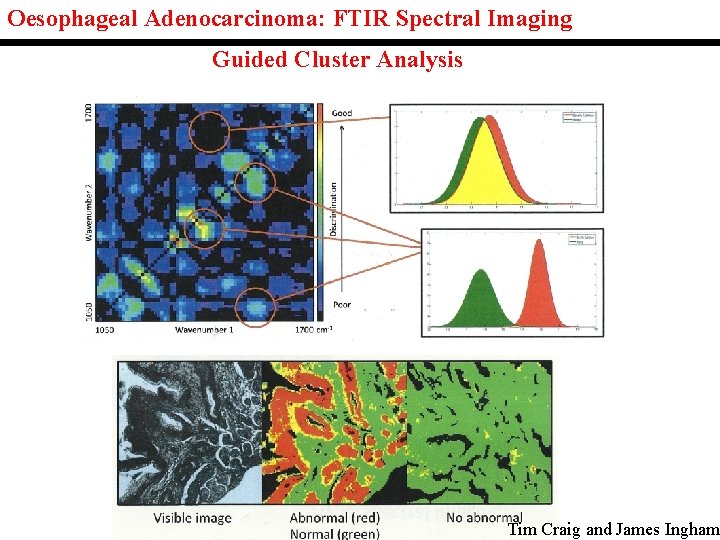
Oesophageal Adenocarcinoma: FTIR Spectral Imaging Guided Cluster Analysis Tim Craig and James Ingham
Conclusions 1) Accelerator based sources of IR and THz have potential for cancer diagnosis. Considerable more work needed on reproducibility and patient variability. 2) If successful need to develop cheap systems for use in hospitals. £ 10 m? 3) Maybe able to develop portable THz instruments for cancer diagnosis. Endoscopes? 4) Intense THz radiation as a cancer therapy? ? ? Very controversial but makes sense theoretically. Needs a lot more research. ALICE accelerator: tissue facility ideal environment.
Acknowledgements University of Liverpool. Prof. S. Chattopadhyay, Prof. A. Wolski, Dr S. D. Barrett, Dr. D. S. Martin, Dr M. S, King, Physics, Mr T. Craig, Mr J. Ingham, Physics. Dr. Y Shen, Electrical Engineering and Electronics. Prof. A. Cricenti and Dr. M. Luca, CNR (Rome). Prof. M. Pritchard, Gastroenterology, Royal Liverpool Hospital. Prof. A. Varro, Physiology, Royal Liverpool Hospital. University of Lancaster. Dr. O. V. Kolosov, Physics, Prof. D. Allsop, Neuroscience. Prof. F. L. Martin, Biological Chemistry, Prof P. L Martin-Hirsch, Gynaeoncology and Obstetrics, Lancashire Hospital, Dr H. F. Stringfellow, Pathology, Lancashire Hospital. University of Manchester. Prof. P. Gardener, Chemical Engineering and Analytical Science. Prof. N. Clarke, Urological Oncology, Christie Hospital. University of Cardiff. Prof. P. Ade, Physics and Astronomy, Dr C. Tucker, Physics and Astronomy. Staff from ASTe. C and Cockcroft Institute of Daresbury Laboratory.