A HISTORY OF DNA Discovery of the DNA


 Bases 3 o 5 o P o")


 would form 2 hydrogen bonds only with thymine (T) •")


- Slides: 36
A HISTORY OF DNA • Discovery of the DNA double helix A. Frederick Griffith – Discovers that a factor in diseased bacteria can transform harmless bacteria into deadly bacteria (1928) B. Rosalind Franklin - X-ray photo of DNA. (1952) C. Watson and Crick - described the DNA molecule from Franklin’s X-ray. (1953)
Watson & Crick proposed • DNA had specific pairing between the nitrogen bases: ADENINE – THYMINE CYTOSINE - GUANINE • DNA was made of 2 long stands of nucleotides arranged in a specific way called the “Complementary Rule”
The Watson and Crick DNA Double helix • The correct structure of DNA was first deduced by J. D. Watson and F. H. C. Crick in 1953. • Their double helix model of DNA structure was based on two major kind of evidence. 1. Chargaff’s rule 2. X – ray diffraction patterns.
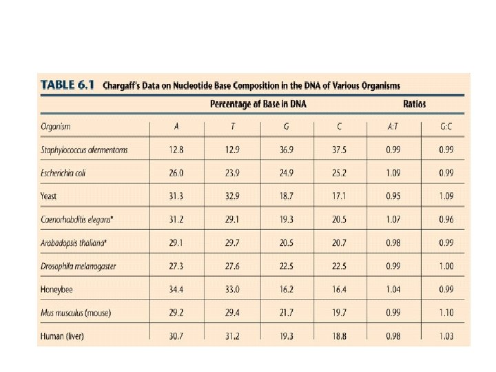
Chargaff’s rule • The composition of DNA from many different organisms was analyzed by E. Chargaff and his colleagues. • It was observed that concentration of thymine was always equal to the concentration of adenine (A = T) • And the concentration of cytosine was equal to the concentration of guanine (G = C). • This strongly suggest that thymine and adenine as well as cytosine and guanine were present in DNA with fixed interrelationship. • Also the total concentration of purines (A +G) always equal to the total concentration of pyrimidine (T +C). However, the (T+ A)/ (G+C) ratio was found to vary widely in DNAs of different species.
The bases always pair up in the same way Adenine forms a bond with Thymine Adenine Thymine and Cytosine bonds with Guanine Cytosine Guanine
X ray diffraction • When X rays are focused through isolated macromolecules or crystals of purified molecules, the X ray are deflected by the atom of the molecules in specific patterns called diffraction patterns. • It provides the information about the organization of the components of the molecules. • Watson and Crick had X ray crystallographic data on DNA structure from the studies of Wilkins and Franklin and their coworkers. • These data indicated that DNA was a highly ordered, multiple stranded structure with repeating sub structures spaced every 3. 4 Ao (1 Angstrom = 10 -10 m )
X-ray diffraction patterns of DNA – Rosalind Franklin and Maurice Wilkins The central cross shaped pattern as indicative of a helical structure. The heavy dark patterns (top and bottom) indicate that the bases are stacked perpendicular to the axis of the molecule.
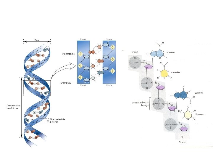
Double Helix • Watson and Crick proposed that DNA exists as a double helix in which two polynucleotide chains are coiled above one another in a spiral. • Each polynucleotide chain consists of a sequence of nucleotide linked together by Phosphodiester bonds. • The two polynucleotide strands are held together in their helical configurations by hydrogen bonding. • The base pairing is specific • That is, adenine is always paired with thymine and guanine is always paired with cytosine • Thus, all base-pairs consists of one purine and one pyrimidine. • Once the sequence of bases in one strand of DNA double helix is known, it is possible to know the other strand sequence of base because of specific base pairing.
• In their most structural configuration, adenine and thymine form two hydrogen bonds, where as guanine and cytosine form three hydrogen bonds. • The two strands of a DNA are complementary (not identical) to each other. It is this property, that makes DNA uniquely suited to store and transmitting the genetic information. • The base-pairs in DNA are stacked 34 Ao apart with 10 base-pairs per turn (3600) of the double helix • The sugar – phosphate backbones of the two complementary strands are antiparallel, that is they have opposite chemical polority.
• As one move unidirectionally along a DNA double helix, the phosophodiester bonds in one strand go from a 3’Carbon of one nucleotide to a 5’Carbon of the adjacent nucleotide. • Where as those in complementary strand go from 5’Carbon to a 3’carbon. • This opposite polarity of the complementary strands is very important in considering the mechanism of replication of DNA. • The high degree of stability of DNA double helices results in part from the large number of hydrogen bonds between base pairs.
• Although each hydrogen bond by itself quite weak, since no. of hydrogen bonds are more, it can withstand. • The planar sides of the base pair are relatively non polar and thus tend to be water insoluble (hydrophobic). • The hydrophobic core stacked base-pairs contributes considerable stability to DNA molecules present in the aqueous protoplasms of living cells.
DNA • The amino acid sequence of a polypeptide is programmed by a gene. • A gene is a small region in the DNA. • Nucleic acids store and transmit hereditary information ﺍﻟﻤﻌﻠﻮﻣﺎﺕ ﺍﻟﻮﺭﺍﺛﻴﺔ. • There are two types of nucleic acids: ribonucleic acid (RNA) and deoxyribonucleic acid (DNA). • DNA also directs m. RNA synthesis, thus, controls protein synthesis. • Organisms inherit ﺗﺘﻮﺍﺭﺙ DNA from their parents. – Each DNA molecule is very long and usually consists of hundreds to thousands of genes. – When a cell divides ﺗﻨﻘﺴﻢ , its DNA is copied and passed to the next generation of cells. • The m. RNA interacts with ribosomes to direct the synthesis of amino acids in a polypeptide (protein)
• The flow of genetic information is from DNA m. RNA protein. – Protein synthesis occurs in ribosomes. – In eukaryotes, DNA is located in the nucleus, but most ribosomes are in the cytoplasm with m. RNA as an intermediary ﻭﺳﻴﻂ.
NUCLEIC ACID STRUCTURE • DNA and RNA are large macromolecules with several levels of complexity – 1. Nucleotides form the repeating units – 2. Nucleotides are linked to form a strand – 3. Two strands can interact to form a double helix – 4. The double helix folds, bends and interacts with proteins resulting in 3 -D structures in the form of chromosomes
Three-dimensional structure
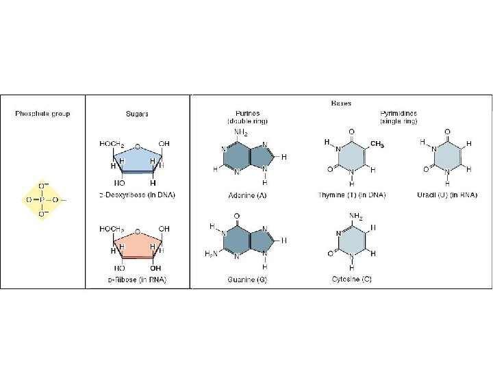
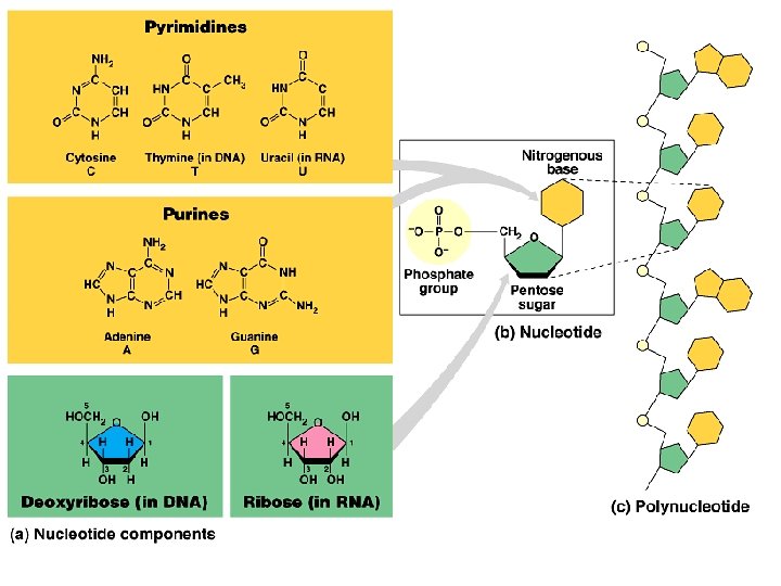
Nucleotides n n The nucleotide is the repeating structural unit of DNA and RNA It has three components n A phosphate group n A pentose sugar n A nitrogenous base
n These atoms are found within individual nucleotides n However, they are removed when nucleotides join together to make strands of DNA or RNA A, G, C or T A, G, C or U The structure of nucleotides found in (a) DNA and (b) RNA
n n Base + sugar nucleoside n Example n Adenine + ribose = Adenosine n Adenine + deoxyribose = Deoxyadenosine Base + sugar + phosphate(s) nucleotide n Example n Adenosine monophosphate (AMP) n Adenosine diphosphate (ADP) n Adenosine triphosphate (ATP)
Base always attached here Phosphates are attached there
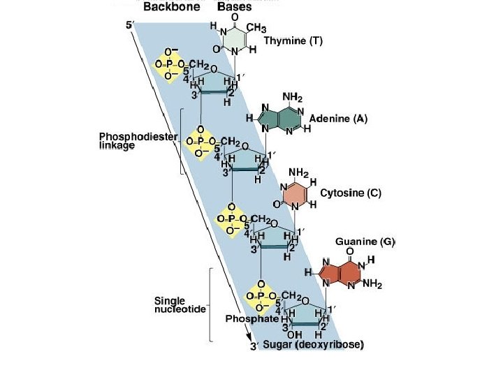
n n n Nucleotides are covalently linked together by phosphodiester bonds n A phosphate connects the 5’ carbon of one nucleotide to the 3’ carbon of another Therefore the strand has directionality n 5’ to 3’ The phosphates and sugar molecules form the backbone of the nucleic acid strand n The bases project from the backbone
Structures of nucleic acids (DNA & RNA) Bases 3 o 5 o P o o Phosphate group o 5 CH 2 4 H H 3 o o DNA nucleotide Base H 1 2 H H Guanine (G) Deoxyribose o P o CH 2 H 5 3 Adenine (A) Purine H 3 Base o Cytosine (C) H H H Sugar-phosphate backbone Thymine (T) Pyrimidine
• The PO 4 group of one nucleotide is attached to the sugar of the next nucleotide in line. • The result is a “backbone” of alternating phosphates and sugars, from which the bases starts.
3 5 Nitrogenous bases ﺍﻟﻘﻮﺍﻋﺪ ﺍﻟﻨﻴﺘﺮﻭﺟﻴﻨﻴﺔ Nitrogenous bases Hydrogen bonds 5 3 Sugar-phosphate backbones Cytosine (C) Guanine (G) Thymine (T) Adenine (A) Uracil (U) Pyrimidines Purine
DNA • Adenine (A) would form 2 hydrogen bonds only with thymine (T) • Guanine (G) would form 3 hydrogen bonds only with cytosine (C).
DNA CH 2 H H o RNA CH 2 H H & H Deoxyribose sugar (O on C 2 is missed) Deoxiribo-Nucleic-Acid Double stranded nucleic acid Bases: A, G, C, T H H o H H OH Ribose sugar (no missed O) Ribo-Nucleic-Acid Single stranded nucleic acid Bases: A, G, C, U
The nucleic acid strand is a polymer of nucleotides • Nucleic acids are polymers of monomers called nucleotides. • Each nucleotide consists of three parts: a nitrogen base, a pentose sugar, and a phosphate group. • The nitrogen bases (rings of carbon and nitrogen) come in two types: Purines and Pyrimidines. • The pentose sugar joined to the nitrogen base is ribose in nucleotides of RNA and deoxyribose in DNA. • The only difference between the sugars is the lack ﻧﻘﺺ of an oxygen atom on carbon 2 in deoxyribose.
• Polynucleotides are synthesized by connecting the sugars of one nucleotide to the phosphate of the next with a phosphodiester link. • This creates a repeating backbone of sugar-phosphate units with the nitrogen bases as appendages. • The sequence of nitrogen bases along a DNA or m. RNA polymer is unique for each gene. • Genes are normally hundreds to thousands of nucleotides long. • The linear order of bases in a gene specifies ﺣﺪﺩ the order of amino acids (the monomers of a protein).
Inheritance is based on replication of the DNA double helix • An RNA molecule is single polynucleotide chain (single strand). • DNA molecules have two polynucleotide strands (double strand) that spiral around to form a double helix.
• The sugar-phosphate backbones of the two polynucleotides are on the outside of the helix. • Pairs of nitrogenous bases (one from each strand) connect the polynucleotide chains with hydrogen bonds. • Most DNA molecules have thousands to millions of base pairs (b. P).
• Because of their shapes, only some bases are compatible ﻣﺘﻮﺍﻓﻘﺔ with each other. – Adenine (A) always pairs with thymine (T) and guanine (G) with cytosine (C). • With these base-pairing rules, if we know the sequence of bases on one strand, we know the sequence on the opposite ﺍﻟﻤﻘﺎﺑﻞ strand. • The two strands are complementary ﻣﻜﻤﻠﻴﻦ ﻟﺒﻌﻀﻬﻤﺎ. • During preparations for cell division each of the strands serves as a template ﻗﺎﻟﺐ ﻧﺴﺦ to order nucleotides into a new complementary strand. • This results in two identical copies ﻧﺴﺨﺘﻴﻦ ﻃﺒﻖ ﺍﻷﺼﻞ of the original doublestranded DNA molecule. – The copies are then distributed ﺗﻮﺯﻉ to the daughter cells. • This mechanism ensures that the genetic information is transmitted to the new cells.
Repeated Sugar - Phosphate DNA backbone Sugar–Phosphate-Base One nucleotide Polynucleotide DNA Molecule DNA Double stranded RNA single stranded DNA A G C T A T C m. RNA TU C G A TU A G