A grade A level students independently study for


- Slides: 16
‘A grade’ A level students independently study for 20 hours per week. What about you? #justsaying
� Stimuli lead to responses � Nervous Coordination � Skeletal Muscles � Homeostasis Miss Marwick Miss Jesusanmi
What are three types of muscle? Cardiac Smooth Skeletal What is an antagonistic muscle pair? Antagonistic pairs of muscles create movement when one contracts and the other relaxes.
Which colour represents the extensor? Which colour represents the flexor?
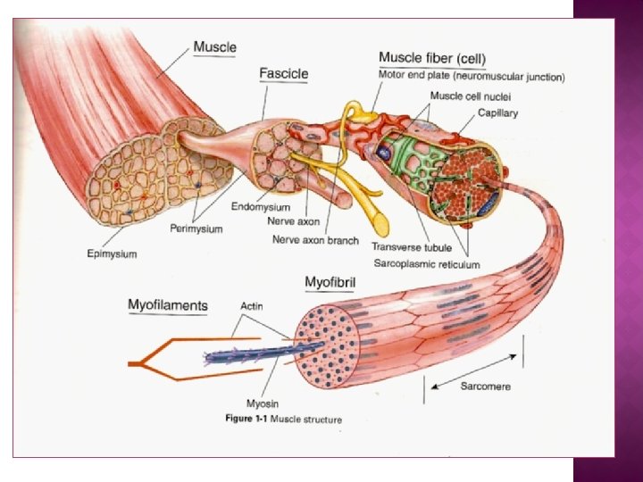
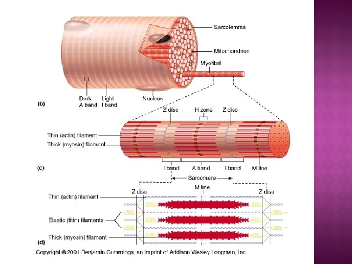
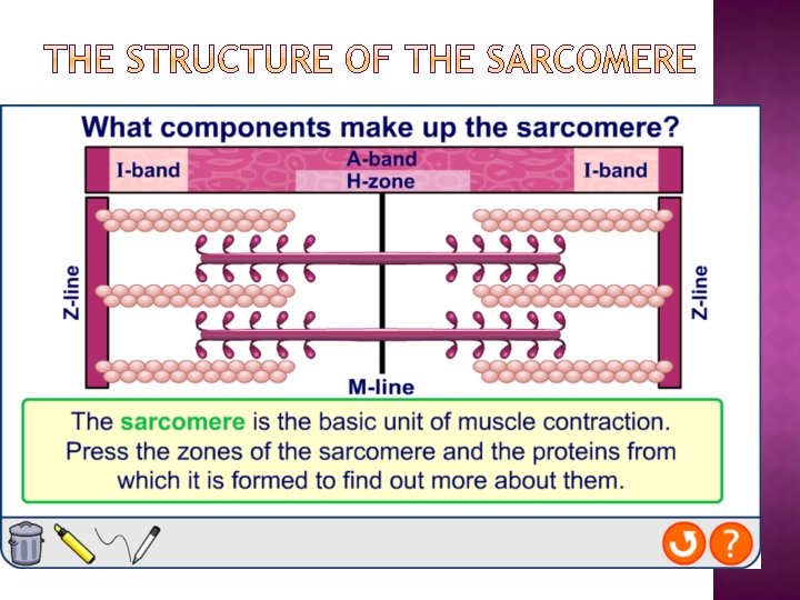
25 October 2020 You should be able to… � Describe the gross structure of skeletal muscles � Explain what is meant by a myofibril � Describe the microscopic structure of skeletal muscle � Explain what is meant by a sarcomere � Explain how actin and myosin are arranged within a myofibril, linked to isotropic bands and anisotropic bands and the thickness of each type of protein filament � Interpret diagrams to identify I bands, A bands, the H zone and the Z line on a diagram.
Some context…
Some context…
What might you expect to find a lot of in the sarcoplasm? Glycogen Mitochondria Myoglobin Why?
Myofibrils contain two different types of filaments: thin filaments made predominantly of actin, and thick filaments made of myosin. These filaments are arranged in an interlocking pattern within the sarcomere, producing the characteristic banding thin filament pattern of the myofibrils. (actin) thick filament (myosin) Z-line
The myosin filament is formed from a number of myosin proteins wound together. Each ends in a myosin head, which contains an ATPase. actin binding site myosin filament ATP binding site ATPase head myosin neck myosin head
The actin filament is formed from a helix of actin sub-units. Each contains a binding site for the myosin heads. troponin tropomyosin actin sub-unit myosin head binding site Two other proteins are attached to the actin fibre: l tropomyosin is wound around the actin l troponin molecules are bound to tropomyosin and contain calcium ion binding sites.
Use the laptops to access ‘Animation: Muscle contraction’ on kerboodle. This will give a recap of the skeletal muscle structure (up to 2 mins 50). Read about fast and slow twitch muscle fibres (page 369) and use the information to fill in the table provided.