
A comparison of pelvic magnetic resonance imaging transvaginal




- Slides: 69
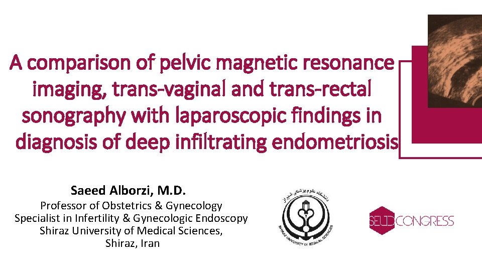
A comparison of pelvic magnetic resonance imaging, trans-vaginal and trans-rectal sonography with laparoscopic findings in diagnosis of deep infiltrating endometriosis Saeed Alborzi, M. D. Professor of Obstetrics & Gynecology Specialist in Infertility & Gynecologic Endoscopy Shiraz University of Medical Sciences, Shiraz, Iran
Introduction • Endometriosis is defined as an ectopic presence of endometrial tissue outside the uterus. • Affects 5 -10% of women of reproductive age • Superficial endometriotic implants are mostly silent and asymptomatic • lesions lying deeper than 5 mm within the peritoneum (DIE) are associated with more severe symptoms. • Common locations for DIE are uterosacral ligaments, posterior vaginal wall and anterior rectal wall • 15 -30% of patients with endometriosis suffer from DIE
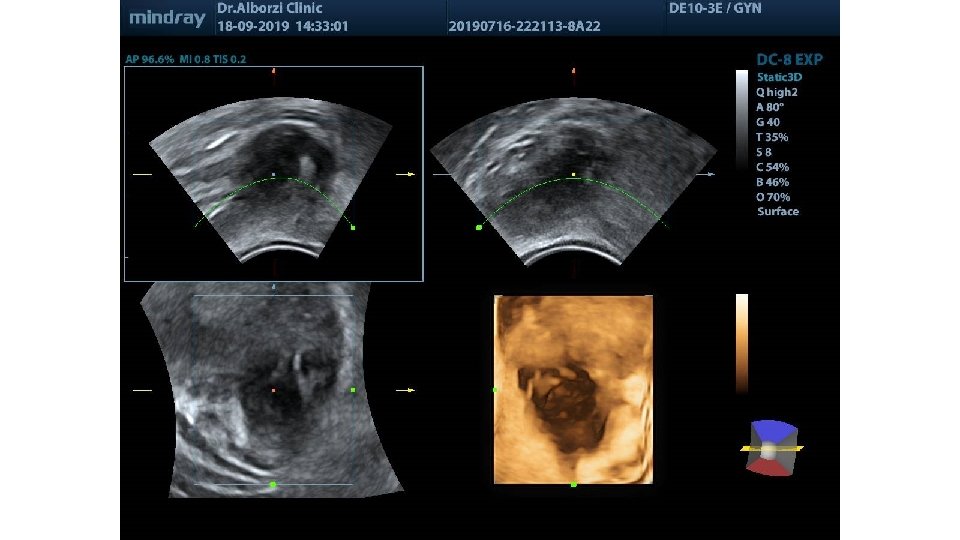
• Diagnosis of DIE is an uphill challenge for gynecologists. • Direct visualization and biopsy of lesions through laparoscopy or laparotomy remains the gold standard method for definitive diagnosis. • Various imaging modalities such as MRI, TVS, TRS as well as 3 D ultrasound are currently available for the diagnosis of DIE. • Considering the cost-effectiveness and accessibility issue, TVS is usually the first diagnostic imaging modality in females with symptoms of DIE. When performed by an expert radiologist, TVS is reported to have a sensitivity of 98. 1% and 95. 1% for diagnosis of rectal and retrocervical DIE, respectively. • MRI is a more expensive and meanwhile non-invasive modality for pre-operative assessment of DIE which provides more accurate data about the extension, localization and penetration of the lesions. The sensitivity of MRI for diagnosis of rectal and retrocervical DIE has been reported 83. 3% and 76%, respectively.
choosing the best imaging modality for diagnosis and pre- operative assessment is still a challenging issue. The aim of this study was to compare the accuracy and define sensitivity and specificity of three different imaging modalities (TVS, MRI and TRS) in a large population of DIE patients referred to a tertiary gynecological surgery centers As far as we reviewed the literature, this study included one of the largest population of patients with endometriosis for whom three different imaging techniques were used to assess preoperative diagnosis of DIE.
Materials and methods üFrom March 2010 to December 2014 ü in a prospective manner üPatients referred to private clinics and hospitals affiliated to Shiraz University of medical sciences ü With symptoms of chronic pelvic pain, dyspareunia and dysmenorrhea, who were candidate for operative laparoscopy with suspicion to DIE were enrolled in this study üOver 500 patients underwent laparoscopic surgery , 317 cases (only nonvirgins) were enrolled in our study (due to lack of possibility of TVS evaluation in virgin subjects) ü All patients were asked to sign the informed consent ü Patients were included from all age groups
Exclusion criteria 1 - Claustrophobia from MRI tunnel 2 - Renal failure or any other contraindication for gadolinium contrast medium injection 3 - History of having metallic implants or prosthetics in the body which prohibits the use of MRI 4 - Structural anomalies of the reproductive system 5 - Patients’ refusal or incompliance with TVS or TRS
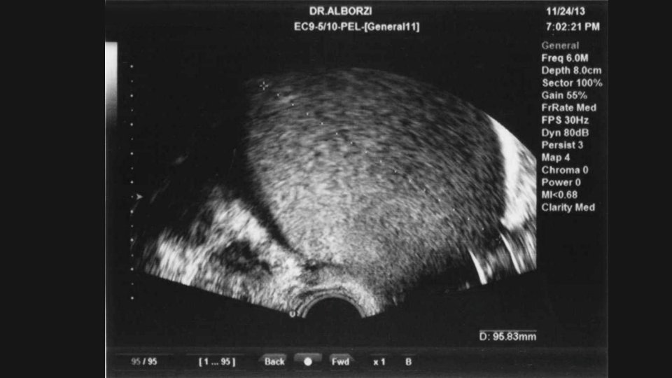
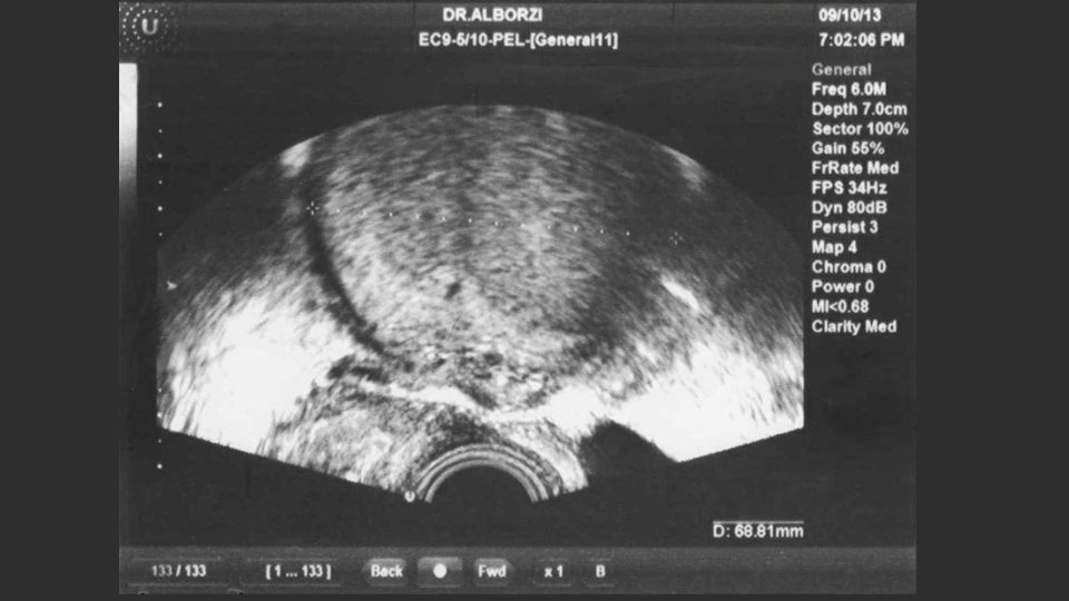
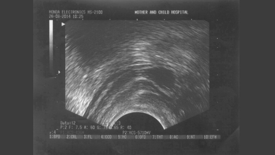
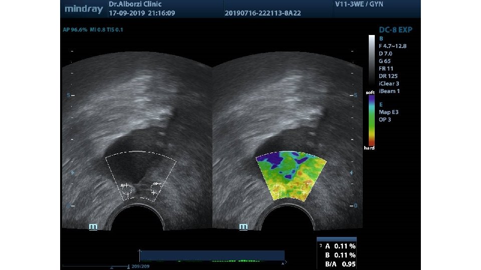
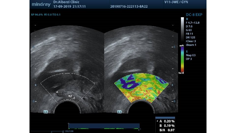
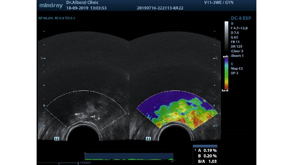
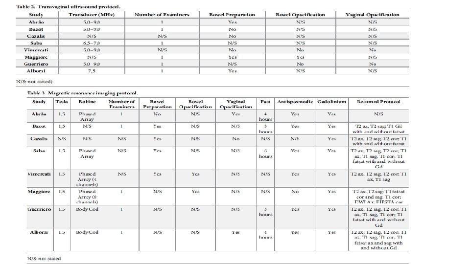
Trans-vaginal sonography § Using 7. 5 MHz probe in non-menstrual days of the cycle for non-virgin pts. § Patients were asked to have empty bladder and simple bowel prep for better visualization of pelvic organs. § Examination protocol included visualization of peritoneum and structures in anterior and posterior compartments, as well as uterus and ovaries. § Nodular, hypoechoic solid lesions with and without cystic components, in different structures of pelvic cavity were considered to be highly suggestive for DIE. § Also hyper-echogenic abnormal thickening of the peritoneum over structures in pelvic cavity was considered as a sign of DIE.
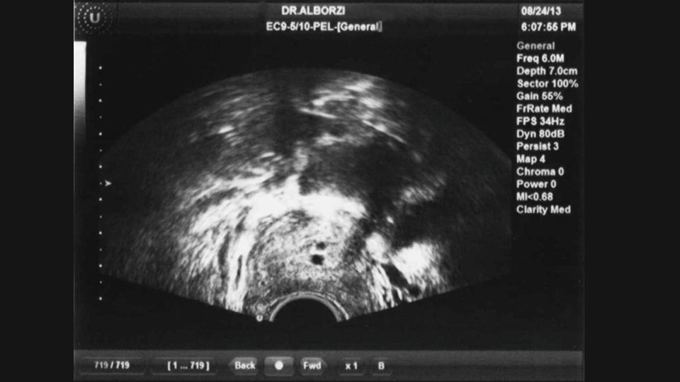
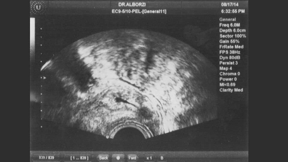
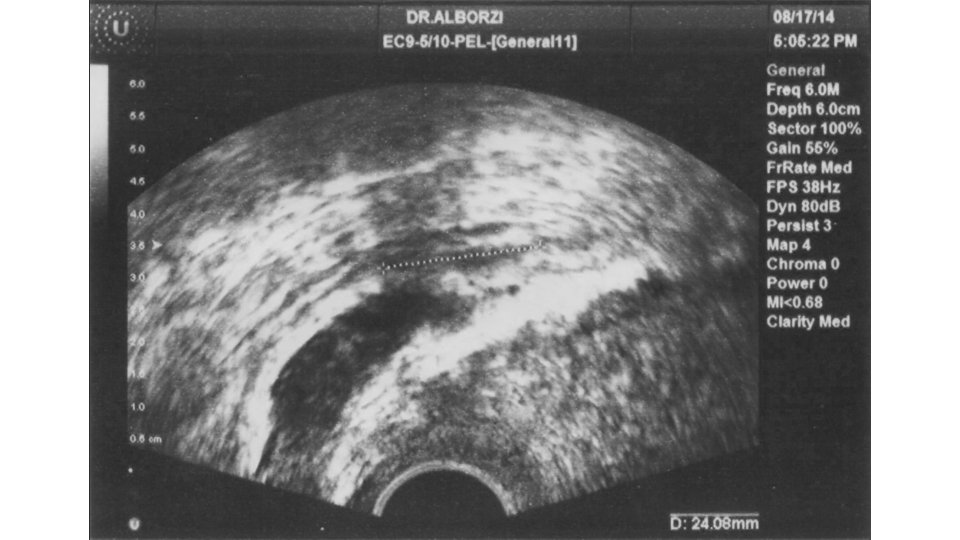
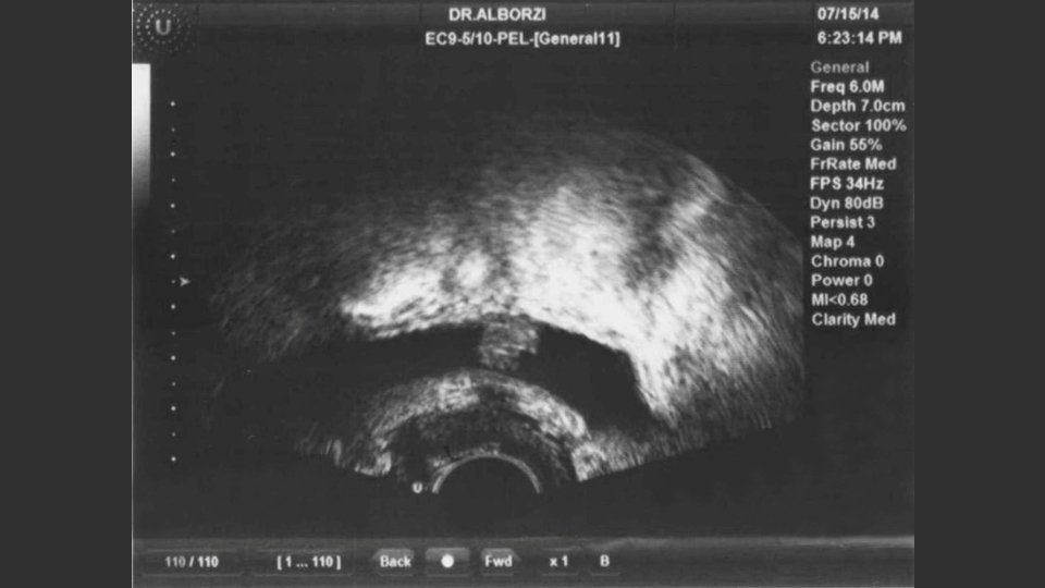
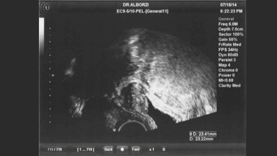
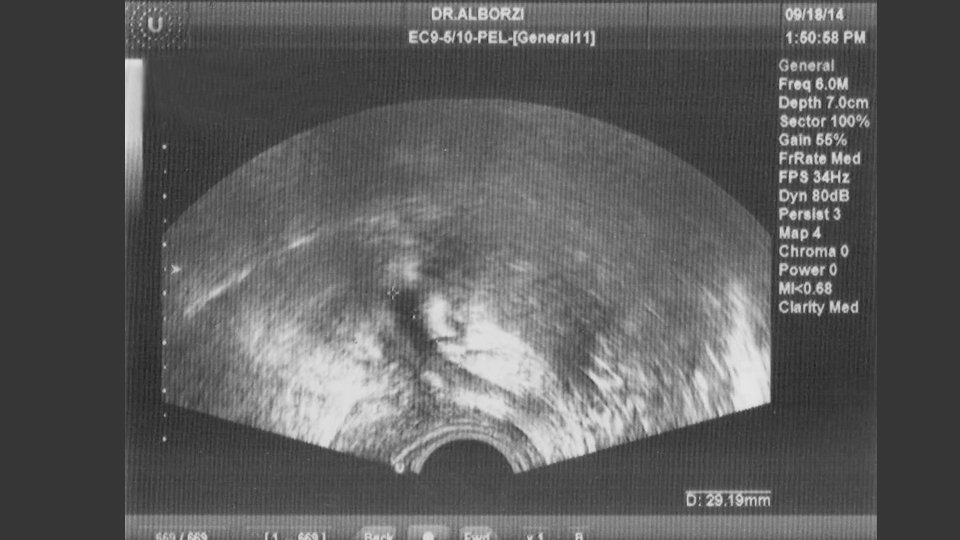
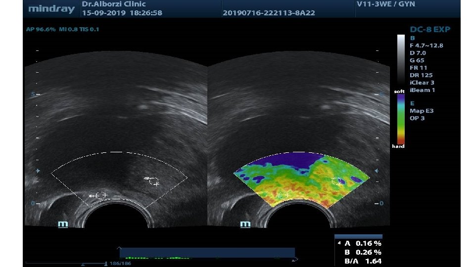
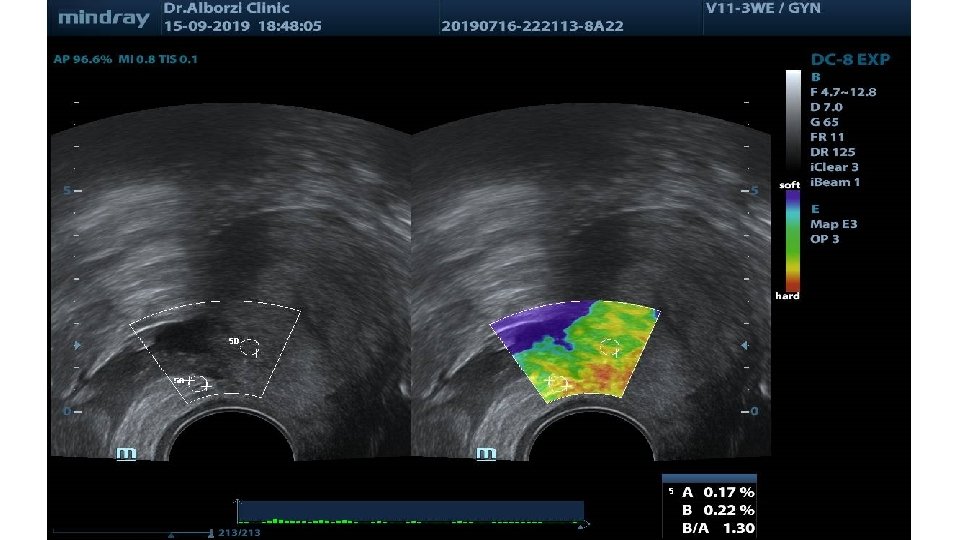
Transrectal sonography § TRS was performed on the same day that TVS was done, by the same attending gynecologist using 7. 5 MHz probe after bowel preparation § Bowel prep is achieved by asking the individuals to have a soft diet in the day before sonography, having 2 spoonfuls of MOM orally after lunch and by rectal administration of bisacodyl (2 suppositories TID on the day before the procedure). They were asked not to have breakfast and also take 2 bisacodyl suppositories at 6 am in the next morning § The procedure was performed with empty bladder and by using lubricant gel without administration of sedatives § Examination protocol included the same structures which were examined in TVS and the same diagnostic criteria were used
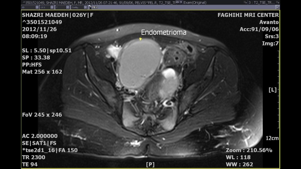
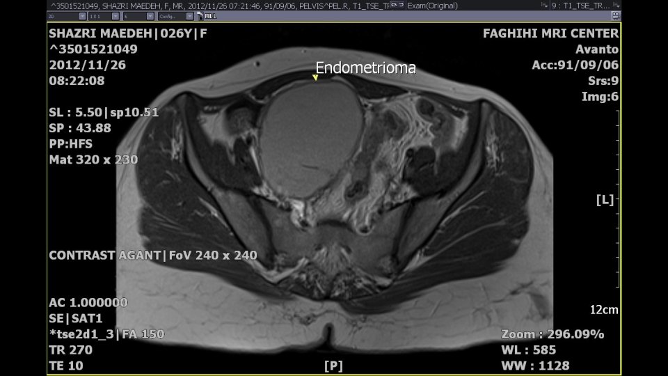
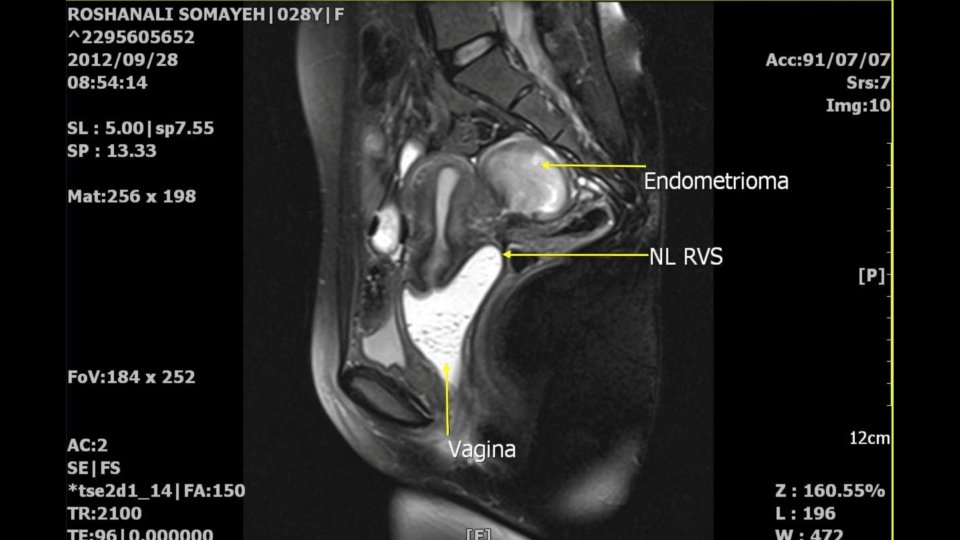
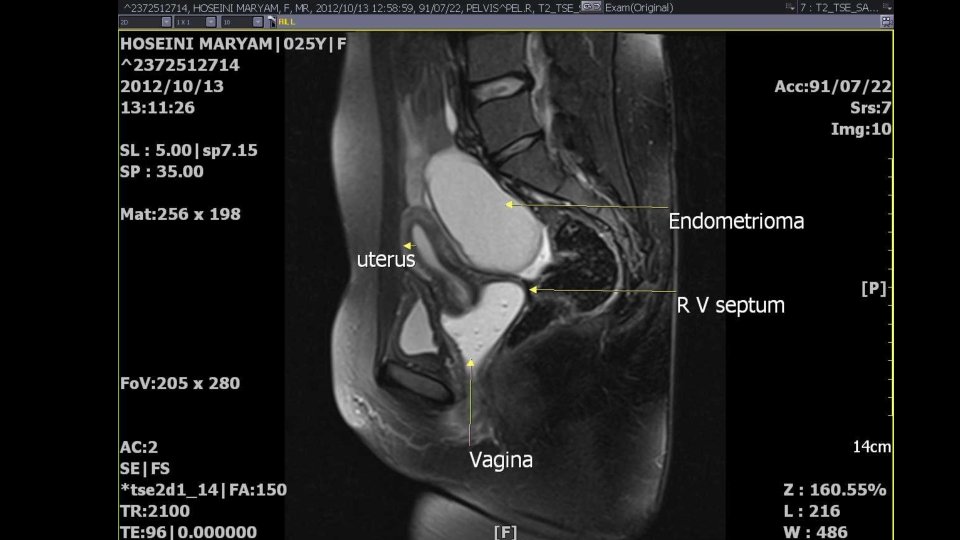
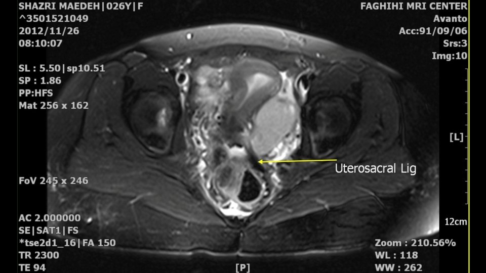
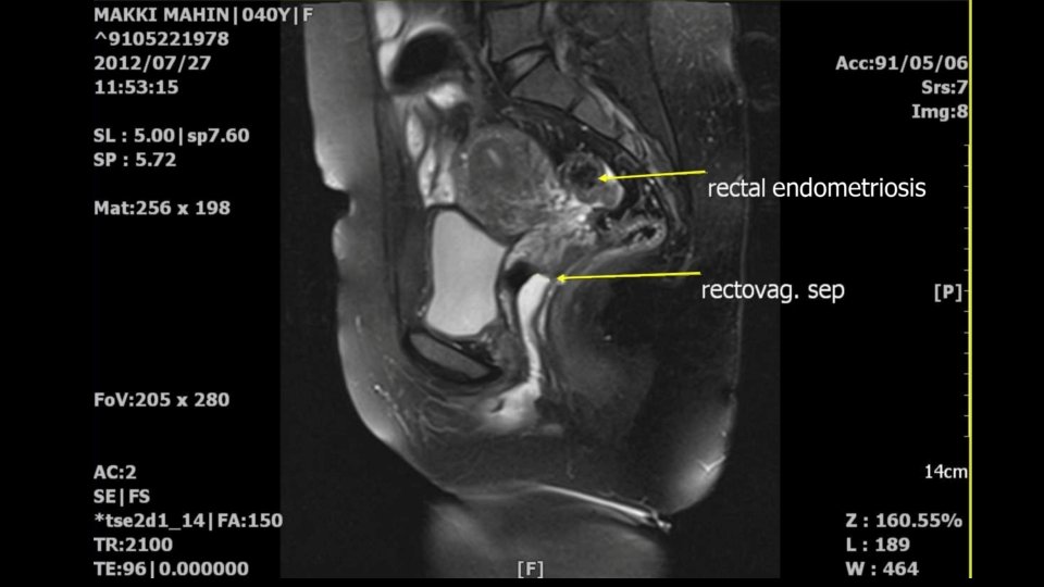
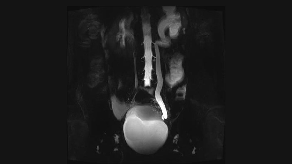
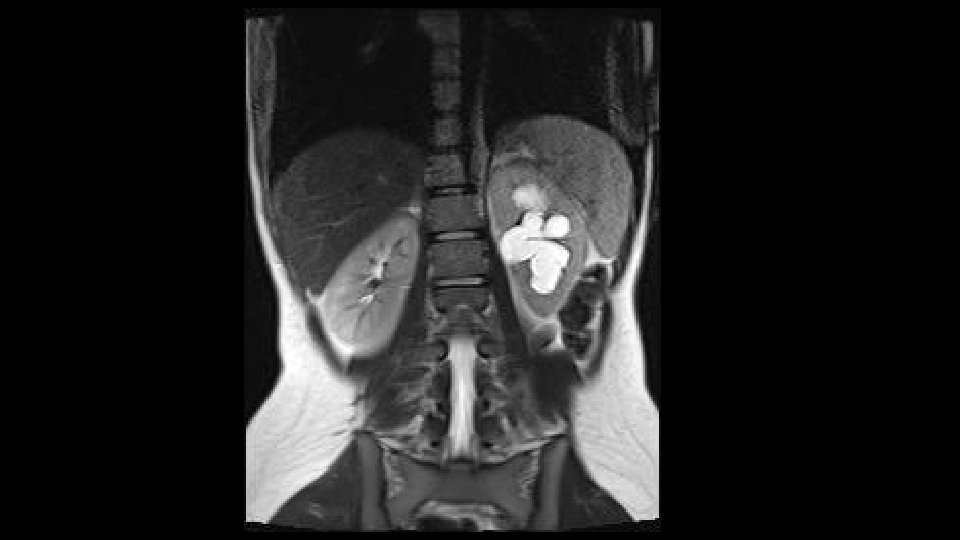
Magnetic resonance imaging § MRI was performed for all patients after 4 -hour fasting with semi-filled bladder, before and after injection of gadolinium contrast medium, with dosage of 0. 01 mmol/kg, using 1. 5 Tesla using body pelvic coil without endovaginal coil § 60 cc lubricant gel was inserted into the vaginal cuff for better delineation of rectal and vaginal walls, and hyoscine 1 Amp IM injected § Protocols include Axial, Coronal and Sagittal T 1 -and T 2 -weighted images to delaminate the anatomy and pathology § T 1 Axial and Sagittal fat saturation technique with and without contrast is also performed § The bladder wall and rectovaginal septum are evaluated in T 2 Sagittal and Axial image § Uterosacral ligaments and rectal wall mostly in coronal and Axial T 2 weighted image § Endometriomas were high signal in T 1 and low signal in T 2 weighted images § DIE were low signal or signal void in T 2 weighted images § Thickening of the walls were in favor of involvements
Laparoscopy üOperative laparoscopy was performed by the same gynecologist in all patients after whole bowel prep under general anesthesia. ü Two 10 mm and two 5 mm ports were used. ü The pelvic cavity was explored and endometriosis was classified according to the revised American Society for Reproductive Medicine (r. ASRM) classification (1985) ü According to different locations, pararectal and paravesical and rectovaginal spaces were dissected when necessary ü All adhesions were released with sharp dissection and all lesions suspicious to DIE were resected to the extent to restore normal anatomic relations and sent for pathologic investigation
• For rectal lesions, pararectal and rectovaginal spaces were dissected and inspected for suspicious areas, and the suspicious lesions were excised, disk resection or segmental resection and reanastomosis of bowel was performed as necessary. • For those patients with extrinsic ureteral lesions, ureterolysis, and for those with intrinsic ureteral lesions, excision and reanastomosis were performed. • In patients who presented with bladder lesions, either shaving or partial cystectomy was done according to the depth of lesions.
Results • Our study population comprised 317 women with signs and symptoms of endometriosis in the age range of 15 to 50 yrs with a mean age of 31 ± 5. 4 ys. • All patients who enrolled in this study underwent laparoscopy and 350 DIE lesions were detected. • In 65 out of 317 patients (20 %), laparoscopy yielded negative results for DIE through the patients might have had endometrioma or superficial endometriosis. • Among 350 DIE lesions, 151 were located within the uterosacral ligaments (43%) as the most frequent site of involvement followed by ovarian fossa (n=59, 16. 9%), rectal wall (n=52, 14. 9%) and rectovaginal septum (n=44, 12. 6%), respectively.
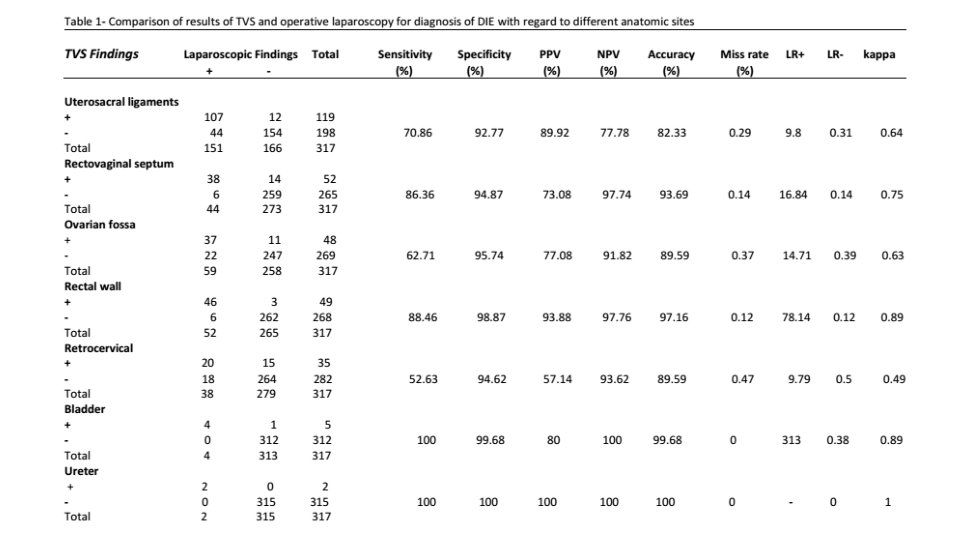
• With regard to TVS, 310 lesions were visualized of which 254 were approved by laparoscopy. • The comparison between TVS and laparoscopic findings for DIE lesions are presented in next table.
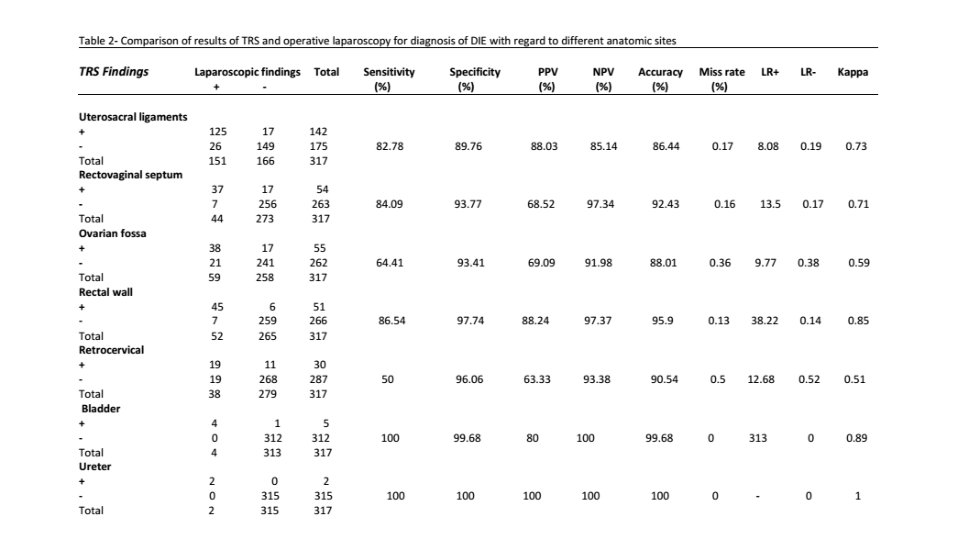
• For TRS, out of 339 diagnosed DIE lesions, 270 were confirmed by laparascopy. • The achieved results are outlined in next table.
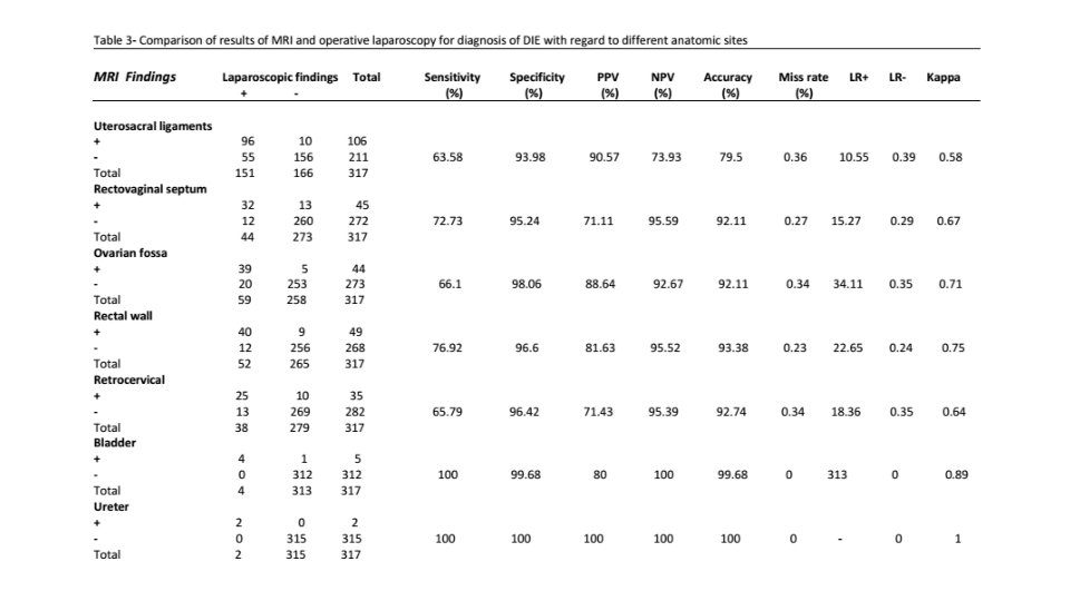
• With respect to pelvic MRI, 286 lesions were detected out of which 238 lesions were confirmed by laparascopy. • Detailed comparison is provided in next table.
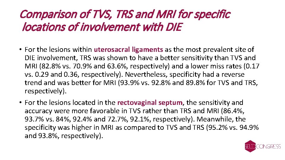
Comparison of TVS, TRS and MRI for specific locations of involvement with DIE • For the lesions within uterosacral ligaments as the most prevalent site of DIE involvement, TRS was shown to have a better sensitivity than TVS and MRI (82. 8% vs. 70. 9% and 63. 6%, respectively) and a lower miss rates (0. 17 vs. 0. 29 and 0. 36, respectively). Nevertheless, specificity had a reverse trend and was better for MRI (93. 9% vs. 92. 8% and 89. 8% for TVS and TRS, respectively). • For the lesions located in the rectovaginal septum, the sensitivity and accuracy were more favorable in TVS rather than TRS and MRI (86. 4%, 93. 7% vs. 84%, 92. 4% and 72. 7%, 92. 1%, respectively). Meanwhile, the specificity was higher in MRI as compared to TVS and TRS (95. 2% vs. 94. 9% and 93. 8%, respectively).
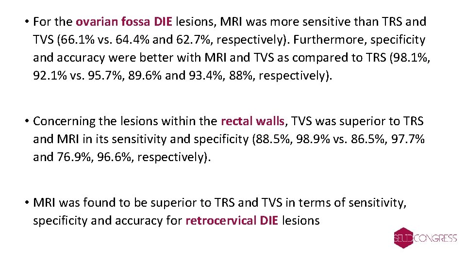
• For the ovarian fossa DIE lesions, MRI was more sensitive than TRS and TVS (66. 1% vs. 64. 4% and 62. 7%, respectively). Furthermore, specificity and accuracy were better with MRI and TVS as compared to TRS (98. 1%, 92. 1% vs. 95. 7%, 89. 6% and 93. 4%, 88%, respectively). • Concerning the lesions within the rectal walls, TVS was superior to TRS and MRI in its sensitivity and specificity (88. 5%, 98. 9% vs. 86. 5%, 97. 7% and 76. 9%, 96. 6%, respectively). • MRI was found to be superior to TRS and TVS in terms of sensitivity, specificity and accuracy for retrocervical DIE lesions
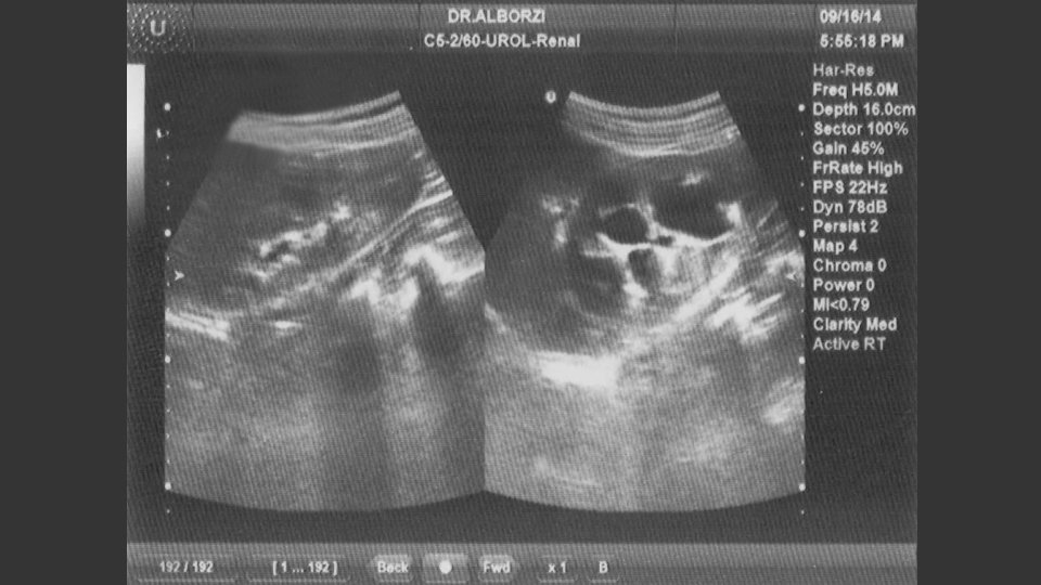
• For bladder and uretral DIEs however, the three modalities were observed to have the same results for the diagnosis of DIE. The sensitivity, specificity and accuracy for the three tests were 100%, 99. 68%, and 99. 68%, respectively
Comparison of TVS, TRS and MRI regarding total lesions • Regardless of anatomical location, TRS possessed marginally higher sensitivity for the diagnosis of DIE lesions than TVS and MRI (81. 1% vs. 80. 1% and 77. 9%, respectively). • Meanwhile, the specificity was slightly higher for MRI as compared to TVS and TRS (97. 1% vs. 96. 6% and 95. 8%, respectively). • TRS held a similar accuracy compared to TVS and MRI (93. 3% vs. 93. 1% and 92. 8%, respectively).
Discussion • Once the diagnosis of DIE is suspected in a woman of child-bearing age, the next question would be “Which imaging modality is appropriate for confirming the diagnosis of DIE? ” • In our tertiary clinic, three options are readily available which were used to identify these lesions prior to operation. These options include TVS, TRS and pelvic MRI. • Our findings demonstrated the superiority of TRS over TVS and MRI for the pre-operative diagnosis of DIE in most anatomical locations such as uterosacral ligaments and rectovaginal septum. • However, TVS was found to be superior in the diagnosis of rectal wall DIEs. • For certain locations such as ovarian fossa and retro cervical DIEs, MRI yielded better results. This might have attributed to the to poor ultrasound view of these locations. • On the other hand, for bladder and uretral DIEs; TVS, TRS and MRI were shown to have same results.
• We defined the exact anatomical site of involvement without merging subclasses together. As such, the most frequent sites of DIE was the uterosacral ligaments (43%). • Other common sites were in turn the ovarian fossa (16. 9%), rectal wall (14. 8%), rectovaginal septum (12. 5%) and retrocervical wall (10. 9%). • Bladder and uretra were only involved in 4 (1. 1%) and 2 (0. 6%) patients, respectively. We did not arrive at any reasonable explanation for such a low frequency as compared to the previous studies
• We compared the performance of each diagnostic modality with the results of operative laparoscopy, for each anatomic location and total lesions. • For lesions located in uterosacral ligaments which consisted 43% of lesions as the most common site of involvement, TRS demonstrated a higher sensitivity than TVS and MRI (82. 8% vs. 70. 9% and 63. 6%, respectively) and a lower miss rate (0. 17 vs. 0. 29 and 0. 36 for TVS and MRI, respectively). Additionally, TRS had a better performance to rule out the lesions were the study reported negative results (NPV of 85. 1 vs. 77. 8 and 73. 9% and LR- of 0. 19 vs. 0. 31 and 0. 39, for TVS and MRI, respectively). Our findings were in line with an earlier report (Grasso, Di Giacomo et al. 2010) on the performance of MRI in detecting DIE lesions within uterosacral ligaments (sensitivity of 63. 5%, specificity of 93. 9%, PPV 90. 5 and NPV 73. 9%, miss rate of 0. 36, accuracy of 79. 5%, LR+ of 10. 55 and LR- of 0. 39). These results were in agreement with another study regarding the MRI performance for the detection of DIE of uterosacral ligaments, in terms of sensitivity and likelihood ratios (Bazot, Lafont et al. 2009).
• With respect to retrocervical lesions, according to Abrao et al. , TVS owns a better sensitivity, specificity, NPV, PPV and accuracy than MRI (Abrao, Goncalves et al. 2007). • In contrast, our study found MRI to have a better diagnostic performance than TVS (sensitivity of 65. 8% and 52. 6%, specificity of 96. 4 and 94. 6%, PPV of 71. 4 and 57. 1%, NPV of 95. 4 and 93. 6%, miss rate of 0. 34 and 0. 47 and accuracy of 92. 7 and 89. 6%, respectively). • Such a disparity in results may at least partly be attributed to a notable difference in sample size.
• For DIE lesions within the rectovaginal septum, unlike a previous study which found MRI to be superior to TVS( Bazot, Lafont et al. 2009) our findings indicated a higher sensitivity, accuracy and likelihood ratios with lower miss rates, for TVS than MRI for the detection of DIE (sensitivity of 86. 4% and 72. 7%, PPV of 73% and 71. 1%, NPV of 97. 7% and 95. 6%, miss rates of 0. 14 and 0. 27, accuracy of 93. 7% and 92. 1% , LR+ of 16. 8 and 15. 27, LR- of 0. 14 and 0. 29, respectively). • Nevertheless, specificity was shown to be superior in MRI as compared to TVS (95. 2% vs. 94. 9%, respectively). • Based on a recent investigation on the ultrasound mapping system for the surgical managements of DIEs, TVS was found to have a sensitivity, specificity, PPV and NPV of 73. 9%, 86. 2%, 80. 9% and 80. 7% for such lesions, respectively (Exacoustos, Malzoni et al. 2014)
• For rectal DIE, similar to Bazot et al. we found the sensitivity and specificity of TVS superior to MRI (88. 46%, 98. 87% for TVS vs. 76. 92%, 96. 6% for MRI)
• Concerning the bladder DIE, our study suggested similar results for the three modalities. • Fedele et al. reported TVS superiority to MRI and trans-abdominal sonography in determining site of the lesions; however, they studied a small series of patients and their investigation dates back to the time when the use of these modalities for the diagnosis of DIE were in their prime • Meanwhile, our findings were in agreement with other reports (Bazot, Detchev et al. 2003, Bazot, Thomassin et al. 2004, Bazot, Lafont et al. 2009) introducing TVS as an accurate method in the diagnosis of DIE within bladder. • An earlier study by Grasso et al. found the sensitivity of 83. 3, specificity of 100% , PPV of 100% and NPV of 92. 5% for MRI in detection of bladder DIE, while our investigation reported a higher sensitivity (100%) and lower precision (PPV=80%) for the same.
• According to Balleyguier et al. MRI provides more accurate results than TVS in detecting the DIE lesions of bladder, especially in deeplyextended posterior DIEs and in small lesions missed by TVS. • Our study supported the findings from a recent investigation which reported the sensitivity of 100%, specificity of 96. 8%, PPV of 72. 7% and NPV of 100% for TVS ( Exacoustos, Malzoni et al. 2014)
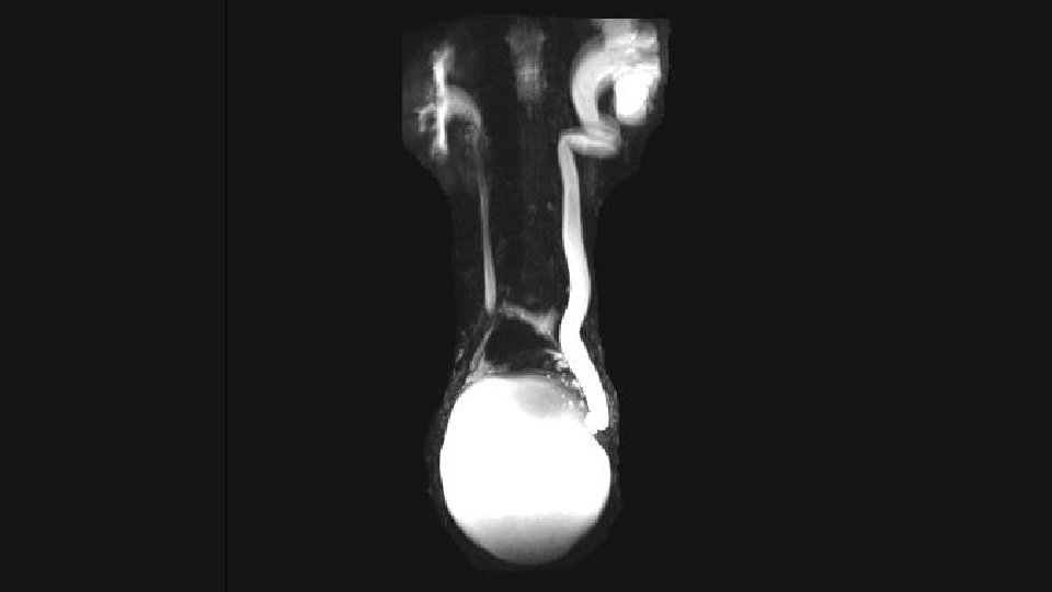
• The observed TVS, TRS and MRI sensitivity of 100% for the detection of ureteral DIE in our study, was not aligned with the results from Chamie and Grasso (MRI sensitivity of 66. 6% and 50%, respectively)
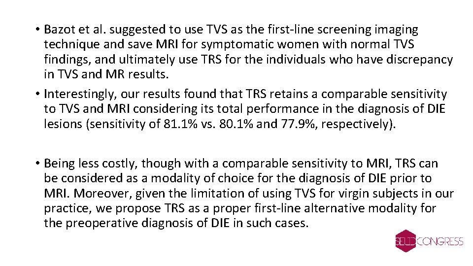
• Bazot et al. suggested to use TVS as the first-line screening imaging technique and save MRI for symptomatic women with normal TVS findings, and ultimately use TRS for the individuals who have discrepancy in TVS and MR results. • Interestingly, our results found that TRS retains a comparable sensitivity to TVS and MRI considering its total performance in the diagnosis of DIE lesions (sensitivity of 81. 1% vs. 80. 1% and 77. 9%, respectively). • Being less costly, though with a comparable sensitivity to MRI, TRS can be considered as a modality of choice for the diagnosis of DIE prior to MRI. Moreover, given the limitation of using TVS for virgin subjects in our practice, we propose TRS as a proper first-line alternative modality for the preoperative diagnosis of DIE in such cases.
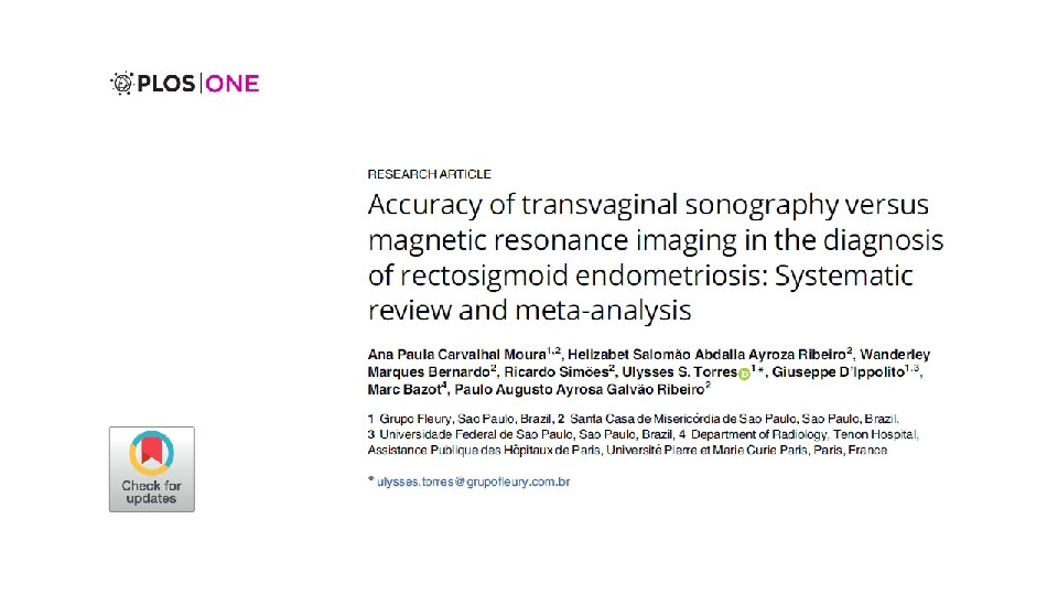
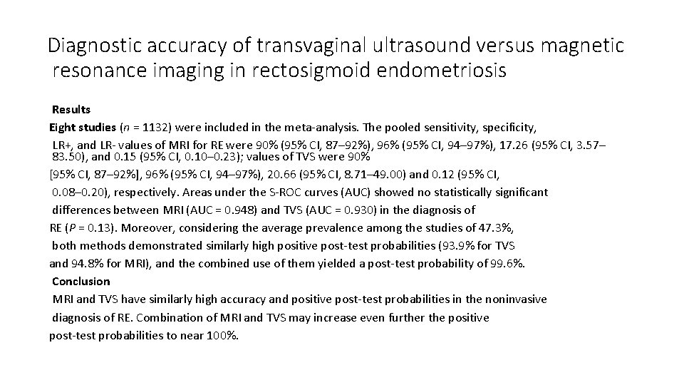
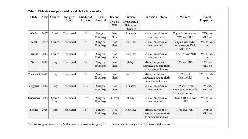
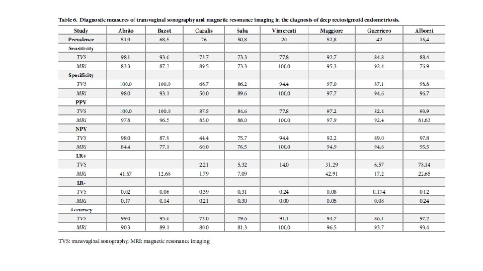
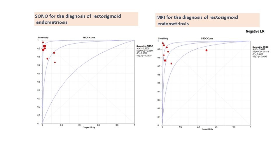
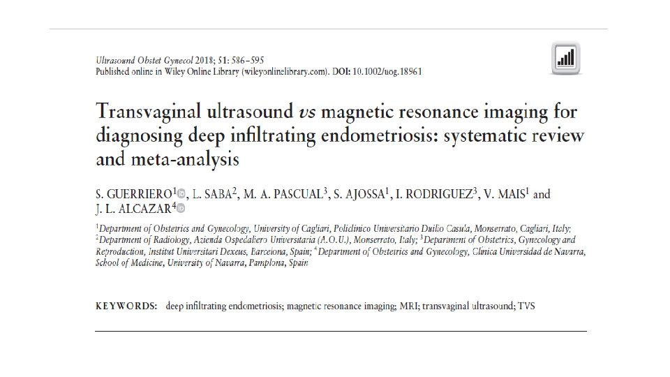
Diagnostic accuracy of transvaginal ultrasound versus magnetic resonance imaging in rectosigmoid endometriosis Results Eight studies (n = 1132) were included in the meta-analysis. The pooled sensitivity, specificity, LR+, and LR- values of MRI for RE were 90% (95% CI, 87– 92%), 96% (95% CI, 94– 97%), 17. 26 (95% CI, 3. 57– 83. 50), and 0. 15 (95% CI, 0. 10– 0. 23); values of TVS were 90% [95% CI, 87– 92%], 96% (95% CI, 94– 97%), 20. 66 (95% CI, 8. 71– 49. 00) and 0. 12 (95% CI, 0. 08– 0. 20), respectively. Areas under the S-ROC curves (AUC) showed no statistically significant differences between MRI (AUC = 0. 948) and TVS (AUC = 0. 930) in the diagnosis of RE (P = 0. 13). Moreover, considering the average prevalence among the studies of 47. 3%, both methods demonstrated similarly high positive post-test probabilities (93. 9% for TVS and 94. 8% for MRI), and the combined use of them yielded a post-test probability of 99. 6%. Conclusion MRI and TVS have similarly high accuracy and positive post-test probabilities in the noninvasive diagnosis of RE. Combination of MRI and TVS may increase even further the positive post-test probabilities to near 100%.
SONO for the diagnosis of rectosigmoid endometriosis MRI for the diagnosis of rectosigmoid endometriosis
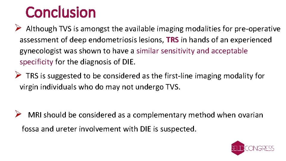
Conclusion Ø Although TVS is amongst the available imaging modalities for pre-operative assessment of deep endometriosis lesions, TRS in hands of an experienced gynecologist was shown to have a similar sensitivity and acceptable specificity for the diagnosis of DIE. Ø TRS is suggested to be considered as the first-line imaging modality for virgin individuals who do may not undergo TVS. Ø MRI should be considered as a complementary method when ovarian fossa and ureter involvement with DIE is suspected.