
A clinical longterm radiographic evaluation of graft height

- Slides: 113
A clinical long-term radiographic evaluation of graft height changes after maxillary sinus floor augmentation with a 2: 1 autogenous bone /xenograft mixture and simultaneous placement of dental implants Naoki Hatano, Yoshinaka Shimizu, Kiyoshi Ooya Clin. Oral Impl. Res. 2004; 15: 339 -345
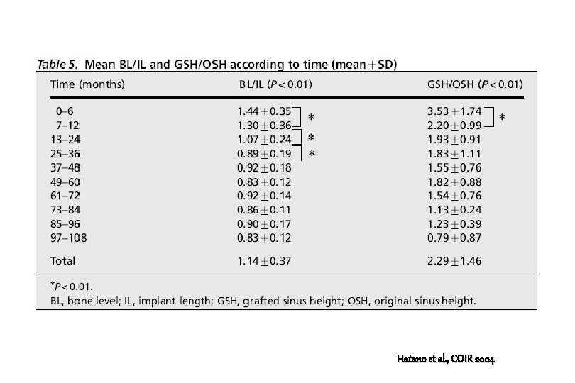
The aim of this study 1. To assess long-term changes in sinus-graft height after maxillary sinus floor augmentation and simultaneous placement of implants. Hatano et al. , COIR 2004
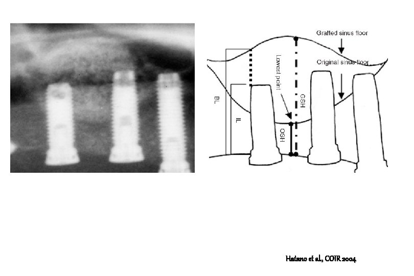
Materials and Methods 1. A total of 191 patients who underwent maxillary sinus floor augmentation were radiographically (using only panoramic x-ray) followed for up to about 10 years. 2. A 2: 1 mixture of autogenous bone and bovine xenograft (Bio. Oss) was used as the graft material. 3. Changes in sinus-graft height were calculated with respect to implant length and original sinus height. Hatano et al. , COIR 2004
Hatano et al. , COIR 2004
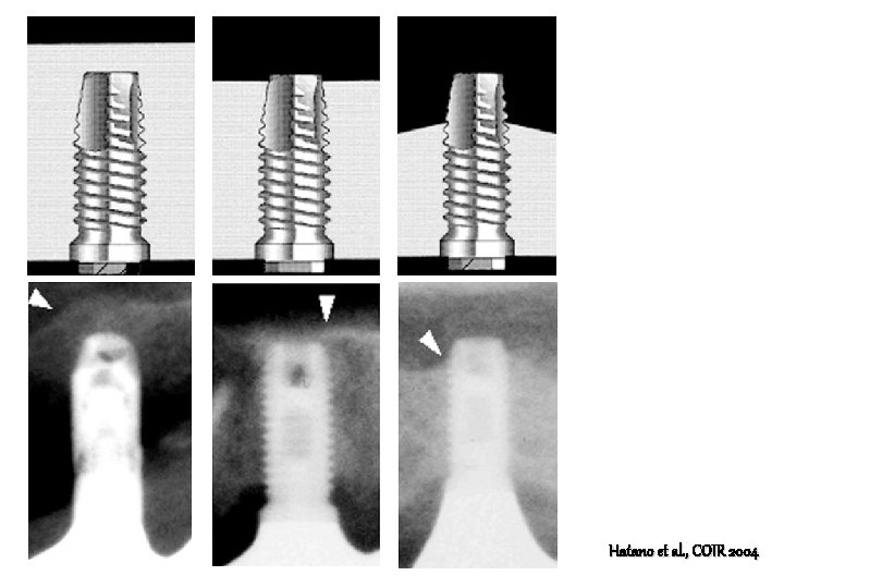
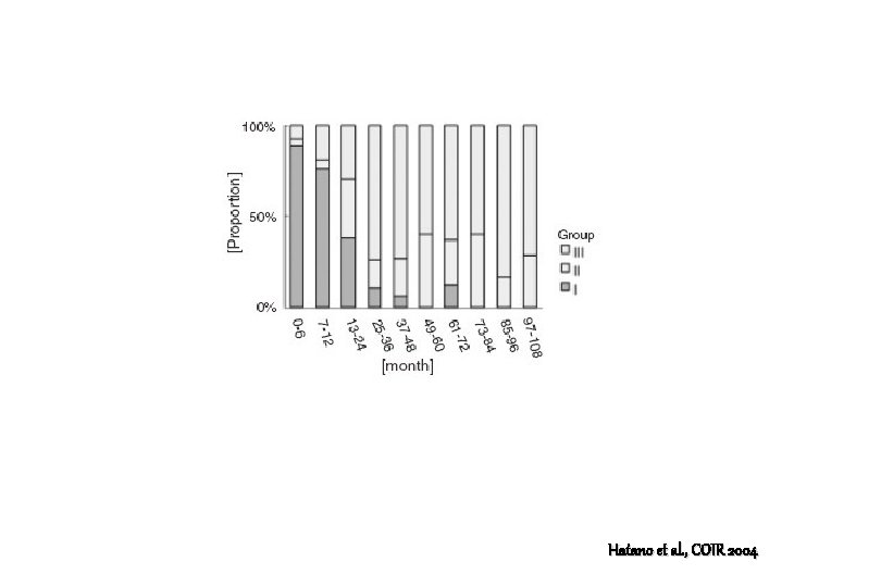
Three groups based on the height of the grafted sinus floor 1. Group I, in which the grafted sinus floor was above the implant apex 2. Group II, in which the implant apex was level with the grafted sinus floor 3. Group III, in which the grafted sinus floor was below the implant apex Hatano et al. , COIR 2004
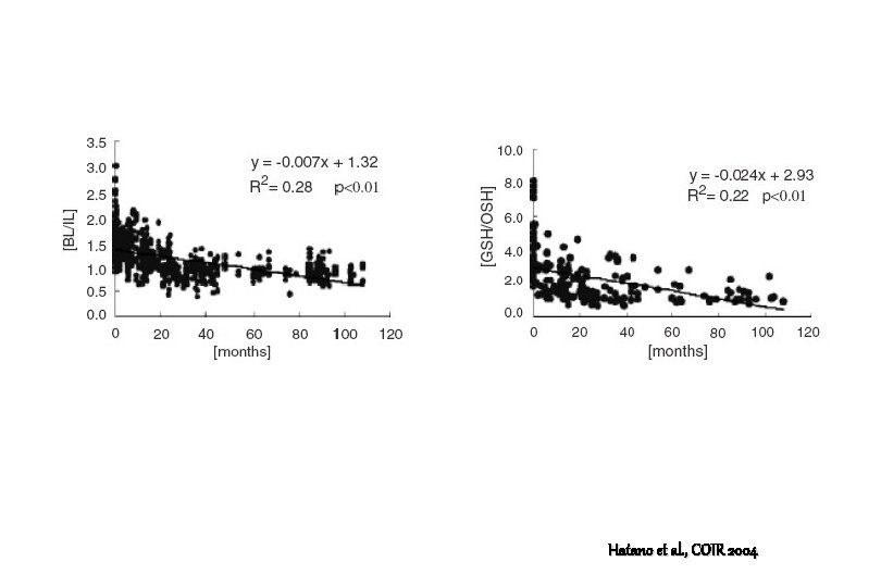
Results 1. After augmentation, the grafted sinus floor was consistently located above the implant apex. 2. After 2 -3 years, the grafted sinus floor was level with or slightly below the implant apex. This relationship was maintained over the long term. 3. Sinus graft height decreased significantly and approached original sinus height. Hatano et al. , COIR 2004
Results 4. The proportion of patients classified as belonging to Group III reached a maximum from year 3 onwards. 5. The clinical survival rate of implants was 94. 2%. All implant losses occurred within 3 years after augmentation. Hatano et al. , COIR 2004
Hatano et al. , COIR 2004
Hatano et al. , COIR 2004
Hatano et al. , COIR 2004
Hatano et al. , COIR 2004
Discussion 1. Changes in the sinus floor in our study might reflect adaptation to implant loading and maxillary sinus ventilation (Asai et al. 2002) 2. The overall height of bone graft decreased during the fist 2 -3 years after augmentation. Therefore, only minor changes occurred. 3. However, graft height up to 96 months after augmentation was higher than that observed preoperatively. Hatano et al. , COIR 2004
Discussion 4. These findings suggest that implant loading promotes osteogenesis over the long term (Nyström et al. 1993; Keller et al. 1994) 5. Implant loading may exert a stabilizing effect on the maintenance of bone graft height, consistent with the findings of Listrom & Symington (1988) Hatano et al. , COIR 2004
Discussion 6. Autopsy studies of retrieved implants placed together with FDBA about 1 year previously showed that the implant apex was covered by sinus mucosa, not by bone (Whittaker et al. 1989; Ga. Rey et al. 1991) 7. In animal studies using autogenous bone as grafting material, augmented bone surrounding implants gradually thinned over time, eventually exposing the implant within the sinus cavity. (Coombs et al. 1995) Hatano et al. , COIR 2004
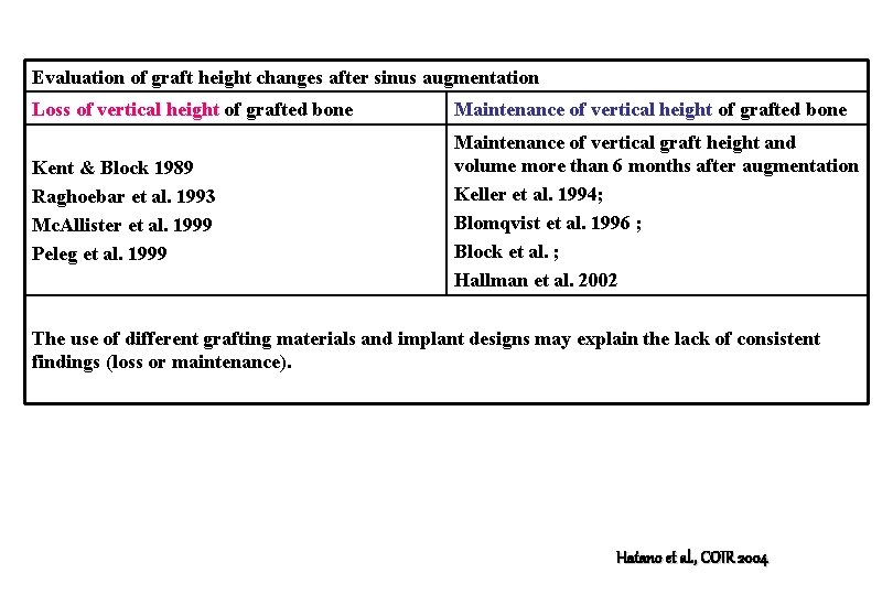
Evaluation of graft height changes after sinus augmentation Loss of vertical height of grafted bone Maintenance of vertical height of grafted bone Kent & Block 1989 Raghoebar et al. 1993 Mc. Allister et al. 1999 Peleg et al. 1999 Maintenance of vertical graft height and volume more than 6 months after augmentation Keller et al. 1994; Blomqvist et al. 1996 ; Block et al. ; Hallman et al. 2002 The use of different grafting materials and implant designs may explain the lack of consistent findings (loss or maintenance). Hatano et al. , COIR 2004
Pneumatization and alveolar bone resorption 1. Pneumatization may be caused by positive intra-sinus air pressure due to respiration (Chanavaz 1990), and this pressure might promote resorption and pneumatization after sinus augmentation. (Hürzeler et al. 1996) 2. Pressure-induced degradation and resorption in the grafted sinus floor may lead to loss of vertical bone height. Hatano et al. , COIR 2004
Evaluation method for confirming the results of sinus grafting 1. Panorama : two dimensional and poor visualization 2. CT : recommended to identify the outline of the rafted sinus floor to measure the height and volume of bone available for implant placement 3. Magnetic resonance imaging; facilitating accurate evaluation of grafted sinus floor 4. Naso-endoscope : reliable diagnostic method providing detailed information Hatano et al. , COIR 2004
Sinus mucosa and healing 1. Sinus mucosa has been shown experimentally to undergo physiological adaptation with the development of chronic sinusitis after maxillary sinus floor augmentation (Bravetti et al. 1998). 2. Penetration of the sinus by implants may increase the risk of infection and is regarded as a contraindication (Pedersen et al. 1979) Hatano et al. , COIR 2004
Sinus mucosa and healing 3. However, Brånemark et al. (1984) reported that sinus penetration by implants caused no undesirable effects. 4. Moreover, Peleg et al. (1999) found no correlation between incomplete bone coverage on computed tomography and implant failure, consistent with the results of our study. Hatano et al. , COIR 2004
Conclusion 1. Progressive sinus pneumatization occurs after augmentation with a 2: 1 autogenous bone/xenograft mixture, and long-term stability of sinus-graft height represents an important factor for implant success. Hatano et al. , COIR 2004
Sinus augmentation utilizing anorganic bovine bone (Bio. Oss) with absorbable and nonabsorbable membranes placed over the lateral window : Histomorphometric and clinical analyses Stephan S. Wallace, Sang-Choon Cho, Nicholas Elian, Diogo Monteiro, Byung Soo Kim, Dennis P. Tarnow J. Clin. Periodontol. 2003; 30: 261 -270
Materials and Methods 1. Sinus lift were performed on 51 patients ( 38 unilateral, 13 bilateral) with the delayed placement of 135 implants 2. 64 sinus augmentation procedures 3. Bio. Oss with and without autogenous bone 4. The grafting material used in these cases was either Bio. Oss alone or a composite grafting consisting of Bio. Oss with less than 20% intra-orally harvested bone. Wallace et al. , IJPRD 2005
Materials and Methods 5. Without membrane vs with Bio. Gide (resorbable) or e-PTFE (non-resorbable, Gore-Tex) 6. Histomorphometric data were obtained at the time of implant placement, 6 to 10 months following the grafting procedure. Wallace et al. , IJPRD 2005
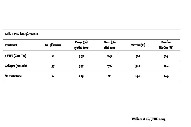
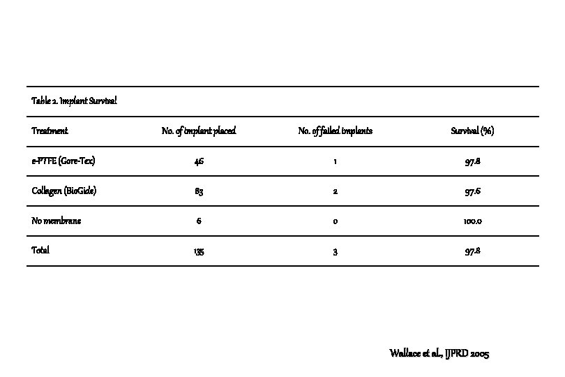
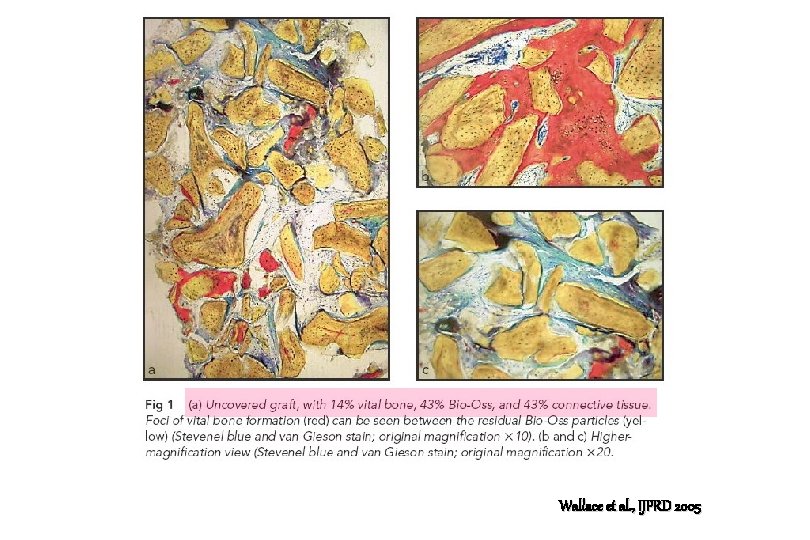
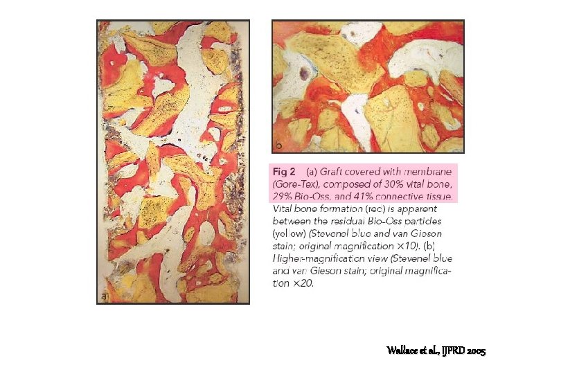
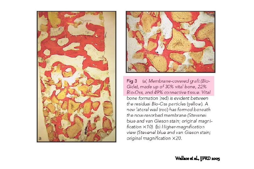
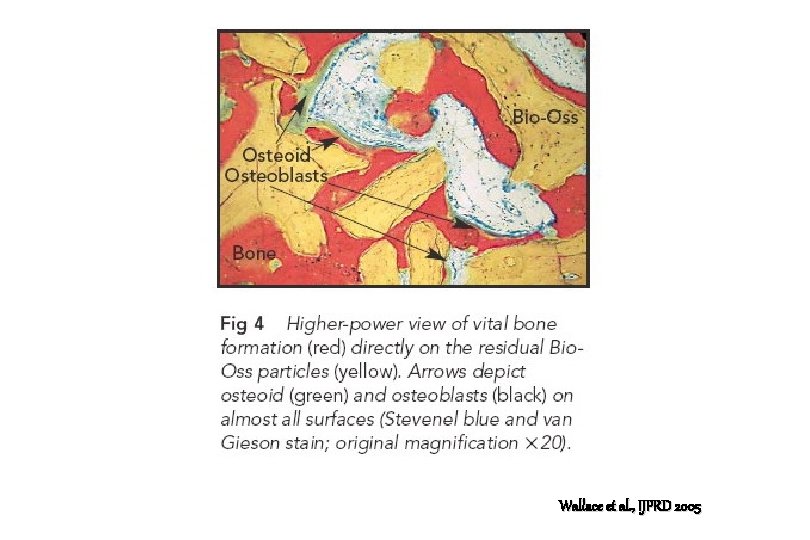
Results 1. Vital bone formation was 17. 6%, 16. 9% and 12. 1%, respectively, for the Bio. Gide, Gore-Tex, and no membrane groups. 2. Of the 135 implants placed there were 3 failures (2 Bio. Gide, 1 Gore-Tex). 3. There were no significant difference between the membrane groups as to vital bone formation and implant survival. Wallace et al. , IJPRD 2005
Table 1. Vital bone formation No. of sinuses Range (%) of vital bone Mean (%) vital bone Marrow (%) Residual Bio-Oss (%) e-PTFE (Gore-Tex) 21 3 -53 16. 9 51. 2 31. 9 Collagen (Bio. Gide) 37 3 -51 17. 6 56. 0 26. 4 No membrane 6 1 -29 12. 1 63. 6 24. 3 Treatment Wallace et al. , IJPRD 2005
Table 2. Implant Survival Treatment No. of implant placed No. of failed implants Survival (%) e-PTFE (Gore-Tex) 46 1 97. 8 Collagen (Bio. Gide) 83 2 97. 6 No membrane 6 0 100. 0 Total 135 3 97. 8 Wallace et al. , IJPRD 2005
Wallace et al. , IJPRD 2005
Wallace et al. , IJPRD 2005
Wallace et al. , IJPRD 2005
Wallace et al. , IJPRD 2005
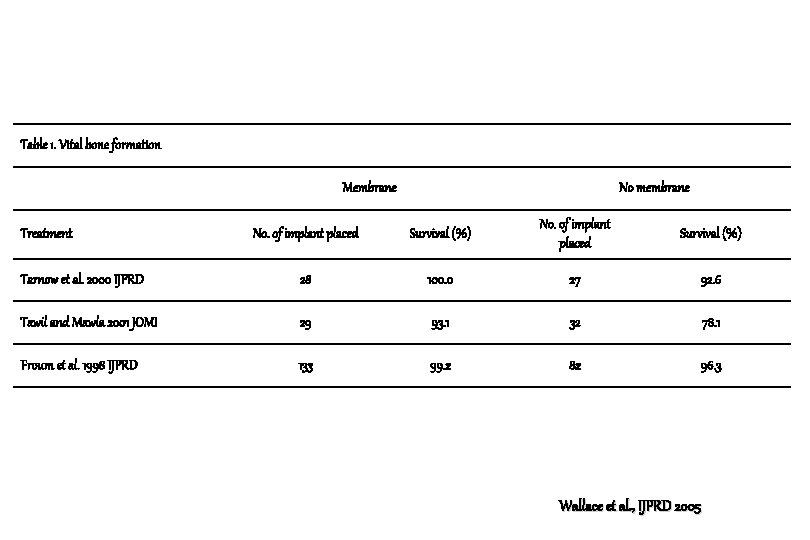
Table 1. Vital bone formation Membrane No membrane No. of implant placed Survival (%) Tarnow et al. 2000 IJPRD 28 100. 0 27 92. 6 Tawil and Mawla 2001 JOMI 29 93. 1 32 78. 1 Froum et al. 1998 IJPRD 133 99. 2 82 96. 3 Treatment Wallace et al. , IJPRD 2005
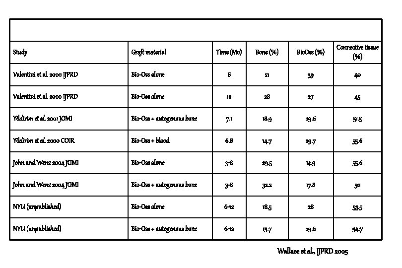
Study Graft material Time (Mo) Bone (%) Bio. Oss (%) Connective tissue (%) Valentini et al. 2000 IJPRD Bio-Oss alone 6 21 39 40 Valentini et al. 2000 IJPRD Bio-Oss alone 12 28 27 45 Yildirim et al. 2001 JOMI Bio-Oss + autogenous bone 7. 1 18. 9 29. 6 51. 5 Yildirim et al. 2000 COIR Bio-Oss + blood 6. 8 14. 7 29. 7 55. 6 John and Wenz 2004 JOMI Bio-Oss alone 3 -8 29. 5 14. 9 55. 6 John and Wenz 2004 JOMI Bio-Oss + autogenous bone 3 -8 32. 2 17. 8 50 NYU (unpublished) Bio-Oss alone 6 -12 18. 5 28 53. 5 NYU (unpublished) Bio-Oss + autogenous bone 6 -12 15. 7 29. 6 54. 7 Wallace et al. , IJPRD 2005
Sinus window and membrane coverage 1. There is sufficient evidence today to justify the placement of a barrier membrane over the lateral window. 2. Clinically, the ingrowth of connective tissue into sinus grafts not protected by a membrane (encleftation) has been reported by Mc. Allister et al. Wallace et al. , IJPRD 2005
“ Caging effect “ 1. When a membrane was placed over a grafted bone defect, completely sealing the defect from the outside environment, corticalization of the wound surface, contiguity of the graft particles, and increased vascularity of the graft were noted. Murray et al. , Am. J. Surg. 1957
Sinus lifting with grafting and new bone formation 1. These histologic wound healing studies all show that bone formation initiates from the walls and floor of sinus after the schneiderian membrane had been lifted and displaced. 2. This displacement would expose the vascular supply in the lateral wall, along with the surrounding perivascular osteoblasts, to the placed graft material, allowing for the “angiogenesis that must precede osteogenesis”. Wallace et al. , IJPRD 2005
Sinus lifting with grafting and new bone formation 3. Bone formation in the grafted sinus is certainly aided by the displacement of the schneiderian membrane, which was shown by Hass et al. (JOMI 2002) to be avascular and by Hürzeler et al. (COIR 1997) to offer minimal osteogenic potential in sinus grafts in an animal model. Wallace et al. , IJPRD 2005
Sinus lifting with grafting and new bone formation 4. Bone formation also appears to be aided by the exclusion of the periosteum from the regenerating sinus graft. 5. Once lifted and replaced, the periosteum loses its osteogenic potential and becomes fibrogenic, which would explain the soft tissue encleftation that is seen when a membrane is not used over the window. Wallace et al. , IJPRD 2005
Sinus lifting with grafting and new bone formation 6. This is quite similar to the soft tissue ingrowth that occurs into unprotected (no membrane coverage ) periodontal and ridge augmentation sites. Wallace et al. , IJPRD 2005
Membrane for sinus window coverage. Bio. Gide or e. PTFE ? 1. There was no statistical difference in the vital bone formation of grafted sinuses between the membrane groups of the present study. 2. Zitzmann et al. showed no statistical difference in implant survival rates following GBR procedures with either Bio. Oss/Gore-Tex (97. 8% implant survival rates) or Bio. Oss/Bio-Gide (97. 6% implant survival rates) Wallace et al. , IJPRD 2005
Bio. Oss and favorable results : Combination effect ? 1. Osteoconductive capacity of the cancellous xenograft, a feature that may be dependent on the pore size. 2. Supplier of minerals that are necessary for the bone formation; 3. Their density provides stability to the graft and the implants placed in them. 4. Their density persists long-term because these grafts do not completely resorb. Wallace et al. , IJPRD 2005
Delayed implantation and biological advantage 1. When implants are placed in these matured bone grafts, the placement procedure creates a secondary wound with subsequent bleeding. 2. Implant surfaces particulaly those that are textured, retain blood clot for the eventual boneimplant contact (BIC) that we call osseointegration. Wallace et al. , IJPRD 2005
Bio-Oss and implant interface 1. They do not show xenograft material in direct contact with the implant surface. 2. This would mean that the residual xenograft in effect increases the mineral content of the resulting matured graft while not interfering with the possibility for BIC. Rosenlicht and Tarnow et al. , Int. J. Oral Implantol. 1999; Valentini et al. COIR 1998; Scarano et al. JP 2004;
Grafted sinus and bone density 1. % of vital bone, connective tissue, and residual xenograft to be in order of 25%, 50% and 25%, respectively, at a time interval of 6 to 12 months. 2. Because of the residual 25% xenograft volume, a mature graft that is classified as type 3 and 4 by nature of it vital bone content may, in fact , function like bone of type 2 density NYU. , unpublished 2005
Grafted sinus and bone density 3. These observed histologic and wound healing phenomena may explain why mineralized particulate bone replacement grafts used for sinus augmentation surgery have been shown to perform as well or better than grafts of 100% autogenous bone Wallace & Froum Ann Periodontol. 2003; del Fabbro et al. IJPRD 2004
Conclusion 1. Vital bone formation in the sinus grafts is improved when a membrane is placed over the window. 2. Vital bone formation is similar with non-resorbable Gore-Tex and bio-absorbable Bio. Gide membranes. 3. Implant survival is similar with Gore-Tex and Bio. Gide membranes. Wallace et al. , IJPRD 2005
Conclusion 4. Bio. Oss demonstrates excellent osteoconductive properties in the grafted sinuses. 5. Bio. Oss alone, or as a composite graft with minimal amounts of autogenous bone, results in an extremely high implant survival rate in the grafted sinus (97. 8%) Wallace et al. , IJPRD 2005
Deproteinized cancellous bovine bone (Bio-Oss ) as bone substitute for sinus floor elevation : A retrospective, histomorphometrical study of five cases Tadjoedin E. S. De Lange G. L. Bronckers ALJJ Lyaruu D. M. Burger E. H. J. Clin. Periodontol. 2003; 30: 261 -270
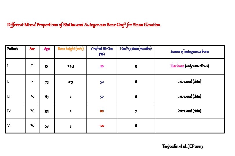
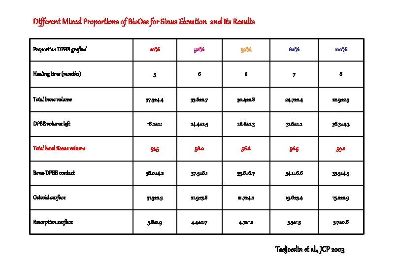
Different Mixed Proportions of Bio. Oss and Autogenous Bone Graft for Sinus Elevation Patient Sex Age Bone height (min) Grafted Bio. Oss (%) Healing time(months) I F 54 2. 5 -3 20 5 Iliac bone (only cancellous) II F 73 2 -3 50 6 Intra oral (chin) III M 63 2 50 6 Intra oral (chin) IV M 59 3 80 7 Intra oral (chin) V M 59 3 100 8 Source of autogenous bone Tadjoedin et al. , JCP 2003
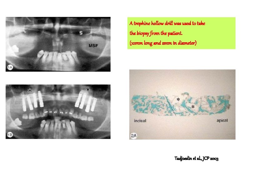
A trephine hollow drill was used to take the biopsy from the patient. (10 mm long and 2 mm in diameter) Tadjoedin et al. , JCP 2003
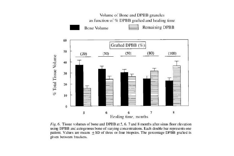
Different Mixed Proportions of Bio. Oss for Sinus Elevation and Its Results Proportion DPBB grafted 20% 50% 80% 100% 5 6 6 7 8 Total bone volume 37. 3± 4. 4 33. 6± 2. 7 30. 4± 2. 8 24. 7± 2. 4 22. 9± 2. 5 DPBB volume left 16. 2± 2. 1 24. 4± 2. 5 26. 6± 2. 3 31. 8± 2. 2 36. 3± 4. 3 53. 5 58. 0 56. 8 56. 5 59. 2 Bone-DPBB contact 38. 0± 4. 2 37. 5± 8. 1 35. 6± 6. 7 34. 1± 6. 6 33. 5± 4. 5 Osteoid surface 31. 5± 2. 3 21. 9± 3. 8 21. 7± 4. 2 19. 6± 3. 4 15. 2± 2. 9 Resorption surface 5. 8± 1. 9 4. 4± 0. 7 4. 7± 1. 2 3. 3± 1. 3 3. 7± 0. 6 Healing time (months) Total hard tissue volume Tadjoedin et al. , JCP 2003
Results ; 20% Bio. Oss + 80 AB, 5 months healing 1. Total bone volume amounted to 37% of the tissue volume which is in the range of normal, unaffected maxillary bone. (Tadjoedin et al. 2000 COIR) 2. In the grafted areas (close to the incisal part) where no DPBB granules were seen, this newly formed trabecular bone was predominantly of the lamellar type with Haversian and Volkmann canals. Tadjoedin et al. , JCP 2003
Results ; 20% Bio. Oss + 80 AB, 5 months healing 3. In other areas where DPBB granules were still present, the bone surrounding the granules was mostly of the woven type. 4. Many trabeculae demonstrated very active bone remodeling with a thick layer of osteoid surface at one side and osteoclastic resorption at the other side. Tadjoedin et al. , JCP 2003
Results ; 50% Bio. Oss + 50 AB, 6 months healing 1. Woven and lamellar type of trabecular bone had formed throughout the graft during the 6 -month healing period. 2. Many active osteoblasts lined the layers of osteoid, and bone remodeling was apparent by the presence of osteoclasts in Howship’s lacunae. Interestingly, osteoclasts could also be seen at the periphery of the DPBB granules. Tadjoedin et al. , JCP 2003
Results ; 80% Bio. Oss + 20 AB, 6 months healing 1. Trabecular bone had formed in the incisal part of the grafted area where DPBB granules were hardly observed. 2. In the middle and the apical region of the graft, many DPBB granules were observed, surrounded and connected to each other by bone tissue, most of the woven type and some of the lamellar type. Tadjoedin et al. , JCP 2003
Results ; 80% Bio. Oss + 20 AB, 7 months healing 3. Many trabeculae were in the process of being remodeled, as shown by the thick layer of osteoid layer lined at one side while osteoclastic resorption at the opposite. Tadjoedin et al. , JCP 2003
Results ; 100% Bio. Oss –incisal part 1. Bone formation had not yet reached the most apical part of the grafted area. 2. The incisal part of the graft contained vital new trabecular bone, mostly of the woven type and sometimes of the lamellar type, but only very few DPBB granules. Tadjoedin et al. , JCP 2003
Results ; 100% Bio. Oss –middle part 3. In the middle part, DPBB granules were clearly present, and had been encapsulated by newly formed woven bone. 4. Osteoclasts were seen in resorption lacunae of both new bone and DPBB granules. Tadjoedin et al. , JCP 2003
Results ; 100% Bio. Oss –apical part 4. In the most apical part of the grafted area , there was no bone present and the grafted DPBB granules were surrounded by soft connective tissue granules. 5. Impending bone formation in this area was however suggested by the presence of osteoid-like (red) material. Tadjoedin et al. , JCP 2003
Two different mechanisms for bone augmentation in sinus 1. Firstly, the growth of bone extends “upward” from the pre-existing bone surface at the sinus floor into the graft area. 2. Secondly, bone formation is initiated anywhere within the graft where autogenous bone particles are present. Tadjoedin et al. , JCP 2003
Conclusion 1. The amount of mineralized bone formed during the healing period correlated inversely with the proportion of grafted DPBB granules, a higher proportion of DPBB leading to less bone volume. Tadjoedin et al. , JCP 2003
Conclusion 2. However, even without any co-grafted autogenous bone particles a substantial amount of trabecular bone had still formed in the patient grafted with 100% DPBB, occupying 23% of the total tissue volume after 8 months of healing. Tadjoedin et al. , JCP 2003
Conclusion 3. The presence of many TRAP-positive multinucleated cells against the DPBB granules (often located in shallow resorption lacunae) suggests that DPBB granules were gradually degraded and resorbed by the activity of osteoclasts. Tadjoedin et al. , JCP 2003
Conclusion 4. The inclusion of autogenous bone particles in our study may be responsible for the higher rate of DPBB resorption that we found. 5. Higher amount of remodeling suggests a high amount of osteoclast recruiting and activity. Tadjoedin et al. , JCP 2003
Conclusion 6. The value of total mineral mass in our study was between 53% and 59%, independent of the amount of DPBB used. This total mass may also be important for providing implant stability (Moy et al. 1993) Tadjoedin et al. , JCP 2003
Maxillary sinus grafting with anorganic bovine bone : A clinical report of longterm results Pascal Valentini, David Abensur Int. J. Oral Maxillofac. Implants 2003; 18 : 556 -560
Materials and Methods 1. 1 -stage technique : residual ridge height of 5 mm or more, simultaneous implant placement and uncovered after 9 months 2. 2 -stage technique : less than 5 mm; implant placement after 6 months and 6 months of healing before being placed into function 3. 59 patients Valentini & Abensur. , IJOMI 2003
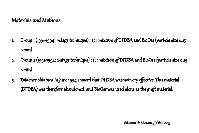
Materials and Methods 1. Group 1 (1991 -1994; 1 -stage technique) : 1 mixture of DFDBA and Bio. Oss (particle size 0. 25 -1 mm) 2. Group 2 (1991 -1994; 2 -stage technique) : 1 mixture of DFDBA and Bio. Oss (particle size 0. 25 -1 mm) 3. Evidence obtained in June 1994 showed that DFDBA was not very effective. This material (DFDBA) was therefore abandoned, and Bio. Oss was used alone as the graft material. Valentini & Abensur. , IJOMI 2003
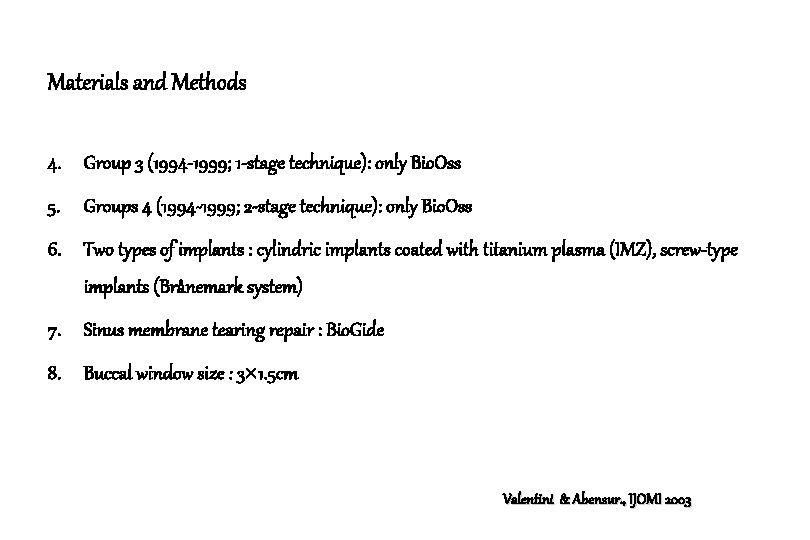
Materials and Methods 4. Group 3 (1994 -1999; 1 -stage technique): only Bio. Oss 5. Groups 4 (1994 -1999; 2 -stage technique): only Bio. Oss 6. Two types of implants : cylindric implants coated with titanium plasma (IMZ), screw-type implants (Brånemark system) 7. Sinus membrane tearing repair : Bio. Gide 8. Buccal window size : 3 1. 5 cm Valentini & Abensur. , IJOMI 2003
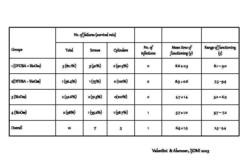
No. of failures (survival rate) Total Screws Cylinders No. of infections Mean time of functioning (y) Range of functioning (y) 1 (DFDBA + Bio. Oss) 5 (82. 1%) 3 (50%) 2 (90. 9%) 0 8. 6 ± 0. 3 8. 1 – 9. 0 2(DFDBA + Bio. Oss) 1 (96. 4%) 1 (75%) 0 (100%) 0 8. 9 ± 0. 6 7. 5 - 9. 4 3 (Bio. Oss) 2 (92. 6%) 2 (91. 3%) 0(100%) 0 4. 7 ± 1. 4 3. 2 – 6. 9 4 (Bio. Oss) 2 (98%) 1 (95. 2%) 1 (98. 7%) 1 5. 7 ± 1. 0 3. 7 – 7. 2 10 7 3 1 6. 5 ± 1. 9 2. 3 - 9. 4 Groups Overall Valentini & Abensur. , IJOMI 2003
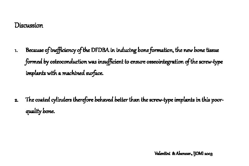
Discussion 1. Because of inefficiency of the DFDBA in inducing bone formation, the new bone tissue formed by osteoconduction was insufficient to ensure osseointegration of the screw-type implants with a machined surface. 2. The coated cylinders therefore behaved better than the screw-type implants in this poorquality bone. Valentini & Abensur. , IJOMI 2003
Discussion 3. One third of the cylindric implants used showed vertical bone loss that could be visualized radiographically; this was always accompanied by suppuration (peri-implantitis). 4. no pathology of this type was observed with the screw-type implants. Valentini & Abensur. , IJOMI 2003
Discussion 5. Analysis of the results showed survival rates that were 96. 8% when the Bio. Oss was used alone and 90% in the case of the mixture with DFDBA. 6. These results compare favorably with the conclusions of the 1996 Sinus Consensus Conference on the use of DFDBA in combination with other biologic materials. Valentini & Abensur. , IJOMI 2003
Conclusion 1. Screw-type implants with a machined surface showed survival rates slightly lower than those for cylindric implants, but they presented fewer long-term complications. 2. These complications were not associated with the grafting procedure. Valentini & Abensur. , IJOMI 2003
Sinus grafting with porous bone mineral (Bio-Oss ) for implant placement : A 5 -year study on 15 patients. Pascal Valentini, David Abensur, Birgit Wenz, Michael Peetz Robert Schenk Int. J. Periodontics Restorative Dent. 2000
Material and Method 1. 15 patients, 20 cases, 57 fixtures 2. Elliptically shaped window preparation (3 cm× 1. 5 cm) 3. Bio. Oss moistened with sterile saline. 4. Allowed to heal for 6 months, after sinus elevation the implants were inserted and remained submerged for 6 months. 5. Progressive loading for 3 months. 6. Mean loading period : 4. 0± 0. 5 years Valentini et al. , IJPRD 2000
Results at 6 months 1. Bio. Oss particles were homogenously distributed and interconnected by bony bridges. 2. Newly formed bone was undergoing remodeling , ranging from woven to well-organized lamellar bone. Valentini et al. , IJPRD 2000
Results at 6 months 3. Bio. Oss surfaces exposed to bone marrow and in contact with osteoclasts exhibits clear seams. 4. The preexisting host bone in the nongrafted areas consists of low-density spongiosa with well-vascularized, fatty-type bone marrow. Valentini et al. , IJPRD 2000
Results at 12 months 1. In comparison to the 6 month biopsies, the interconnecting bone between the Bio. Oss particles had matured by 12 months. 2. Intense remodeling activity, visible as osteoclastic resorption and new bone formation, continued. 3. Large surfaces of Bio. Oss were exposed to bone marrow. These surfaces regularly exhibited clear seams that were frequently covered by osteclasts. Valentini et al. , IJPRD 2000
Conclusion 1. With a mean loading period of 4. 0± 0. 5 years, the overall survival rate was 98. 1%. 2. Grafted compartments always showed a denser mineral scaffold consisting of Bio. Oss and bone than did the non-grafted sites. Valentini et al. , IJPRD 2000
Conclusion 3. While the amount of newly formed or existing bone in the grafted and non-grafted areas was strikingly similar (on average 21% and 25%, respectively), the implanted Bio. Oss particles increased the total mineral content. 4. However, the % of bone marrow decreased in the regenerated area. Both grafted and non-grafted compartments showed well-vascularized vital bone marrow. Valentini et al. , IJPRD 2000
Characteristics of grafted bone to implant interfaces 1. The degree of osseointegration was also equivalent to that seen in the surrounding implants placed into native, non-grafted bone. 2. As previously reported by Berglundh and Lindhe, in the Bio. Oss grafted areas, only regenerated bone came into direct contact with the implant surfaces ; grafted Bio. Oss particles did not contact the implants. 3. As in that study, Bio. Oss appears to be a good osteoconductive material leading to effective osseointegration of dental implants. Valentini et al. , IJPRD 2000
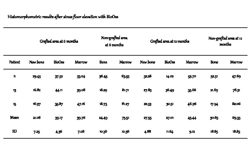
Histomorphometric results after sinus floor elevation with Bio. Oss Grafted area at 6 months Non-grafted area at 12 months Grafted area at 12 months Patient New bone Bio. Oss Marrow Bone Marrow 11 29. 45 37. 52 33. 04 36. 45 63. 55 32. 28 14. 02 53. 70 52. 31 47. 69 13 16. 82 44. 11 39. 08 18. 29 81. 71 27. 85 36. 49 35. 66 21. 69 78. 31 15 16. 97 35. 87 47. 16 18. 73 81. 27 22. 53 30. 51 46. 96 17. 94 82. 06 Mean 21. 08 39. 17 39. 76 24. 49 75. 51 27. 55 27. 01 45. 44 30. 65 69. 35 SD 7. 25 4. 36 7. 08 10. 36 4. 88 11. 64 9. 12 18. 85
Characteristic features of pressure zone of orthodontic moved tooth 1. Arrested root resorption 2. Active bone resorption 3. Disorganized collagen fibers in parts of the periodontal ligament Valentini et al. , IJPRD 2000
Conclusion 1. Bio. Oss particles were frequently observed to reside within the periodontal ligament, but never in direct contact with the root surface. 2. The relative volume of the tissue that was occupied by Bio. Oss particles, however, was much smaller in zone B than in zone A. Valentini et al. , IJPRD 2000
Conclusion 3. This finding, coupled with the fact that the biomaterial never occurred at the tension sidezone C- indicates that when the augmented site was challenged by physical means, i. e. the orthodontic tooth movement, the rate of degradation of Bio. Oss was enhanced. Valentini et al. , IJPRD 2000
Conclusion 4. Bio. Oss particles, which at 12 months occurred in the periodontal ligament of the pressure side (zone B) were either in direct contact with a fibroblast-collagen rich tissue, with lamellar bone or with multinucleated osteoclast-like cells. 5. Although the process including bone resorption and degradation of Bio. Oss may have many features in common, the detailed mechanism behind Bio. Oss degradation and elimination is still not totally understood. Valentini et al. , IJPRD 2000
Histological evaluation of Bio. Oss in a 2 -stage sinus floor elevation and implantation procedure. Valentini P. Abensur D. Densari D. Graziani J. N. Hämmerle C. H. F. Clin. Oral. Impl. Res. 1998
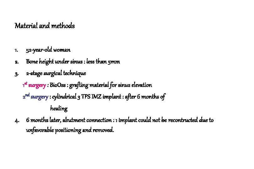
Material and methods 1. 52 -year-old woman 2. Bone height under sinus : less than 5 mm 3. 2 -stage surgical technique 1 st surgery : Bio. Oss : grafting material for sinus elevation 2 nd surgery : cylindrical 3 TPS IMZ implant : after 6 months of healing 4. 6 months later, abutment connection : 1 implant could not be recontructed due to unfavorable positioning and removed.
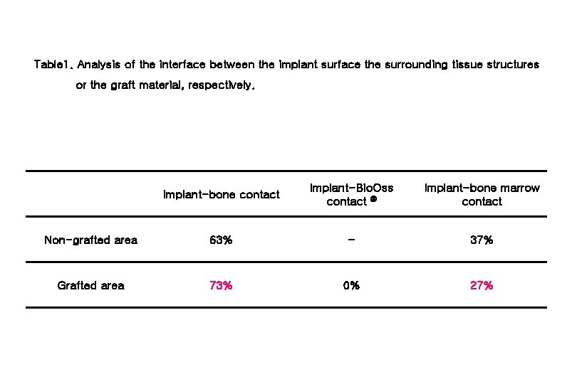
Table 1. Analysis of the interface between the implant surface the surrounding tissue structures or the graft material, respectively. Implant-bone contact Implant-Bio. Oss contact Implant-bone marrow contact Non-grafted area 63% - 37% Grafted area 73% 0% 27%
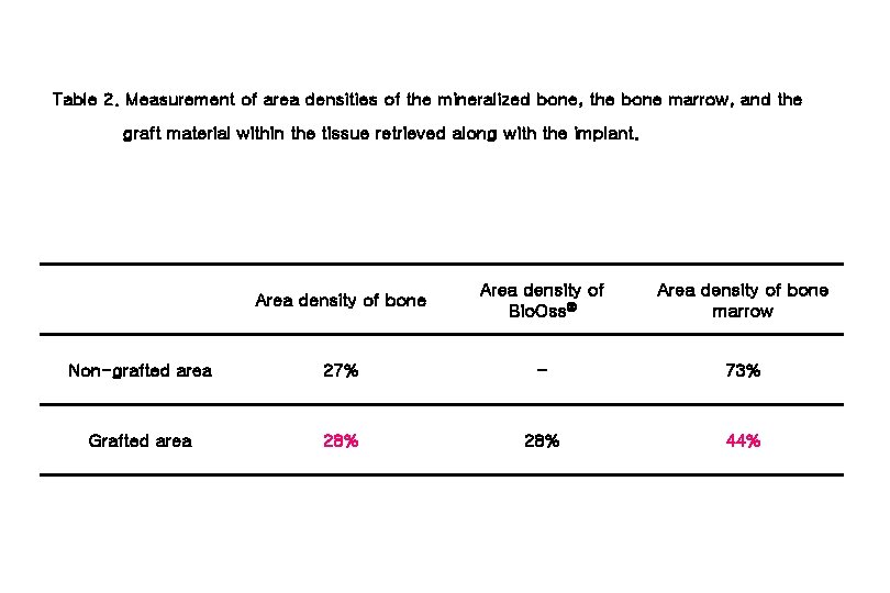
Table 2. Measurement of area densities of the mineralized bone, the bone marrow, and the graft material within the tissue retrieved along with the implant. Area density of bone Area density of Bio. Oss Area density of bone marrow Non-grafted area 27% - 73% Grafted area 28% 44%
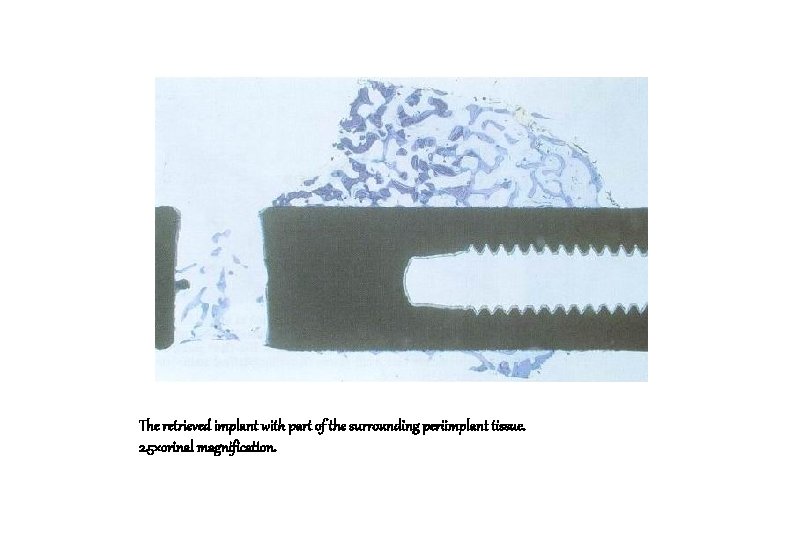
The retrieved implant with part of the surrounding periimplant tissue. 2. 5×orinal magnification.
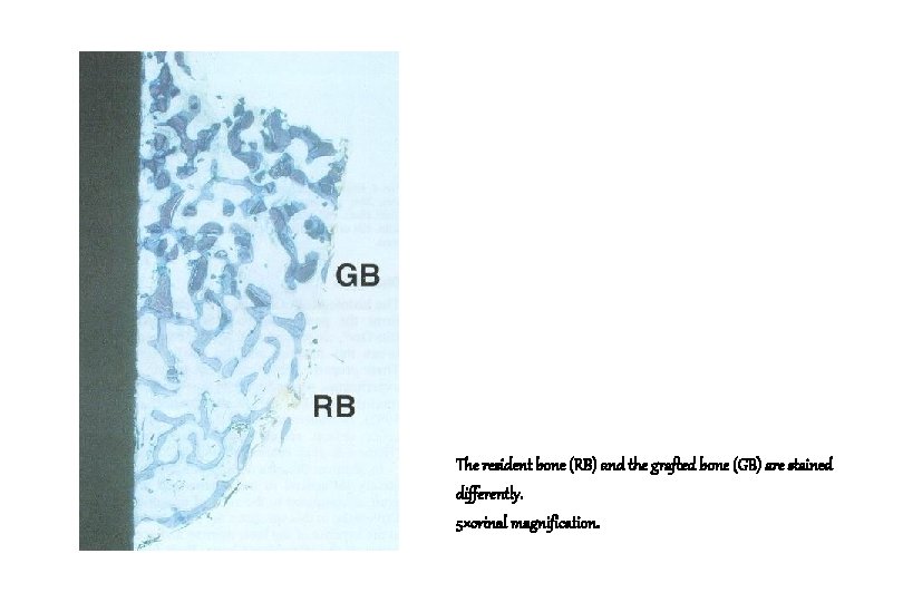
The resident bone (RB) and the grafted bone (GB) are stained differently. 5×orinal magnification.
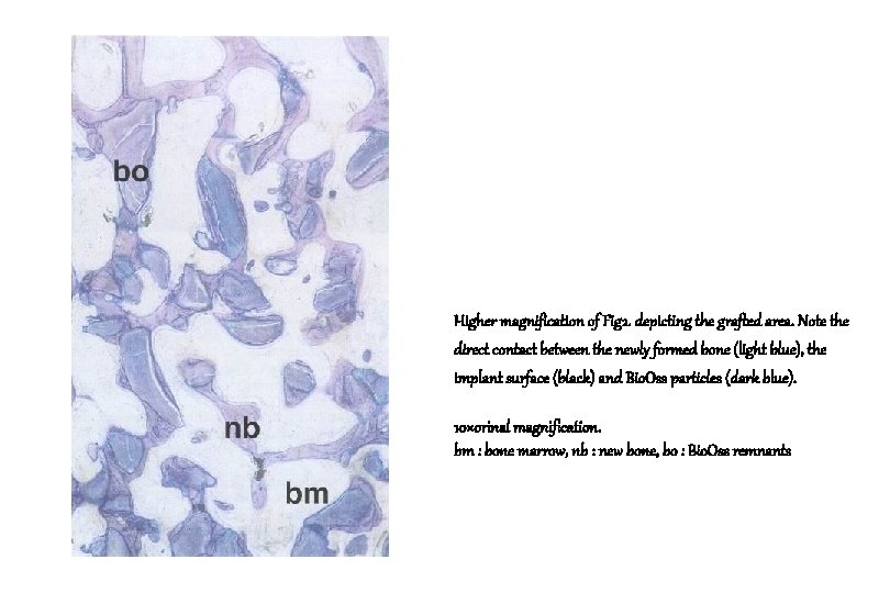
Higher magnification of Fig 2. depicting the grafted area. Note the direct contact between the newly formed bone (light blue), the implant surface (black) and Bio. Oss particles (dark blue). 10×orinal magnification. bm : bone marrow, nb : new bone, bo : Bio. Oss remnants
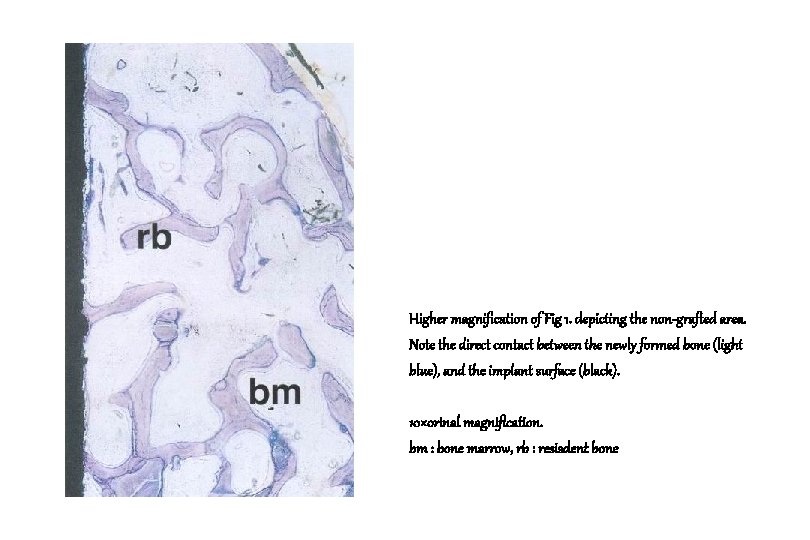
Higher magnification of Fig 1. depicting the non-grafted area. Note the direct contact between the newly formed bone (light blue), and the implant surface (black). 10×orinal magnification. bm : bone marrow, rb : resisdent bone
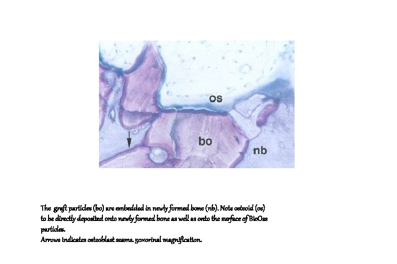
The graft particles (bo) are embedded in newly formed bone (nb). Note osteoid (os) to be directly deposited onto newly formed bone as well as onto the surface of Bio. Oss particles. Arrows indicates osteoblast seams. 50×orinal magnification.
Conclusion 1. Area density of bone was not negatively influenced in the regenerated area by the graft as compared to the zone of pre-existing bone. 2. Grafting with Bio. Oss does not decrease the adaptive capabilities of the new bone to give rise to osseointegration of an implant surface. Valentini et al. COIR 1998; 9: 59 -64
Conclusion 3. The absence of Bio. Oss resorption will not jeopardize the osseointegration of such implant since, no contact between the graft particles and the implant surface. 4. Therefore, Bio. Oss resorption does not seem absolutely necessary to provide predictable osseointegration. Valentini et al. COIR 1998; 9: 59 -64
Maxillary sinus floor elevation for implant placement with Demineralized Freeze-Dried Bone and Bovine bone (Bio-Oss ) : A clinical study of 20 patients Pascal Valentini and David Abensur Int. J. Periodontics Restorative Dent. 1997; 17: 233 - 241
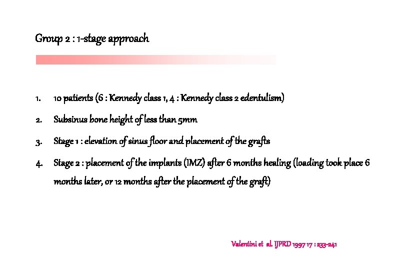
Group 2 : 1 -stage approach 1. 10 patients (6 : Kennedy class 1, 4 : Kennedy class 2 edentulism) 2. Subsinus bone height of less than 5 mm 3. Stage 1 : elevation of sinus floor and placement of the grafts 4. Stage 2 : placement of the implants (IMZ) after 6 months healing (loading took place 6 months later, or 12 months after the placement of the graft) Valentini et al. IJPRD 1997 17 : 233 -241
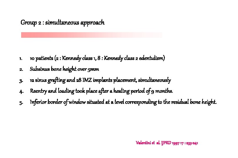
Group 2 : simultaneous approach 1. 10 patients (2 : Kennedy class 1, 8 : Kennedy class 2 edentulism) 2. Subsinus bone height over 5 mm 3. 12 sinus grafting and 28 IMZ implants placement, simultaneously 4. Reentry and loading took place after a healing period of 9 months. 5. Inferior border of window situated at a level corresponding to the residual bone height. Valentini et al. IJPRD 1997 17 : 233 -241
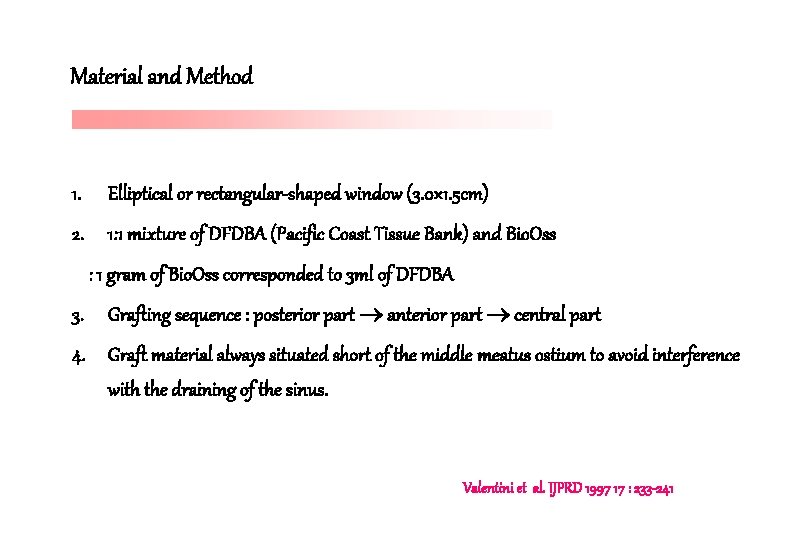
Material and Method 1. Elliptical or rectangular-shaped window (3. 0× 1. 5 cm) 2. 1: 1 mixture of DFDBA (Pacific Coast Tissue Bank) and Bio. Oss : 1 gram of Bio. Oss corresponded to 3 ml of DFDBA 3. Grafting sequence : posterior part anterior part central part 4. Graft material always situated short of the middle meatus ostium to avoid interference with the draining of the sinus. Valentini et al. IJPRD 1997 17 : 233 -241
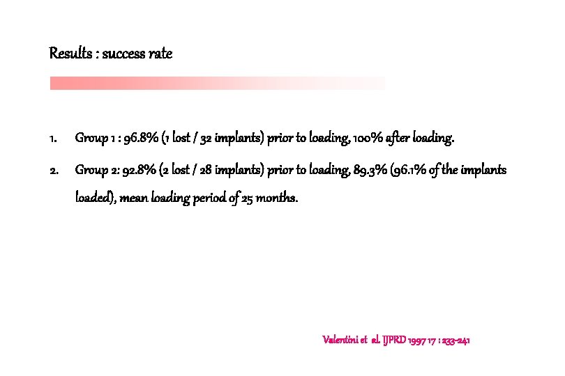
Results : success rate 1. Group 1 : 96. 8% (1 lost / 32 implants) prior to loading, 100% after loading. 2. Group 2: 92. 8% (2 lost / 28 implants) prior to loading, 89. 3% (96. 1% of the implants loaded), mean loading period of 25 months. Valentini et al. IJPRD 1997 17 : 233 -241
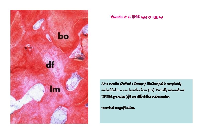
Histological evaluation 1. 6 months : new woven bone in contact with Bio. Oss and the absence of mineralized new bone in contact with DFDBA. 2. 9 months : lamellar bone with osteons were present, as were islands of woven bone 3. 12 months : new bone was totally remodeled into lamellar bone except in the nonresorbed DFDBA area, in which there was no new bone or mineralization. At 9 and 12 months there were no areas of Bio. Oss resorption Valentini et al. IJPRD 1997 17 : 233 -241
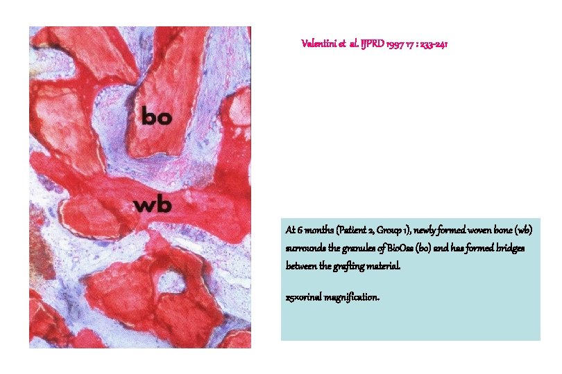
Valentini et al. IJPRD 1997 17 : 233 -241 At 6 months (Patient 2, Group 1), newly formed woven bone (wb) surrounds the granules of Bio. Oss (bo) and has formed bridges between the grafting material. 25×orinal magnification.
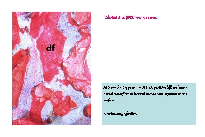
Valentini et al. IJPRD 1997 17 : 233 -241 At 6 months it appears the DFDBA particles (df) undergo a partial recalcification but that no new bone is formed on the surface. 10×orinal magnification.
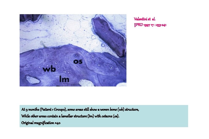
Valentini et al. IJPRD 1997 17 : 233 -241 At 9 months (Patient 1 Group 2), some areas still show a woven bone (wb) structure, While other areas contain a lamellar structure (Im) with osteons (os). Original magnification × 40
Valentini et al. IJPRD 1997 17 : 233 -241 At 12 months (Patient 2 Group 1), Bio. Oss (bo) is completely embedded in a new lamellar bone (Im). Partially mineralized DFDBA granules (df) are still visible in the center. 10×orinal magnification.
Histologic findings of DFDBA 1. Osseoinductive effect of DFDBA is insufficient and that promotion of new bone is a result of the osseoconductive properties of the bovine bone mineral matrix (Bio. Oss) 2. The use of DFDBA (previously accepted to have osseoinductive qualities by the authors and others) seems questionable. Valentini et al. IJPRD 1997 17 : 233 -241
Quality of osseointegration 1. Failures in Group 2, like that of Group 1, can be interpreted as a consequence of the insufficient osseointegration of screw implants with a machined surface in grafts of this type. 2. The bone obtained by means of these composite graft is of type 4, or at best of type 3, and the prognosis for screw implants in this type of bone is well below that of TPS cylinder implants with this bone quality (4 lost / 8 machined implants) Valentini et al. IJPRD 1997 17 : 233 -241
Conclusion 1. Composite grafts represent an important alternative to autografts in sinus floor lift procedures, especially when cylindrical implants are used. 2. Bio. Oss showed excellent osseoconductive properties and could be indicated alone in case of sufficient remaining bone around the sinus. Valentini et al. IJPRD 1997 17 : 233 -241