A 1 Neural Development Animal Models in Neuroscience


 � An area of the ectoderm develops into the neural plate")


 �There")


- Slides: 29
A. 1: Neural Development
Animal Models in Neuroscience �Neuroscience is all about understanding how the nervous system works and is formed from embryo to adults. �Understanding can lead to the treatment of various diseases of the nervous system. �However, because our nervous systems are quite complex and because of ethical reasons, neuroscientists often study the nervous systems of animals
Animal Models �Flatworm – they mature quickly and low, fixed number of cells as an adult have a �Fruit Fly – breed easily, mature quickly, and only 4 chromosomes �Zebrafish – tissues practically transparent
Animal Models �American clawed frog – large eggs, easily manipulated �House mouse – shares many human diseases
Development of the Neural Tube �Neurulation: the process in which chordates* develop a dorsal nerve cord in the early stages of development. �For humans, it occurs during the 1 st month of pregnancy. �*Chordates: a phylum that humans belong to
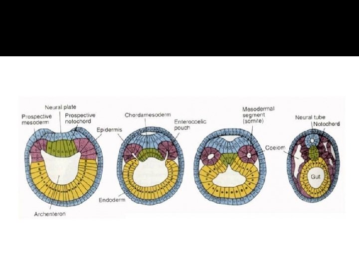
Neurulation (p 514) � An area of the ectoderm develops into the neural plate � The neural plate folds inwards forming a groove along the back of the embryo and separates from the rest of the ectoderm � This is the neural tube that elongates as the embryo grows � The space inside will be the centre of the spinal cord
Neurulation in Xenopus �Annotate a diagram of embryonic tissues in Xenopus laevis (African clawed frog) �See page 515
Spina bifida �A condition caused by the incomplete closure of the embryonic neural tube. �It usually occurs in the lower back. �Can be mild (with no apparent symptoms – severe (with debilitating symptoms that can lower ability to walk, result in problems with bladder and bowel control)
Spina Bifida
Development of Neurons �There are billions of neurons in your body. �As the neural tube is produced, cells undergo mitosis and differentiation occurs leading to cells turning into neurons. �Proliferation continues as the spinal cord and brain develop.
Development of Neurons �MYTH: You are born with all the neurons you will ever have. You do not produce more neurons after birth �Cell division stops in most parts of the nervous system before birth, however, there are may parts of the brain where new neurons are produced during adulthood!
Migration of Neurons �Migration of neurons in important in brain development. �Some neurons are produced in one parts of the developing brain need to migrate to another part. �The cytoplasm and organelles in it are moved from one end of the neuron to anther (similar to how an Amoeba moves with contractile actin filaments) �https: //www. youtube. com/watch? v=ZRF- g. KZHINk
Migration of Neurons �Mature, functional neurons do not normally move, though their axons and dendrites may regrow if damaged
Development of Axons �An immature neurons contains a cell body with a cytoplasm and nucleus. �An axon is a long extension of the cytoplasm from the cell body which carries nerve impulses to another neuron. �Only one axon develops on each neuron (though it can be highly branched at the end plate)
Development of Axons �Chemical stimuli determine neuron differentiation. �(Dendrites are shorter and numerous and bring impulses from other neurons to the cell body)
Growth of Axons �Axons grow at their tips �Within the CNS they are short and make connections between other neurons in the CNS �Some neurons within the PNS develop very long axons (1 m+ in humans) and take impulses to other neurons or effector cells (muscles or glands)
�Axons can me many meters long in some animals
Axons �As long as the cell body of the neuron remains intact, the axon may be able to regrow if damaged or severed outside the CNS. �Can be as rapid as 5 mm /day �Control of muscles and return of sensation may occur with time after the initial injury.
Development of Synapses �A developing neuron forms multiple synapses. �The minimum is 2 (one to bring impulses in at the dendrites, the other to pass them on at the axon end plate) �Most neurons develop multiple synapses (some in the brain develop hundreds) allowing for complex patterns of neural communication.
Development of Synapses
Development of Synapses �Synapse development involves special structures being assembled in the membranes on either side of the synapse and in the synaptic cleft between them
Elimination of Synapses �Synapses often disappear if they are not used. �When synaptic transmission occurs, chemical markers are left that cause the synapse to be strengthened. (ie: increasing the number of receptors on post synaptic membrane) �Inactive synapses will not have these markers and will become weaker and are eventually eliminated.
Neural Pruning �Neural Pruning: the elimination of a neuron (or part of it) �There are more neurons in some parts of newborn babies brains than in adults. �This indicates that some neurons are lost during childhood. �Neurons that are not used destroy themselves by apoptosis.
Did you know? � New born babies have ~11. 2 million neurons in the thalamus � Adult humans have ~6. 43 in the thalamus
Plasticity of the Nervous System �The nervous system changes with experiences �Connections between neurons can be changed by growth of axons and dendrites Formation of new synapses Pruning of dendrites, axons, or whole neurons
Plasticity of the Nervous System �Plasticity continues throughout life, but much higher from birth to age 6. �Plasticity occurs because of experiences and the way the nervous is utilized by the individual �It’s the basis forming new memories and reasoning.
Strokes �During a stroke, part of the brain is deprived of sufficient oxygen and glucose. �Neurons may become irreparably damaged and die. �Minor strokes may be hardly noticed. �Of major strokes, 1/3 will full recover, 1/3 survive with a disability
Strokes �Recovery from strokes will happen within the 6 months following a stroke �It involves parts of the brain taking on new functions to make up for the damaged areas. �New synapses form �In this way, a stroke patient may relearn aspects of speech, writing, motor skills, etc.