9 1 Functions of the Muscular System Movement


 Muscle cells derive from myoblasts The number")

 Skeletal muscle is striated and voluntary. The")


")
 Depolarization: Inside of plasma membrane becomes less")


- Slides: 47
9. 1 Functions of the Muscular System ·Movement of the body ·Maintenance of posture ·Respiration ·Production of body heat ·Communication ·Constriction of organs and vessels ·Contraction of the heart 9 -1 Copyright © Mc. Graw-Hill Education. Permission required for reproduction or display. 1
9. 2 General Properties of Muscle ·Contractility: ability of a muscle to shorten with force ·Excitability: capacity of muscle to respond to a stimulus (from our nerves) ·Extensibility: muscle can be stretched to its normal resting length and beyond to a limited degree ·Elasticity: ability of muscle to recoil to original resting length after stretched 9 -2 Copyright © Mc. Graw-Hill Education. Permission required for reproduction or display. 2
Types of Muscle Tissue There are 3 types of muscle tissue in the muscular system: Skeletal muscle: • Attached to bones of skeleton • Voluntary (consciously controlled) Cardiac muscle: • Makes up most of the wall of the heart • Involuntary (non-consciouslycontrolled) • Responsible for pumping action of the heart Smooth muscle: • Found in walls of internal organs, such as those of digestive tract • Involuntary (non-consciouslycontrolled) 3
Types of Muscle Tissue Skeletal: • About 40% of the body’s weight • Locomotion, facial expression, posture, respirations. Smooth: • Most widely distributed type of muscle • Found in digestive tract, blood vessels, eyes, urinary tract Cardiac: • Found only in the heart • Makes heart contract to pump blood
9. 1: Structure of a Skeletal Muscles: • Attach to bones, and skin of face • Under conscious control (voluntary) • A skeletal muscle is an organ of the muscular system • Skeletal muscles are composed of: • Skeletal muscle tissue • Nervous tissue • Blood • Connective tissues 5
Connective Tissue Coverings Connective tissue coverings over skeletal muscles: Fascia – a sheet of connective tissue that surrounds muscles and separates muscles Tendons Aponeuroses- a sheet of connective tissue that Takes place of tendon 6
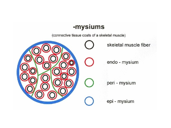
9. 3 Connective Tissue Coverings Muscle coverings: • Epimysium: surrounds whole muscle • Perimysium: surrounds fascicles within a muscle • Endomysium: surrounds muscle fibers (cells) within a fascicle 7
A skeletal muscle is composed of a variety of tissues, including layers of connective tissue. Fascia covers the surface of the muscle, epimysium lies beneath the fascia, and perimysium extends into the structure of the muscle, where it separates muscle cells into fascicles (bundles of muscle fibers (cells)). Endomysium separates individual muscle fibers (cells).
At the attachment points of the muscle all the connective tissue elements combine to form the connective tissue attachment of the muscle to bone or other tissue. If this attachment is round and cord-like it is called a tendon. If the attachment is broad and sheet-like it is called an aponeurosis.
Nerves and Blood Vessels Motor neurons: • Stimulate muscle fibers to contract. Nerve cells with cell bodies in brain or spinal cord; axons extend to skeletal muscle fibers through nerves • Axons branch so that each muscle fiber is innervated • Contact of nerve to muscle fiber is neuromuscular junction Capillary beds surround muscle fibers
Under the microscope: Skeletal Muscle Fibers (cells) Muscle cells derive from myoblasts The number of muscles cells remains about the same after birth, but exercise can make them bigger (hypertophy). Hypertrophy is an increase in size of a muscle cell, not number of muscles cells.
Muscle cells have the same parts as other cells but they have special names
Under the microscope: Skeletal Muscle Fibers (cells) Skeletal muscle is striated and voluntary. The word striated means “striped” and the significance of this term will become apparent when we consider the histology. Skeletal muscle is the only type of muscle that we can consciously control through our nervous system. This is the reason it is also voluntary. Skeletal muscle cells are also long and cylindrical. For this reason a skeletal muscle cell can also be referred to as a skeletal muscle fiber. A skeletal muscle fiber can be up to a foot long! These long, highly specialized cells result from the fusion of many cells and after the cells fuse their individual nuclei are retained. As a result, skeletal muscle fibers are multinucleate.
Muscle fiber: long and cylinrical Multinucleate
Skeletal muscle cells contain similar components and structures as other cells but different terms are used to describe those components and structure in skeletal muscle cells. The cytoplasm is known as sarcoplasm There are many mitochondria The plasma membrane of skeletal muscle is called the sarcolemma The sarcolemma invaginates to form tubes called T tubules The T tubules are associated with the sarcoplasmic reticulum. The endoplasmic reticulum is called the sarcoplasmic reticulum.
All Cells have a cytoskeleton. The cytoskeleton is composed of protein fibers called filaments that serve such functions as: establishing cell shape providing mechanical strength locomotion chromosome separation in mitosis and meiosis intracellular transport of organelles • • Muscle cells are also called muscle fibers. Muscle cells contain filaments called myofibrils Myofibrils are involved in contraction. Two main ones are: Thick filaments made of myosin, and thin filaments made of actin
A thick filament is made up of a chain of myosin molecules
Thin filaments are made up of chains of actin molecules, tropomyosin, and troponin
A muscle contraction involves reactions that bring myosin and actin together. And they lived happily ever after.
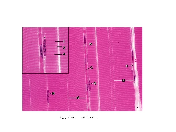
Microscopic Anatomy The plasma membrane of the skeletal muscle fiber is called a sarcolemma. The muscle fiber contains long cylindrical structures, the myofibrils. The myofibrils almost entirely fill the cell and push the nuclei to the outer edges of the cell under the sarcolemma. The many myofibrils each have light and dark bands and are aligned with one another so that the light and dark bands are next to one another. This gives the cell its striated appearance.
The light bands are called I bands and the dark bands are called A bands. In the middle of the I bands there is a line called the Z line (or disc). In the middle of the A bands (or dark bands) there is a light zone called the H zone. In the middle of the H zone there is another line, the M line. The precise arrangement of these features is due to a chain of functional units in the myofibrils, sarcomeres.
The sarcomere consists myofilaments. The two major types of myofilaments are actin and myosin.
Thick myofilaments are made up of proteins molecules called myosin. The myosin molecules are shaped like golf clubs with long shafts. Myosin forms the thick myofilaments by forming bundles in which the heads of the “golf clubs” stick out at either end of the filament and the shafts form a “bare” zone in the middle of the filaments • The heads of the thick myofilaments form attachments with the thin actin myofilaments. • These attachments are called cross bridges. • The heads are also the places on the thick myofilaments that use the energy in the ATP molecule to power the muscle contraction.
The thin myofilaments are composed of the protein actin. The thin myofilaments have the binding sites to which the heads of the thick myofilaments attach
Now we can relate the banding pattern and sarcomere to the myofilaments: Sarcomere – An individual sarcomere extends from one Z line to the next. • I band – The I band corresponds to a region that overlaps two adjacent sarcomeres where there are only thin myofilaments. • Z line or disc – The Z line in the center of the I band is where proteins hold the thin myofilaments in position. • A band – The A band is where thick myofilaments are positioned. • H zone – The H zone is the region in the middle of the sarcomere where thin myofilaments fail to overlap the thick myofilaments. • M line – The M line in the center of the sarcomere and A band is where proteins hold the thick myofilaments in position.
Skeletal Muscle Fibers Striation pattern has 2 main parts: • I Band: Light band, composed of thin actin filaments • A Band: Dark band, composed of thick myosin filaments overlapping with thin actin filaments • H Zone: Center of A band; composed of thick myosin filaments • Z Line: Anchors filaments in place; sarcomere boundary; center of I band • M Line: Anchors thick filaments; center of A band 29
Skeletal Muscle Fibers Thick filaments: Composed of myosin protein; heads form crossbridges Thin filaments: Composed of actin protein; associated with troponin and tropomyosin, which prevent crossbridge formation when muscle is not contracting 30
9. 4 Physiology of Skeletal Muscle Fibers Contraction of a muscle fiber: • Requires interaction from several chemical and cellular components • Results from a movement within the myofibrils, in which the actin and myosin filaments slide past one another, shortening the sarcomeres • Muscle fiber shortens and pulls on attachment points 31
Sliding Filament Model • Actin myofilaments sliding over myosin to shorten sarcomeres – Actin and myosin do not change length – Shortening sarcomeres responsible for skeletal muscle contraction • During relaxation, sarcomeres lengthen because of some external force, like contraction of antagonistic muscles that produce the opposite effect 9 -32
Sarcomere Shortening Copyright © The Mc. Graw-Hill Companies, Inc. Permission required for reproduction or display. Z I (a) Relaxed sarcomere Z H A I In a relaxed muscle, the actin and myosin myofilaments overlap slightly, and the H zone is visible. The sarcomere length is at its normal resting length. As a muscle contraction is initiated, actin myofilaments slide past the myosin myofilaments, the z disks are brought closer together, and the sarcomere begins to shorten. Z I A (b) Fully contracted sarcomere Z I In a contracted muscle, the A bands, which are equal to the length of the myosin myofilaments, do not narrow because the length of the myosin myofilaments does not change, nor does the length of the actin myofilaments. In addition, the ends of the actin myofilaments are pulled to and overlap in the center of the sarcomere, shortening it and the H zone disappears. 9 -33
How does this happen? • Muscle cells, can be stimulated by electricity. • The nervous system controls skeletal contractions through axons of nerve cells. • Electrical signals (axon potentials) travel from brain or spinal cord to muscle fibers and cause them to contract.
9. 4 Skeletal Muscle Fiber Physiology Oscilloscope + + + + + – – – – – 0 m. V– 50 – 85 – – – – – Time + + + + + Nerve or muscle cell Measuring the resting membrane potential • Nervous system controls muscle contractions through action potentials • Resting membrane potentials – Membrane voltage difference across membranes (polarized) • Inside cell more negative due to accumulation of large protein molecules. More K+ on inside than outside. K+ leaks out but not completely because negative proteins hold some back. • Outside cell more positive and more Na+ on outside than inside. • Na+ /K+ pump maintains this situation. – Must exist for action potential to occur 9 -35
Ion Channels • The electrical properties of muscle and nerves depend on the action of channels in the cell membrane. • The inside of the cell membrane is hydrophobic and charged ions need to travel through ion channels
Ion Channels • Types – Ligand-gated. Ligands are molecules that bind to receptors. Receptor: protein or glycoprotein with a receptor site • Example: neurotransmitters • Gate is closed until neurotransmitter attaches to receptor molecule. – Voltage-gated • Open and close in response to small voltage changes across plasma membrane • Each is specific for certain ions 9 -37
Action Potential THE ACTION POTENTIAL • Neurons use all-or-nothing messages called action potentials to relay electrical impulses down the axon to the synaptic bouton. Action potentials ultimately cause the release of neurotransmitters into the synaptic cleft.
• • • Resting Membrane Potential Electrically excitable cells are polarized. The inside of the cell is more negatively charged than the outside The voltage difference between inside and outside of an unstimulated cell is called the resting membrane potential. The resting potential is the result of 3 factors: • The concentration of K+ inside the cell is higher than outside • The concentration of Na+ outside the cell is higher than inside • The plasma membrane is more permeable to K+ than Na+
Resting Membrane Potential. • This means that there is an electrical potential difference (voltage) between the inside of the cell and the extracellular space. • This is about – 70 m. V, with the inside of the cell being negative relative to the outside.
Action Potential Phases (resting cell is polarized) Depolarization: Inside of plasma membrane becomes less negative. If change reaches threshold, depolarization occurs Repolarization: return of resting membrane potential. Note that during repolarization, the membrane potential drops lower than its original resting potential, then rebounds. All-or-none principle: like camera flash system
Propagate: Spread from one location to another. Action potential does not move along the membrane: new action potential at each successive location. Action Potential Frequency: number of action potential produced per unit of time
An action potential can propagate because an action potential produced at one location on the plasma membrane can stimulate the production of another action potential in a location next to it.
Gated Ion Channels and the Action Potential Na+ channel 1 Resting membrane potential. Na+ channels (pink) and some, but not all, K+ channels (purple) are closed. K+ diffuses down its concentration gradient through the open K+ channels, making the inside of the cell membrane negatively charged compared to the outside. K+ concentration gradient Extracellular fluid K+ channel Charge Difference Across the Plasma Membrane + + + + – – – – 1 Cytoplasm K+ Na+ Depolarization. Na+ channels are open. Na+ diffuses down its concentration gradient through the open Na+ channels, making the inside of the cell membrane positively charged compared to the outside. – – –– + + + + 2 Na+ concentration gradient Na+ 2 Na+ diffuse into cell Na+ 3 Repolarization. Na+ channels are closed, and Na+ movement into the cells stops. More K+ channels open. K+ movement out of the cell increases, making the inside of the cell membrane negatively charged compared to the outside, once again. K+ concentration gradient K+ diffuse out of cell + + ++ – – – – 3 K+ K+ channels open K+ Na+ channels close 9 -44
Action Potential Propagation 1 An action potential in a local area of the plasma membrane is indicated by the orange band. Note the reversal of charge across the membrane. + + – – + + – – – – 1 – – + + – – + + + + Stimulus 2 The action potential is a stimulus that causes another action potential to be produced in the adjacent plasma membrane. 3 The action potential propagates along the plasma membrane (orange arrow). + + – – Muscle fiber – – + + + + – – – 2 – – + + + – – – – + + + – – – – – + + + + – – – + + 3 9 -45
Neuromuscular Junction Skeletal muscle fibers contract only when stimulated by a motor neuron Action potentials of neurons (nerve cells) stimulate action potentials in muscle fibers because of events that occur at the neuromuscular junction. Nerve axons (the end of the axon at the NMJ is is the 1. presynaptic terminal) connect with the muscle fiber at the 2. sarcolemma to form a neuromuscular junction or synapse.
Neuromuscular Junction The space between the presynaptic terminal and the muscle fiber is the synaptic cleft. Each presynaptic terminal contains many mitochondria and sacs called 3. synaptic vesicles The vesicles contain acetyl cholineneurotransmitter. The 4. postsynaptic membrane or motor end plate contains receptors to acetylcholine 5. Mitochondria in muscle fiber (cell)