8 MALE REPRODUCTIVE PHYSIOLOGY AND ERECTION Prof Sherif

 are produced in the primary sex")


The secondary sex organs include: A) Epididymis and vasdeferens • sperm removed from the")
 The seminal vesicles • these structure are only secretory glands instead of sperm")

Activin, follistatin, insulin- like growth factor-1, transferrin and cytokines. *Factors affecting spermatogenesis: 1 -Central")
Estrogen: - formed by the Sertoli cells under FSH stimulation. - essential for spermiation.")


: • Mechanism of action of (T): -Like")
 increases proteins synthesis. # (T) increases the quantity")

- Slides: 16
8. MALE REPRODUCTIVE PHYSIOLOGY AND ERECTION. Prof. Sherif W. Mansour Physiology dpt. , Mutah School of medicine 2020 -2021
Male Reproductive System The male germ cells (sperms) are produced in the primary sex organs (two testes). in addition to these primary sex organs, there are secondary sex organs that provide the necessary tract and environment for the sperms. Structure of male reproductive system (I)The primary sex organ (testis): *The testis is a compound organ composed of: A-The seminiferous tubules: -These are long, tortuous, hallow tubules in which spermatogenesis takes place. The seminiferous tubules are lined with spermatogonia. The spermatogonia lie close to the basement membrane and mature into spermatozoa which moves to the center of the tubules. - Sertoli cells which are glycogen containing cells from which the sperms obtain their nourishment. *Blood-testis barrier: Tight junction between adjacent Sertoli cells near the basal lamina form a blood- testis barrier, that protects the germ cells from blood-borne noxious agents, prevents antigenic products of germ cell division and maturation from entering the circulation and generating an autoimmune response. B) The interstitial cells of Leydig: They are located between the seminiferous tubules and produce testosterone.
Male Reproductive System.
*Location of the testis In the scrotum outside the abdominal cavity. The temperature of the scrotum is usually two degree lower than the abdominal cavity due to: 1 The absence of subcutaneous fat in the scrotum. 2 The spermatic arteries in the pampiniform plexus to the testes are tortuous, and blood in them runs parallel but in the opposite direction to blood in the spermatic veins. this anatomic arrangement allow countercurrent exchange for heat and testosterone. 3 Dartos muscle relaxes in hot weather to draw the testes away from the high abdominal temperature, and contracts in cold weather to attract the testes near the abdomen. Cryptorchidism -Failure of testicular descend in the scrotum due to: (a) Obstruction of the inguinal canal. (b) Testosterone deficiency. -Cryptorchidism results in a complete failure of spermatogenesis and impairment of the endocrine function of the testis. Treatment: must start as early as possible by: a-Testosterone administration. b-Surgical.
(II)The secondary sex organs include: A) Epididymis and vasdeferens • sperm removed from the seminiferous tubules are completely nonmotile, and they can not fertilize an ovum. However, after the sperm remain in the epididymis for about 18 hours to 10 days, during this period they develop the capability of motility. The sperm also become capable of fertilizing the ovum, a process called maturation. • A small quantity of sperm can be stored in the epididymis, but most sperm are stored in the vasdeferens and the ampulla’s of the vasdeferens. They can remain stored and fertile for several months.
B) The seminal vesicles • these structure are only secretory glands instead of sperm storage areas. • The seminal vesicles are tortuous, lobulated tube about 7. 5 cm long lined with a secretory epithelium range from columnar to cuboidal that secretes fructose, prostaglandins and fibrinogen. • Prostaglandins are believed to aid fertilization in two ways: 1 - React with the cervical mucous to make it more receptive to sperm. 2 - Cause reverse peristaltic movement in the uterus and fallopian tubes to move the sperm toward the ovaries. • Fructose concentration is 150 – 650 mg/ml, it is the metabolic fuel for the spermatozoa. • The secretion of the seminal vesicles form 60% - 80% of the total volume of semen. C) The prostate gland • It secretes a thin milky alkaline fluid containing citric acid, calcium, acid phosphate and clotting enzymes. The prostatic fluid about 13% - 33% of semen volume. • The prostatic fluid neutralizes the acidity of the fluid of the vasdeferens and that of the vagina helping the process of fertilization by greatly enhances the motility of the spermatozoa. D) Bulbourethral glands (Cowper , s gland) • It secrets mucous that lubricate the distal urethra. E) External genitallia (penis and scrotum).
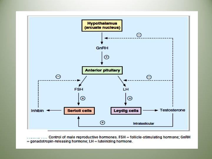
Spermatogenesis -The spermatogonia lie on the tubular membrane are non-motile stem cells, they divide by mitosis to form two cellular pools : a pool of additional stem cells, which undergo continual renewal by mitosis, and a pool of type A spermatogonia, which enter the maturation process called spermatogenesis. -Spermatogenesis requires about 64 – 74 days in man. The role of the Sertoli cells in spermatogenesis • They provide a special environment for germinal cells development. • They secrete a fluid that provide nutrients for the developing sperm. • Spermiation : the conversion of the spermatid to mature sperm. During this process, the spermatids are attached to the Sertoli cells, which secrete digestive enzymes that remove most of the cytoplasm from the spermatids. • They play a physical role in shaping the head and tail of the sperm. • They secrete some hormones: (1)Mullerian inhibitory factor (MIF) during fetal development to inhibit the formation of fallopian tubes from the mullerian ducts in male fetus. (2)Estradiol , as one of the stimulatory factors in spermatogenesis. (3)Inhibin, has a feedback inhibitory effect on the anterior pituitary gland to prevent over secretion of FSH. (4)Androgen – binding protein (ABP), that maintain a high, stable supply of androgen in the tubular fluid.
(5)Activin, follistatin, insulin- like growth factor-1, transferrin and cytokines. *Factors affecting spermatogenesis: 1 -Central nervous system *The hypothalamus contains dopaminergic and noradrenergic neurons, when stimulated → release of releasing factors → ant. Pit. → controlling gonadotropins secretions. * Various psychic stimuli feeding into the hypothalamus → marked excitatory and inhibitory effect on gonadotropin secretion →altering the degree of fertility. 2 - Hormonal factors a)Pituitary gonadotropins *-FSH: - needed for growth and maturation of the testes. -needed for the normal functions of Sertoli cells. - facilitate the last stages of spermatid maturation and promote the production of androgen binding protein (ABP). *- LH: maintains testosterone secretion by the Leydig cells. b)Testosterone: -Responsible for the development and maintenance of the germinal epithelium. - needed for complete meiosis. - needed for spermiogenesis.
c)Estrogen: - formed by the Sertoli cells under FSH stimulation. - essential for spermiation. - excessive estrogen inhibits FSH secretion→ depression of spermatogenesis. d)Thyroxin: Stimulates spermatogenesis via the stimulatory effect on cell metabolism. e)Growth hormone(GH): promotes early division of the spermatogonia, in its absence as in pituitary dwarfs, spermatogenesis is severely deficient or absent. 3 - Temperature: • spermatogenesis requires a temperature lower than that of the interior of the body. The testes are normally maintained at a temperature of about 32 ºC. • when the testes are retained in the abdomen. Degeneration of the tubular wall and sterility result. • Hot baths (45 ºC for 30 min / day) and insulated athletic supporters reduce the sperm count. 4 - Diet: # Complete protein starvation causes arrest of spermatogenesis, probably due to failure of gonadotropin secretion. # Vitamin E deficiency in animals causes irreversible tubular degeneration. This is not proved in man.
# The tubular germ cells require vit. B group as catalysts for metabolic processes. # Vit. A: it , s deficiency causes keratinization and atrophy of the germinal epithelium. # Vit. C: it , s deficiency inhibits spermatogenesis as it is needed in testosterone synthesis. 5 - Irradiation: The germinal epithelium can be destroyed by certain doses of radiations. These doses spare Sertoli cells and the endocrine cells of the testes. 6 - Hypoxia & toxins : (bacterial or chemical) depress spermatogenesis. The Male Sexual Act 1 -Erection: • Erection is initiated by dilation of the arterioles of the penis. As the erectile tissue of the penis fills with blood, the veins are compressed blocking outflow and adding to the turgor of the organ. • The integrating centers in the lumber segments of the spinal cord are activated by impulses through afferents from the genitalia and descending tracts that mediate erection in response to erotic psychic stimuli. • The efferent parasympathetic fibers are in the pelvic nerve(nervi erigentes). • The fibers presumably contain acetylcholine, and vasoactive intestinal peptide(VIP) as cotransmitter.
There also nonadrenergic noncholinergic fibers in the nervi erigentes, and this contain large amount of nitric syntheses, the enzyme that catalyze the formation of nitric oxide (NO). NO activates guanylyl cyclase resulting in increased production of c. GMP, and c. GMP is a potent vasodilator. Thus, it seams clear that NO plays the prominent role in the production of erection • Erection is inhibited by sympathetic vasoconstrictor impulses to the arterioles. 2 - Emission: • Contraction of the vasdeferens and the ampulla causes expulsion of sperm into the internal urethra. Then, contractions of the prostatic capsule and seminal vesicles expel prostatic fluid and seminal fluid, forcing the sperm forward. • All this fluids are mixed in the internal urethra with mucous secreted by the bulbourethral glands forming semen. 3 – Ejaculation: • The propulsion of semen out of the urethra at the time of orgasm. • Orgasm is a pleasurable feeling that usually occurs simultaneously with emission and /or ejaculation. The physiologic mechanisms responsible for orgasm are unknown. • The afferent pathway is mostly fibers from touch receptors in the glans penis that reach the spinal cord through the internal pudendal nerves. • Semen is propelled out of the urethra by contraction of the bulbocavernosus muscle.
SEMEN It is the fluid ejaculated at the time of orgasm. It contains spermatozoa suspended in the secretions of the seminal vesicles, prostate and Cowper , s gland. Fertilization of the ovum requires that the ejaculum contain enough viable motile spermatozoa. * Coagulation and liquefaction of semen: *The clotting enzyme of the prostatic fluid causes the fibrinogen of the seminal vesicles fluid to form a weak coagulum, which then dissolute during the next 15 – 20 min. by the prostatic profibrinolysin. The sperm remain immotile, due to the high viscosity of the coagulum. However, after dissolution of the coagulum, the sperm simultaneously become motile. * Impaired liquefaction may occur in 4 – 9 of infertile men, and artificial liquefaction with amylase may enhance fertility. *Capacitation: Sperm removed from the male genital ducts are not capable of penetrating the layer of granulosa cells that cover the ovum, but they do this a few hours after being deposited in the female genital tract. This may be due to that the acrosomes contain hydrolytic and proteolytic enzymes that could destroy the male genital tract if released prematurely. The fluids in the seminiferous tubules, epididymis and in the vas deferens contain vesicles filled with cholesterol covering the acrosome and prevents the release of this enzymes. After ejaculation, the sperm loss their excess cholesterol so that the enzymes in the acrosome are released and allow the sperm to penetrate the ovum.
Endocrine function of the testicles Testosterone (T): • Mechanism of action of (T): -Like other steroids, T binds to an intracellular receptors, and the receptor-steroid complex then binds to DNA in the nucleus, facilitating transcription of various genes. • Actions of (T): 1)During fetal life - (T) is responsible for the development of the male sex organs e. g. the formation of a penis and a scrotum. - (T) is responsible for descend of the testis into the scrotum during the last 2 months of gestation. 2)At puberty: A-On the primary sex organs (T) is essential for spermatogenesis B-On the secondary sex organs: Seminal vesicles, prostate, bulbourethral glands and the external genitalia are dependent on (T) for their growth and maturation. (T) is responsible for the maintenance of their integrity and function. C- On the secondary sex characteristics: 1)-The distribution of body hair: Beard appears. Hairline on scalp recedes antrolaterally. Pubic hair grows with male pattern(triangle with apex up). General body hair increases. 2)-Baldness: decrease the growth of hair on the top of the head. 3)-Voice: (T) causes hypertrophy of the laryngeal mucosa and enlargement of the larynx �deeper voice. 4)-Skin: (T) increases thickness of the skin. It also increases the rate of secretion of the sebaceous glands → acne formation. 5)-Broaden shoulder and increased muscle bulk.
D- General metabolic effects: # (T) increases proteins synthesis. # (T) increases the quantity of bone matrix, and causes Ca++ retention. # (T) increases basal metabolic rate. # (T) increases food intake. # (T) increases RBCs number, by stimulating erythropoietin synthesis. # (T) increases Na+ reabsorption in the distal convoluted tubules �slight increase in the extracellular fluid. E- Regulation of behavioral effects, including libido. (II) Dihydrotestosterone (DHT): -About 20 % of plasma DHT is synthesized by the testis, probably because of the action of 5 -α -reductase from the Sertoli cells on testosterone secreted by the Leydig cells. The remainder is derived from the peripheral conversion of T to DHT by 5 α -reductase in some target cells. -DHT circulates in blood, with a plasma level that is 10 % of the T level. -T- receptor complexes are less stable than DHT-receptor complexes in the target cells. Thus DHT is more potent than T. Congenital 5 α -reductase deficiency, produces male pseudo-hermaphroditism. (III) -Estrogens # 30 % of estrogens secreted by the testes, some from Leydig cells and some from Sertoli cells. # 70 % of estrogens formed by aromatization of circulating T androstenedione. # very small amount may be secreted by the adrenal cortex. # in men, the plasma estrogen level is 2 ng /d. L. # the peripherally derived estrogens may be released into the blood and transported to other tissues, where they may exert some biological actions. This is particularly evident in certain endocrine disorders, in which plasma (T) levels are depressed causing an elevation in the ratio of plasma estrogens to androgens. Under these condition, some feminization of the male body may occur. Inappropriate breast development, or gynecomastia.
Thank You