
6 Time frame for evolution of the major
- Slides: 35
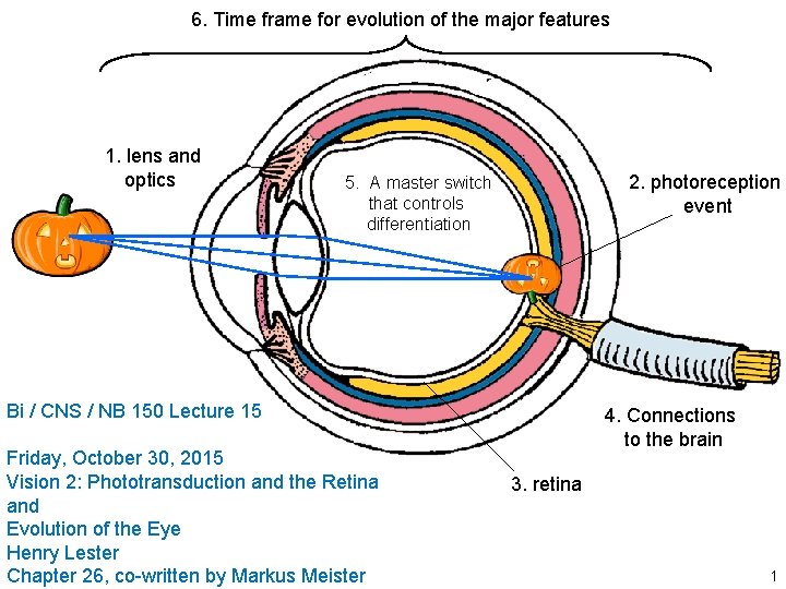
6. Time frame for evolution of the major features 1. lens and optics 2. photoreception event 5. A master switch that controls differentiation Bi / CNS / NB 150 Lecture 15 Friday, October 30, 2015 Vision 2: Phototransduction and the Retina and Evolution of the Eye Henry Lester Chapter 26, co-written by Markus Meister 4. Connections to the brain 3. retina 1
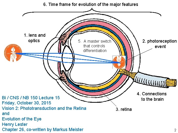
6. Time frame for evolution of the major features 1. lens and optics 2. photoreception event 5. A master switch that controls differentiation Bi / CNS / NB 150 Lecture 15 Friday, October 30, 2015 Vision 2: Phototransduction and the Retina and Evolution of the Eye Henry Lester Chapter 26, co-written by Markus Meister 4. Connections to the brain 3. retina 2
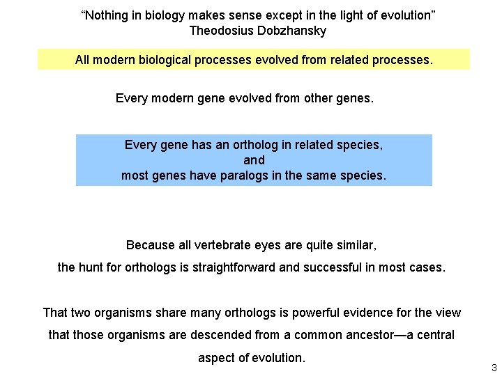
“Nothing in biology makes sense except in the light of evolution” Theodosius Dobzhansky All modern biological processes evolved from related processes. Every modern gene evolved from other genes. Every gene has an ortholog in related species, and most genes have paralogs in the same species. Because all vertebrate eyes are quite similar, the hunt for orthologs is straightforward and successful in most cases. That two organisms share many orthologs is powerful evidence for the view that those organisms are descended from a common ancestor—a central aspect of evolution. 3
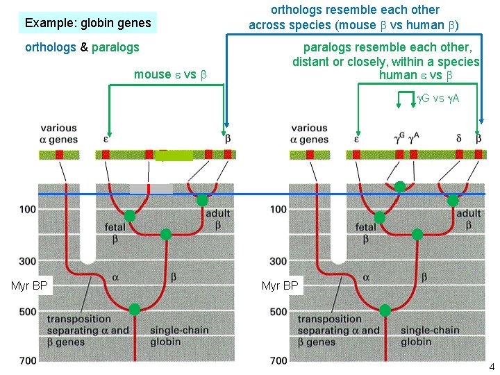
Example: globin genes orthologs & paralogs Hemoglobin paralogs in the mouse genome mouse e vs b orthologs resemble each other across species (mouse b vs human b) paralogs resemble each other, distant or closely, within a species Hemoglobin paralogs in the human genome human e vs b chromosome 7 Myr BP g. G vs g. A Myr BP 4
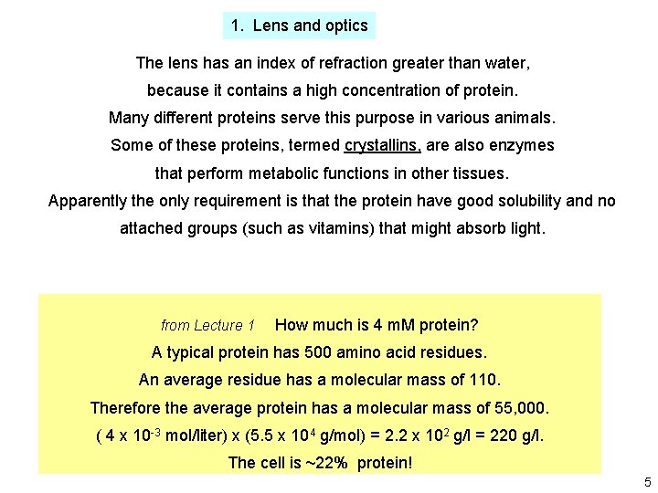
1. Lens and optics The lens has an index of refraction greater than water, because it contains a high concentration of protein. Many different proteins serve this purpose in various animals. Some of these proteins, termed crystallins, are also enzymes that perform metabolic functions in other tissues. Apparently the only requirement is that the protein have good solubility and no attached groups (such as vitamins) that might absorb light. from Lecture 1 How much is 4 m. M protein? A typical protein has 500 amino acid residues. An average residue has a molecular mass of 110. Therefore the average protein has a molecular mass of 55, 000. ( 4 x 10 -3 mol/liter) x (5. 5 x 10⁴ g/mol) = 2. 2 x 10² g/l = 220 g/l. The cell is ~22% protein! 5
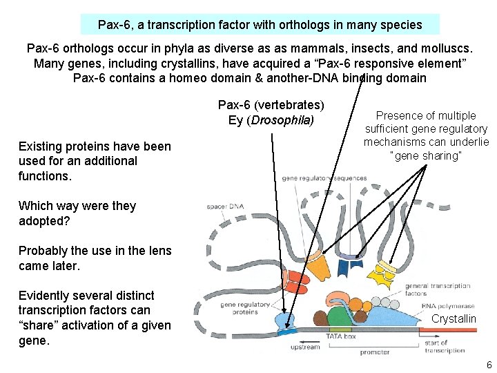
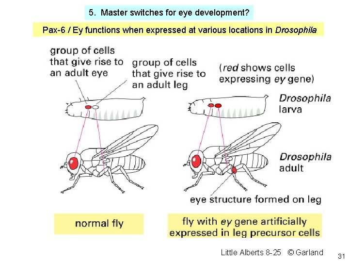
Pax-6, a transcription factor with orthologs in many species Pax-6 orthologs occur in phyla as diverse as as mammals, insects, and molluscs. Many genes, including crystallins, have acquired a “Pax-6 responsive element” Pax-6 contains a homeo domain & another-DNA binding domain Pax-6 (vertebrates) Ey (Drosophila) Existing proteins have been used for an additional functions. Presence of multiple sufficient gene regulatory mechanisms can underlie “gene sharing” Which way were they adopted? Probably the use in the lens came later. Evidently several distinct transcription factors can “share” activation of a given gene. Crystallin 6
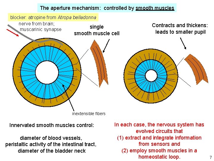
The aperture mechanism: controlled by smooth muscles blocker: atropine from Atropa belladonna nerve from brain; single muscarinic synapse smooth muscle cell Contracts and thickens: leads to smaller pupil inextensible fibers Innervated smooth muscles control: diameter of blood vessels, peristaltic activity of the intestinal tract, diameter of the bladder neck In each case, the nervous system has evolved circuits that (1) extract and integrate information from sensors and (2) employ smooth muscles in a homeostatic loop. 7
2. The photoreception event Photoreceptor organs have evolved independently at least 40 times, each time responding to the visible spectrum and near-UV. How do we explain the use of a limited part of the spectrum? Infrared light is not sufficiently energetic to provoke photochemistry such as cis-trans isomerization. Shorter-wavelength ultraviolet light is too energetic and would destroy organic molecules. 8
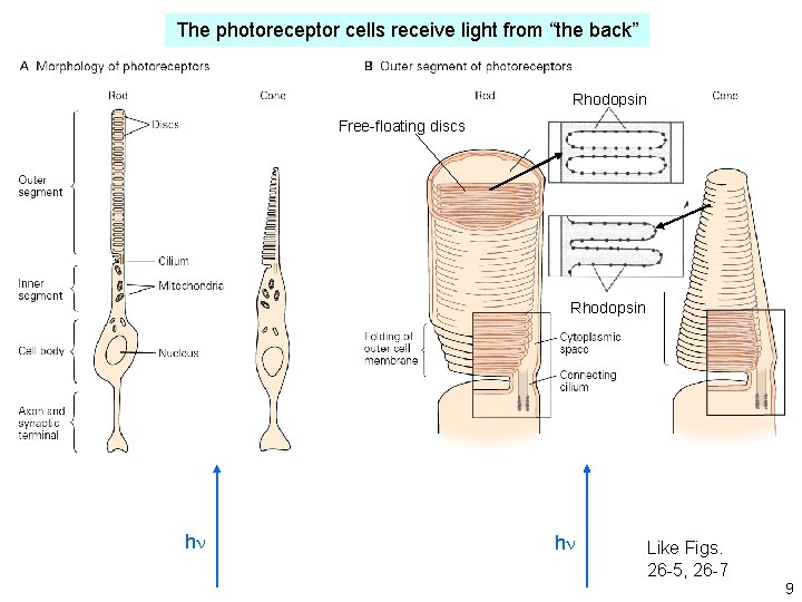
The photoreceptor cells receive light from “the back” Rhodopsin Free-floating discs Rhodopsin hn hn Like Figs. 26 -5, 26 -7 9
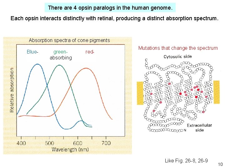
There are 4 opsin paralogs in the human genome. Each opsin interacts distinctly with retinal, producing a distinct absorption spectrum. Absorption spectra of cone pigments Blue- greenabsorbing red- Mutations that change the spectrum Like Fig. 26 -8, 26 -9 10
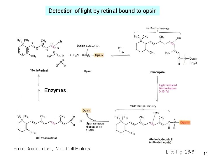
Detection of light by retinal bound to opsin Enzymes From Darnell et al. , Mol. Cell Biology Like Fig. 26 -8 11
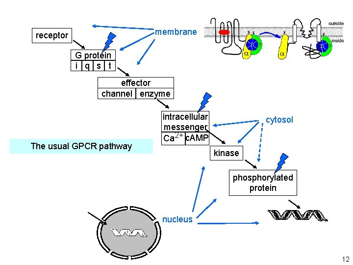
from Lecture 12 membrane receptor G protein i q s t effector channel enzyme The usual GPCR pathway intracellular messenger Ca 2+ c. AMP cytosol kinase phosphorylated protein nucleus 12
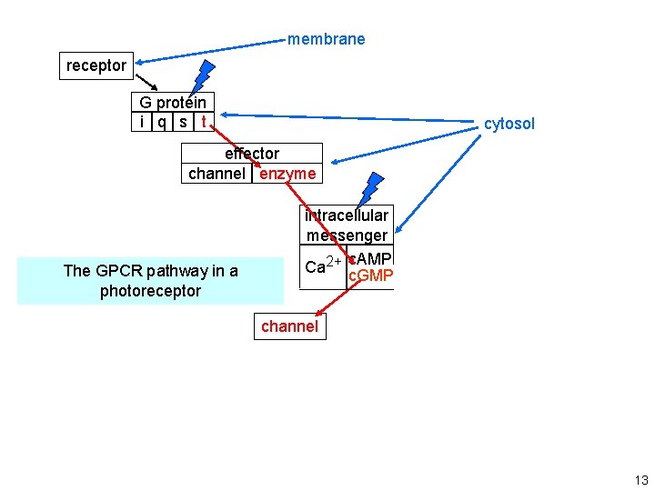
membrane receptor G protein i q s t cytosol effector channel enzyme The GPCR pathway in a photoreceptor intracellular messenger c. AMP Ca 2+ c. GMP channel 13
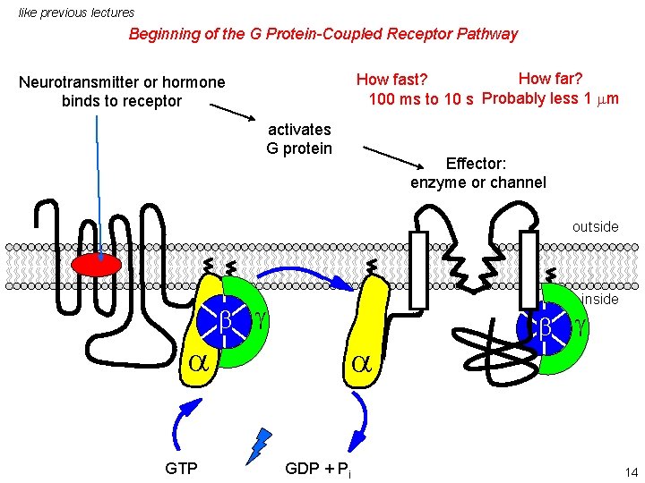
like previous lectures Beginning of the G Protein-Coupled Receptor Pathway How far? How fast? 100 ms to 10 s Probably less 1 mm Neurotransmitter or hormone binds to receptor activates G protein Effector: enzyme or channel outside inside b g a GTP a GDP + Pi b g 14
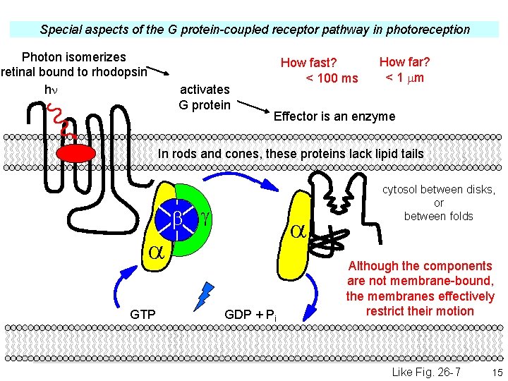
Special aspects of the G protein-coupled receptor pathway in photoreception Photon isomerizes retinal bound to rhodopsin hn activates G protein How fast? < 100 ms How far? < 1 mm Effector is an enzyme In rods and cones, these proteins lack lipid tails b g a a GTP GDP + Pi cytosol between disks, or between folds Although the components are not membrane-bound, the membranes effectively restrict their motion Like Fig. 26 -7 15
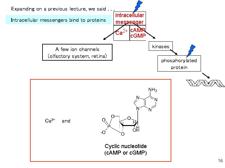
Expanding on a previous lecture, we said. . . Intracellular messengers bind to proteins intracellular messenger c. AMP Ca 2+ c. GMP A few ion channels (olfactory system, retina) Ca 2+ kinases phosphorylated protein and Cyclic nucleotide (c. AMP or c. GMP) 16
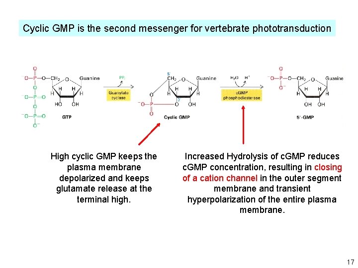
Cyclic GMP is the second messenger for vertebrate phototransduction High cyclic GMP keeps the plasma membrane depolarized and keeps glutamate release at the terminal high. Increased Hydrolysis of c. GMP reduces c. GMP concentration, resulting in closing of a cation channel in the outer segment membrane and transient hyperpolarization of the entire plasma membrane. 17
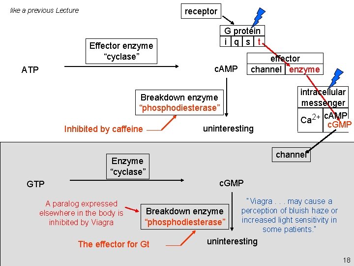
receptor like a previous Lecture G protein i q s t Effector enzyme “cyclase” effector channel enzyme c. AMP ATP Breakdown enzyme “phosphodiesterase” uninteresting Inhibited by caffeine intracellular messenger c. AMP Ca 2+ c. GMP channel Enzyme “cyclase” c. GMP GTP A paralog expressed elsewhere in the body is inhibited by Viagra Breakdown enzyme “phosphodiesterase” The effector for Gt “Viagra. . . may cause a perception of bluish haze or increased light sensitivity in some patients. ” uninteresting 18
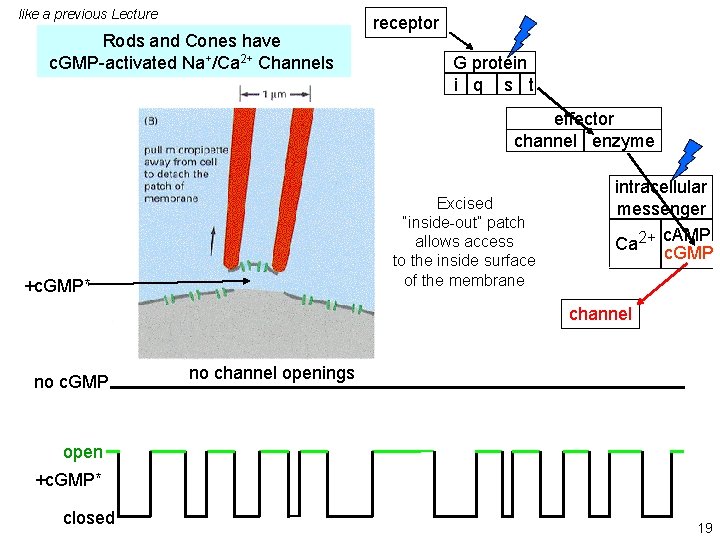
like a previous Lecture Rods and Cones have c. GMP-activated Na+/Ca 2+ Channels receptor G protein i q s t effector channel enzyme Excised “inside-out” patch allows access to the inside surface of the membrane +c. GMP* intracellular messenger c. AMP Ca 2+ c. GMP channel no c. GMP no channel openings open +c. GMP* closed 19
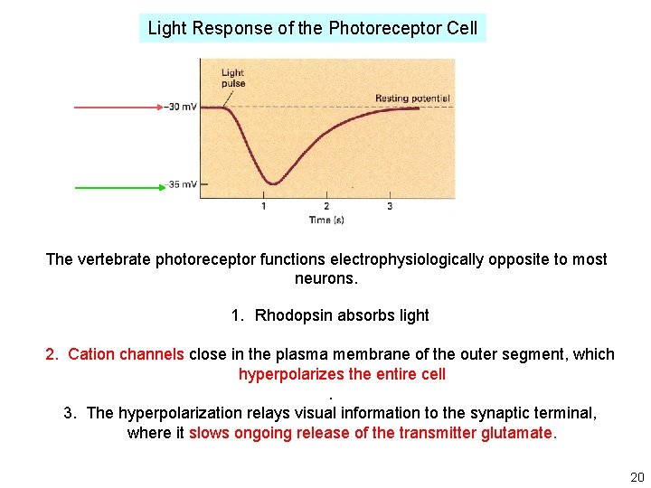
Light Response of the Photoreceptor Cell The vertebrate photoreceptor functions electrophysiologically opposite to most neurons. 1. Rhodopsin absorbs light 2. Cation channels close in the plasma membrane of the outer segment, which hyperpolarizes the entire cell. 3. The hyperpolarization relays visual information to the synaptic terminal, where it slows ongoing release of the transmitter glutamate. 20
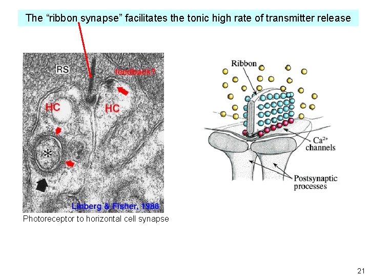
The “ribbon synapse” facilitates the tonic high rate of transmitter release Photoreceptor to horizontal cell synapse 21
The Phototransduction Cascade: 1. Amplification 2. Adaptative/homeostatic mechanisms 1. When fully dark-adapted, many species can detect ~1 photon per photoreceptor cell 2. When fully light-adapted, many species can accurately analyze light at intensities ~1010 fold brighter Many adaptive and homeostatic mechanisms underlie these phenomena. Note: it is incorrect to explain that your favorite process (memory, learning, addiction) occurs “because of” homeostasis or adaptation. Homeostasis and adaptation are not, by themselves, mechanisms. There are homeostatic and adaptive mechanisms. 22
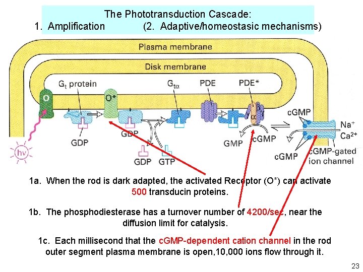
The Phototransduction Cascade: 1. Amplification (2. Adaptive/homeostasic mechanisms) 1 a. When the rod is dark adapted, the activated Receptor (O*) can activate 500 transducin proteins. 1 b. The phosphodiesterase has a turnover number of 4200/sec, near the diffusion limit for catalysis. 1 c. Each millisecond that the c. GMP-dependent cation channel in the rod outer segment plasma membrane is open, 10, 000 ions flow through it. 23
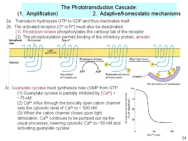
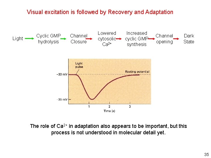
The Phototransduction Cascade: (1. Amplification) 2. Adaptive/homeostatic mechanisms 2 a. Transducin hydrolyses GTP to GDP and thus inactivates itself. 2 b. The activated receptor (O* or R*) must also be deactivated. (1) Rhodopsin kinase phosphorylates the carboxyl tail of the receptor (2) The phosphorylation permits binding of the inhibitory protein, arrestin 3 c. Guanylate cyclase must synthesize new c. GMP from GTP (1) Guanylate cyclase is partially inhibited by [Ca 2+] > ~75 n. M. (2) Ca 2+ influx through the tonically open cation channel sets the cytosolic level of Ca 2+ to ~ 500 n. M. (3) When the cation channel closes upon light stimulation, Ca 2+ continues to be pumped out via the usual processes, lowering cytosolic Ca 2+ to ~50 n. M and activating guanylate cyclase 24
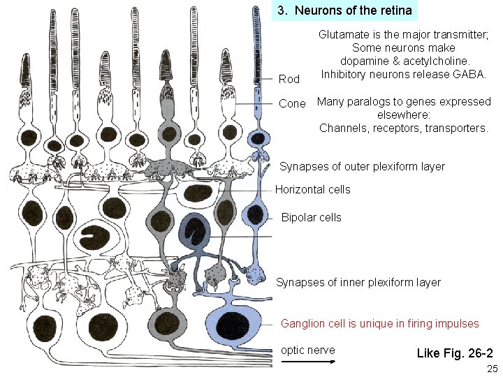
3. Neurons of the retina Rod Cone Glutamate is the major transmitter; Some neurons make dopamine & acetylcholine. Inhibitory neurons release GABA. Many paralogs to genes expressed elsewhere: Channels, receptors, transporters. Synapses of outer plexiform layer Horizontal cells Bipolar cells Synapses of inner plexiform layer Ganglion cell is unique in firing impulses optic nerve Like Fig. 26 -2 25
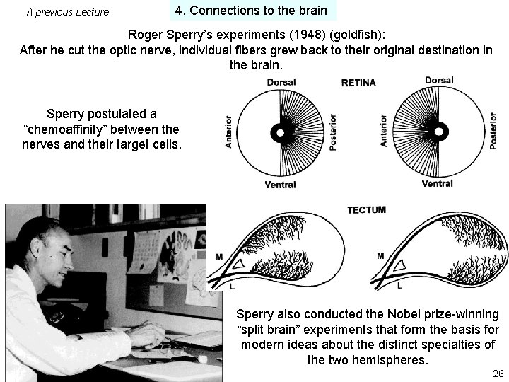
A previous Lecture 4. Connections to the brain Roger Sperry’s experiments (1948) (goldfish): After he cut the optic nerve, individual fibers grew back to their original destination in the brain. Sperry postulated a “chemoaffinity” between the nerves and their target cells. Sperry also conducted the Nobel prize-winning “split brain” experiments that form the basis for modern ideas about the distinct specialties of the two hemispheres. 26
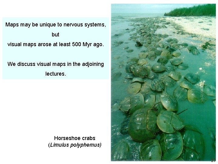
Maps may be unique to nervous systems, but visual maps arose at least 500 Myr ago. We discuss visual maps in the adjoining lectures. Horseshoe crabs (Limulus polyphemus) 27
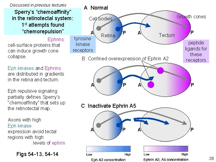
Discussed in previous lectures Sperry’s “chemoaffinity” in the retinotectal system: 1 st attempts found “chemorepulsion” Ephrins: cell-surface proteins that can induce growth cone collapse. A Normal Growth cones Cell bodies A tyrosine kinase receptors Retina P A B Confined overexpression of Ephrin A 2 Eph kinases and Ephrins are distributed in gradients in the retina and tectum. A Eph repulsive signaling partially defines Sperry’s “chemoaffinity” that sets up the retinotectal map. Axons with high Eph kinase expression avoid tectal regions with high levels of ephrin Figs 54 -13, 54 -14 Tectum P P peptide ligands for these receptors A P C Inactivate Ephrin A 5 A P 28
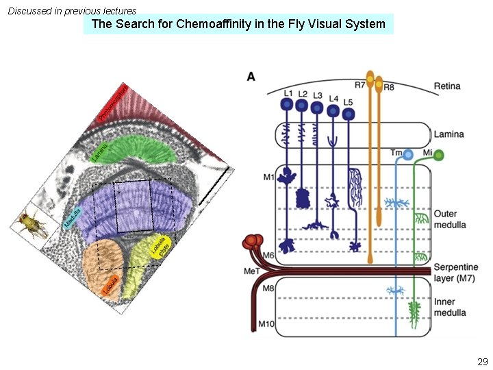
Discussed in previous lectures The Search for Chemoaffinity in the Fly Visual System 29
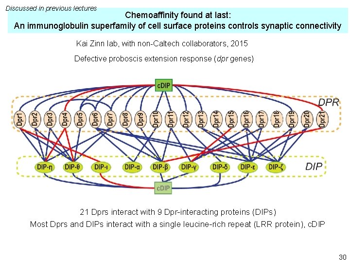
Discussed in previous lectures Chemoaffinity found at last: An immunoglobulin superfamily of cell surface proteins controls synaptic connectivity Kai Zinn lab, with non-Caltech collaborators, 2015 Defective proboscis extension response (dpr genes) 21 Dprs interact with 9 Dpr-interacting proteins (DIPs) Most Dprs and DIPs interact with a single leucine-rich repeat (LRR protein), c. DIP 30
5. Master switches for eye development? Pax-6 / Ey functions when expressed at various locations in Drosophila Little Alberts 8 -25 © Garland 31
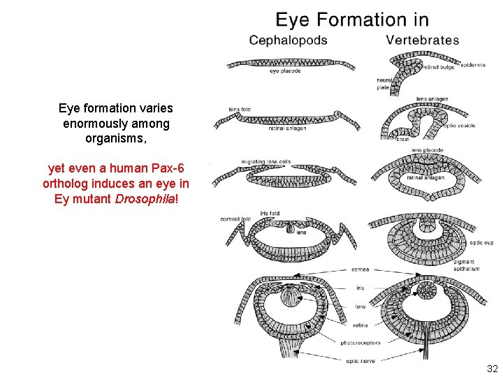
Eye formation varies enormously among organisms, yet even a human Pax-6 ortholog induces an eye in Ey mutant Drosophila! 32
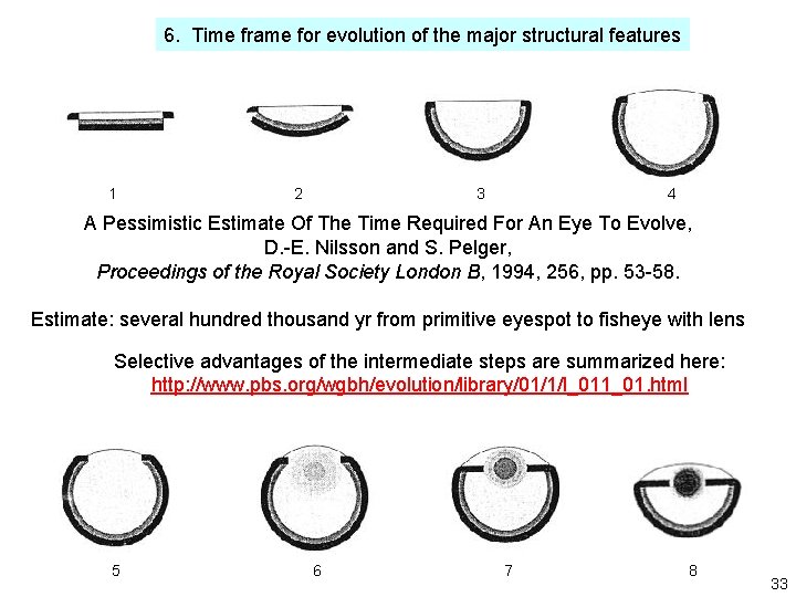
6. Time frame for evolution of the major structural features 1 2 3 4 A Pessimistic Estimate Of The Time Required For An Eye To Evolve, D. -E. Nilsson and S. Pelger, Proceedings of the Royal Society London B, 1994, 256, pp. 53 -58. Estimate: several hundred thousand yr from primitive eyespot to fisheye with lens Selective advantages of the intermediate steps are summarized here: http: //www. pbs. org/wgbh/evolution/library/01/1/l_011_01. html 5 6 7 8 33
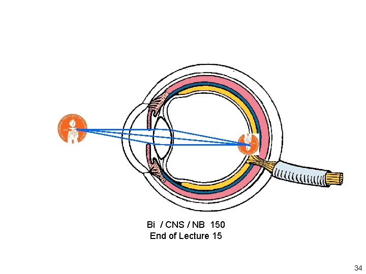
Bi / CNS / NB 150 End of Lecture 15 34
Visual excitation is followed by Recovery and Adaptation Light Cyclic GMP hydrolysis Channel Closure Lowered cytosolic Ca 2+ Increased cyclic GMP synthesis Channel opening Dark State The role of Ca 2+ in adaptation also appears to be important, but this process is not understood in molecular detail yet. 35