6 FUNCTION OF AUTONOMIC NERVOUS SYSTEM1 PNS MODULE


![DISTRIBUTIONS OF THE SYMPATHETIC SYSTEM There are 4 sympathetic distributions: [A] Cervical division: (which](https://slidetodoc.com/presentation_image_h2/b5131616e8516b992be43ee586cf98b7/image-5.jpg "DISTRIBUTIONS OF THE SYMPATHETIC SYSTEM There are 4 sympathetic distributions: [A] Cervical division: (which")

![[B] Cardiopulmonary (thoracic) division : (to thoracic structures) Preganglionic fibers arise from the upper](https://slidetodoc.com/presentation_image_h2/b5131616e8516b992be43ee586cf98b7/image-8.jpg "[B] Cardiopulmonary (thoracic) division : (to thoracic structures) Preganglionic fibers arise from the upper")

![[D] Somatic division (sympathetic supply to skin and skeletal muscles. ) -To the upper](https://slidetodoc.com/presentation_image_h2/b5131616e8516b992be43ee586cf98b7/image-10.jpg "[D] Somatic division (sympathetic supply to skin and skeletal muscles. ) -To the upper")


- Slides: 12
6. FUNCTION OF AUTONOMIC NERVOUS SYSTEM-1 PNS MODULE Prof. Sherif W. Mansour Physiology dpt. , Mutah school of medicine. 2020/2021
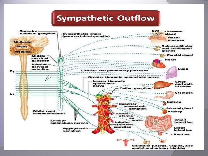
-The nervous system can be classified into: 1 -Central nervous system: which is protected by bone (skull-vertebral column), and includes, the brain (cerebral cortex - brain stem "medulla, pons, midbrain"- cerebellum) and the spinal cord which is formed of 31 segments : 8 cervical (C) , 12 thoracic, 5 lumbar (L) , 5 sacral (S), 1 coccygeal. 2 -Peripheral nervous system: which is divided into somatic (motor nerves and sensory nerves) and autonomic nervous system. -The somatic nervous system that controls the voluntary skeletal muscles contraction. (formed of 12 pairs of cranial nerves, and 31 pairs of spinal nerves) -The autonomic nervous system which controls the involuntary activities of the heart, exocrine glands and the smooth muscles all over the body. -The functions of the A. N. S : 1 - Prepare the body to face emergencies (stresses). 2 -Regulate the process of food digestion. 3 -Regulation of the body temperature. 4 -Regulation of heart rate, blood pressure. 5 -Control some hormonal secretion as catecholamines. 6 -Regulation of vital excretory processes as micturition & defecation. -The autonomic nervous system includes two types of fibers: A)Afferent fibers: which carry sensations from viscera to the C. N. S. This system is widely distributed. B) Efferent fibers: which emerges from the C. N. S. to reach visceral organs and the smooth muscle. *The efferent autonomic nervous system: - The A. N. S reaches its target organs via the autonomic nerve. Each autonomic nerve is an efferent axon of two nerve cells: The first one lies inside either the brain or the spinal cord (C. N. S) and it's axon is called the pre-ganglionic fiber. The second cell lies outside C. N. S (in what is called the autonomic ganglia) and it's axon which reaches the organ is called the post-ganglionic fiber.
Autonomic ganglia Definition: A ganglion is a collection of nerve cells outside the C. N. S surrounded by connective tissue capsule. It contains the nerve fibre of the pre-ganglionic neurons and cells of the post-ganglionic neurons. Function of autonomic ganglia: 1 - Relay stations for the preganglionic fibers. 2 - Site of action of autonomic drugs. 3 - Distribution centers as the ratio of pre to postganglionic fibers is 1: 8. In this way, postganglionic impulses are expanded into several postganglionic neurons. Postganglionic nerves arise in the ganglia and are distributed to various organs. Types of autonomic ganglia: 1 -Lateral (Paravertebral ): Which form the sympathetic chains lying on both sides of the vertebral column. Each chain is formed of 23 ganglia connected to each other by nerve fibers i. e. 3 cervical (superior, middle and inferior), 12 thoracic, 4 lumbar and 4 sacral ganglia. Sometimes, the inferior cervical ganglia fuses with the upper thoracic one to form the "stellate ganglion". Lateral ganglia are only sympathetic. 2 -Collateral (Prevertebral): These are the celiac, the renal, the superior and inferior mesenteric ganglia (sympathetic) and otic, ciliary, sphenopalatine and submandibulare ganglia (parasympathetic). Prevertebral ganglia are interconnected by intermediary neurons. 3 -Terminal (Peripheral) : They are present near or in the wall of the autonomic organ, e. g. the eye, the heart, the stomach and the urinary bladder. Terminal ganglia are only parasympathetic.
The Sympathetic nervous System A-The sympathetic system arises from the LHC of all the thoracic and the upper 2 lumbar segments of the spinal cord. (Thoraco-Lumbar) B-Preganglionic fibers leave the spinal cord to run with the spinal nerves. And then leave the spinal nerve to enter into the lateral ganglia in the white ramus comunicans (mylinated fibers). In the lateral ganglia, the Preganglionic fibers take one of three courses: 1 Relay in the lateral ganglion. The postganglionic fibers go to join the spinal nerve as the gray rami comunicans (unmylinated fibers). 2 - Go up or down the sympathetic chain before relay. 3 Leave the sympathetic chain without relay to relay in a collateral ganglia or reach the adrenal medulla as preganglionic fibers. C- The actions of the sympathetic system prepare the body to face stresses or emergencies and is usually catabolic i. e. leads to energy loss.
DISTRIBUTIONS OF THE SYMPATHETIC SYSTEM There are 4 sympathetic distributions: [A] Cervical division: (which supplies structures in the head and neck) Preganglionic fibers arise from the upper 2 thoracic segments and relay in the superior cervical sympathetic ganglion. Postganglionic fibers follow the course of carotid arteries to: 1 -Eye: -Motor to dilator pupillae muscle→ mydriasis (dilatation of the pupil) -Motor to the superior and inferior tarsal muscles → widening of the palpebral fissure. Thus widening the field of vision. -Motor to Muller's muscle (in animals) → exophthalmos (forward protrusion of the eye ball) -Relaxation of the ciliary muscle, decreasing the power of the lens to prepare the eye for far vision. 2 -Glands: -Lacrimal glands : little secretion of tears and vasoconstriction. -Salivary glands: trophic secretion (small in amount, viscid and concentrated) from the submaxillary gland. 3 -Skin : -Sweat glands : copious secretion eccrine glands (cholinergic fibers). -Erector pilae muscles→ erection of hair. -Bloods vessels vasoconstriction. 4 -Cerebral vessels: -Mild vasoconstriction. Still during sympathetic excitement, cerebral blood flow increase due to the rise in arterial blood pressure.
Horner’s Syndrome This occurs due to damage of the superior cervical sympathetic ganglion either experimentally or by disease. This leads to: 1 -Ptosis: drop of upper eye lid, due to paralysis of superior tarsal muscle. 2 -Miosis: pupillo-constriction, due to paralysis of the dilator pupillae muscle. The constrictor pupillae muscle acts unantagonized. 3 -Enophthalmos: inward sinking of the eyeball in the orbit due to paralysis of Muller's muscle (in animals). 4 -Anhydrosis: dryness of the skin due to absence of sweat secretion on the affected side. 5 -Vasodilatation: in the affected side, so appearing more red than the normal side. This is due to the absence of the sympathetic vasoconstrictor tone to the skin vessels.
[B] Cardiopulmonary (thoracic) division : (to thoracic structures) Preganglionic fibers arise from the upper 4 thoracic segments. They relay in the cervical and upper 4 thoracic ganlglia. Postganglionic fiber form the superficial and deep cardiopulmonary plexuses which supply: 1 -The heart: a- They stimulate all the properties of the cardiac muscle (contractility, rhythmicity, conductivity and excitability) and increase its metabolism & 02 consumption b- Coronary vessels: Direct effect is vasoconstriction (alpha l adrenergic effect), but coronary vessels dilate due to increased metabolism of the heart that decrease O 2 concentration (indirect effect ). The metabolites itself cause direct dilatation. 2 -The Lung a-Bronchi: Bronchodilation and inhibition of bronchial secretions. b-Pulmonary vessels: vasoconstriction. This widens the air passages lead to better ventilation. [C] Splanchnic division : (to abdominal and pelvic viscera) A-Abdominal division: Preganglionic fibers arise from the lower 8 thoracic segments and pass without relay to form the greater splanchnic nerves which relay in celiac, renal and superior mesenteric ganglia. . The postganglionic fibers supply: 1 -Gastrointestinal tract (GIT): relaxation of the wall, but constriction of the sphincters. Leading to delayed evacuation of food. 2 -GIT secretions: General inhibition of GIT secretions. 3 -The splanchinic vessels vasoconstrictor (via α receptors) and vasodilator (β-receptors) fibers (the effect is mainly vasoconstriction).
4 -The spleen motor to smooth muscle fibers of the capsule and trabeculae → 250 m. L of stored blood is poured into the circulation. This action is more prominent in animals. 5 -The liver to stimulate metabolism , glycogenolysis with increase blood glucose level , lipolysis with elevation of the blood lipid level and dilatation to it's vessels. 6 -The endocrine pancreas: usually inhibition of insulin secretion. 7 - The Kidney: Vasoconstriction of renal blood vessels, decreased renal blood flow, decreased urinary output and stimulation of renin secretion. 8 -The adrenal medulla (preganglionic cholinergic fibers) secretion of catecholamines, adrenaline (80%) and noradrenaline (20%) hormones. The released adrenaline stimulates lipolysis, thermogenesis and enhances blood clotting by releasing blood clotting factors from the liver. It stimulates the reticular formation of the brainstem → increased alertness with lack of sleep (insomnia). B- Pelvic division : The preganglionic fibres arise from upper 2 lumbar segments and pass without relay to form the lesser splanchnic nerve. The lesser splanchinic nerves on both sides unit to form the presacral nerve which relay in the inferior mesenteric ganglion. The postganglionic fibers supply: 1 -The urinary bladder : inhibitory to the wall (detrusor muscle ) and motor the internal urethral sphincter → retention of urine. It prevents semen to flow back into the bladder during ejaculation. 2 -The rectum: inhibitory to the wall and motor to the internal anal sphincter → retention of feaces. Desire of micturition and defecation disappear. 3 -The sex organs: In female- It is mainly inhibitory on uterus and fallopian tubes, but late in pregnancy it is excitatory to the uterus. In male - It is mainly excitatory on the smooth muscles of epididymis, vas deferens, seminal vesicles and prostate motor fibers with emission of semen during sexual intercourse leading to ejaculation.
[D] Somatic division (sympathetic supply to skin and skeletal muscles. ) -To the upper limbs: Preganglionic fibers arise from the 4 th to 8 th thoracic segments, relay in the lower cervical and upper 4 thoracic ganglia. Postganglionic fibers join the brachial plexus. -To the lower limbs: Preganglionic fibers arise from 10 th thoracic to 2 nd lumbar segment, relay in the lumbar and sacral ganglia. Postganglionic fibers join the lumbar plexus. -Fibers going to the skin supply: 1 - Sweat glands: . Eccrine copious secretion (cholinergic). i. e. found in skin all over the body. Apocrine thick odoriferous secretion. . i. e. found in axilla and genital areas. 2 -Cutaneous blood vessels → vasoconstriction. 3 -Erector pilea muscles → piloerection, i. e. hair erection. This is more prominent in animals like cats during fighting or cold whether. -Fibers going to the skeletal muscles supply: 1 -Blood vessels of skeletal muscles causing vasodilatation (cholinergic effect). 2 -This vasodilatation increases the blood flow and stimulates metabolic processes needed for energy production leading to increase power of contraction, delay of fatigue and early recovery after exhaustion. This effect is known as “Orbelli phenomenon”.
*General function of sympathetic N. S: I-During rest: It causes sympathetic tone on the blood vessels leading to continuous mild vasoconstriction to maintain the blood pressure. Also the heart has week sympathetic tone. II-In emergency conditions (The alarm or Stress response): In cases of fight, flight, muscular excitement occurs to help the person to face the emergency situation with a better performance. 1) Acceleration of the heart: to supply blood to active tissues. 2) Vasoconstriction in inactive regions, skin and splanchinic area to divert more blood to active regions e. g. muscles, heart and brain. 3) Dilation of bronchi; facilitating pulmonary ventilation. 4) Contraction of spleen: to give more RBCs to carry more oxygen to the active tissues. 5)Sweat secretion: to get rid of the excess heat by evaporation. 6)Delay muscle fatigue (Orbelli phenomenon). 7) Glycogenolysis: supplying glucose to the active tissues for energy production. Also, lipolysis. 8) Adrenal medulla is stimulated to secrete adrenaline and noradrenaline to aid and intensify all the above reaction. Adrenaline stimulates the brain to increase alertness and shorten response time. 9) Increase field of vision 10) Clotting of the blood is enhanced for more effective hemostasis (stoppage of bleeding). 11) Inhibition of gastrointestinal activities, defecation, micturition and erection take place. All these factors lead to shift of blood from inactive areas as the skin and the gastrointestinal tract to active contracting muscles and heart to enable the body to face emergencies.
Thank You