6 1 Human Vision Parts of the Eye


 Mc. Graw Hill Ryerson 2007")


 Mc.")


 Mc. Graw Hill Ryerson 2007")


 Mc. Graw Hill Ryerson 2007")


 Mc. Graw Hill Ryerson 2007")


- Slides: 32
6. 1 Human Vision Parts of the Eye
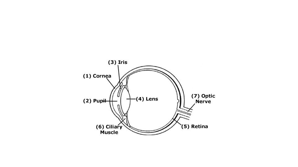
Parts of the Eye • Pupil – the hole that lets light in. • Iris - coloured circle of muscle surrounding the pupil. • controls the amount of light entering the eye. • Sclera - white part of the eye surrounding the iris. (c) Mc. Graw Hill Ryerson 2007 See pages 202 - 203
Iris Controls amount of light entering pupil (c) Mc. Graw Hill Ryerson 2007
Pupil shape depends on your way of life • How do you think a predator s eye placement is different from prey? • What if an animal was active at night?
Predators need to focus Prey need to watch out
• If you have a vertical slit, you're very likely to be an ambush predator (the kind of animal who lies in wait and then leaps out to kill). These predators need to accurately judge the distance to their prey, and the vertical slit has optical features that make it ideal for that. Moslty applied to shorter animals, if its eyes aren't too high off the ground. • foxes, have vertical pupils, but wolves have round pupils • small pet cat has vertical slits, but the larger predators, like lions and tigers, have round pupils.
• round pupils seem to be common in taller hunters that actively chase down their prey • Meanwhile, if you're the kind of animal that gets hunted, you're very likely to have a horizontal pupil and to have your eyes on the side of your head to offer prey animals a panoramic view, so they can best scan all directions for danger.
Whose eyes are these ?
cuttlefish domestic cat lion horse goat gecko
Cornea-Lens-Retina System Cornea - transparent tissue covering the iris and the pupil. (c) Mc. Graw Hill Ryerson 2007
Cornea-Lens-Retina System Lens - behind the pupil • focuses light onto the retina located in back of the eye. • Flexible, Convex (c) Mc. Graw Hill Ryerson 2007
Cornea-Lens-Retina System Retina – lining of back of eye, covered with light sensitive cells that convert light energy into electrical energy. (c) Mc. Graw Hill Ryerson 2007
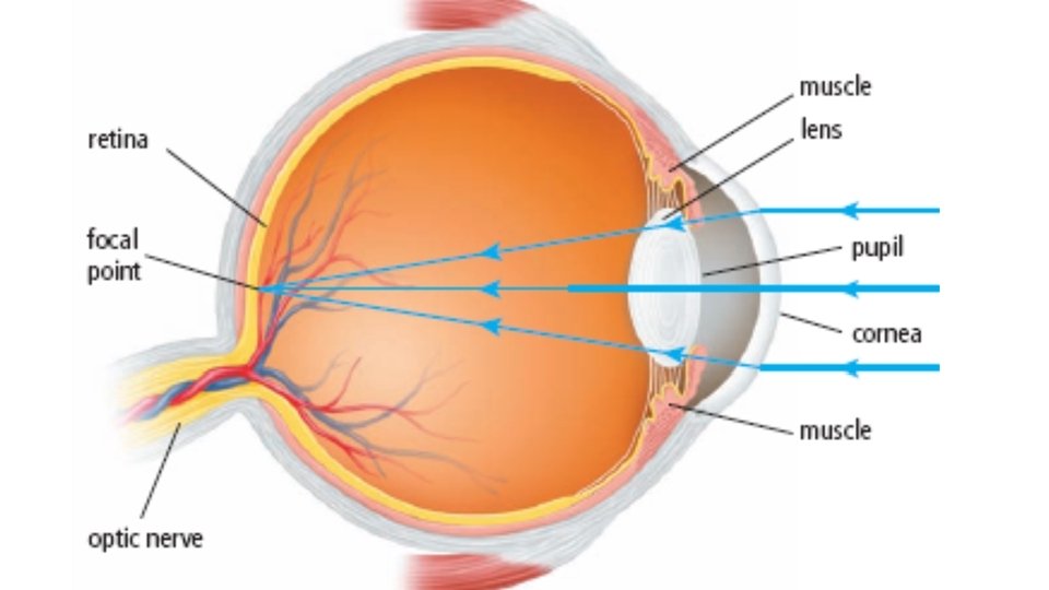
The Cornea-Lens-Retina System • Light rays first entering the eye are refracted by the cornea so that they converge toward the retina. • Cornea actually does most of the focussing. See pages 204 - 205 (c) Mc. Graw Hill Ryerson 2007
The Cornea-Lens-Retina System • Light then passes through the lens which fine-tunes the focus by changing shape. • Muscles attached to the lens contract, causing lens to be thicker. A thicker lens can focus on near objects. This is why you may get eye strain from reading. • When looking at a distant object, the muscles relax, increasing tension on the lens, making it thinner.
The Cornea-Lens-Retina System • When the light rays hit the back of the eye, the cells in the retina absorb and convert the light to electrochemical impulses which are transferred along the optic nerve and then to the brain.
The image that forms on the retina is inverted. See pages 204 - 205 (c) Mc. Graw Hill Ryerson 2007
Macula – central part of retina • Where sharp, clear, straightahead vision is processed • The macula provides the sharp, central vision we need for reading, driving, and seeing fine detail.
Fovea • a tiny pit located in the macula of the retina that provides the clearest vision of all. • Only in the fovea are the layers of the retina spread aside to let light fall directly on the cones, the cells that give the sharpest image. • Also called the central fovea or fovea centralis.
The Cornea-Lens-Retina System • The area where the optic nerve enters the retina is called the blind spot. • This area has no light-sensing cells. See pages 204 - 205 (c) Mc. Graw Hill Ryerson 2007
Locate your blind spot See page 205 (c) Mc. Graw Hill Ryerson 2007
Optic Nerve • carries electrical signals from eye to brain.
What is inside the eye • Aqueous Humour: • Fluid between the lens and cornea, • supports cornea and lens, • provides nutrients to cornea (cornea has no blood vessels). • Vitreous Humour: • Fluid behind lens, • supports lens and gives shape to eye (c) Mc. Graw Hill Ryerson 2007
What’s happening here? (c) Mc. Graw Hill Ryerson 2007
Tapetum Lucidum • Reflective surface behind the retina, enhancing ability to see in low light. • provides the light-sensitive cells in retina with a second opportunity for photon-photoreceptor stimulation. • can be white, green, blue, yellow, orange or red.
Even in some spiders
Check your understanding • Explain how human vision depends on: • reflection, • refraction, • absorption and • transmission of light. (c) Mc. Graw Hill Ryerson 2007
Check your understanding 1. What happens to light rays after they enter the eye through the pupil? 2. Where does most of the focussing in the eye occur? 3. How does the lens change to focus on object that are close? 4. How does the lens change to focus on object that are distant? 5. Why is the image of an object inverted when it strikes the retina? (c) Mc. Graw Hill Ryerson 2007
Compound Eyes (c) Mc. Graw Hill Ryerson 2007
How others sense light • Sci Show: • https: //www. youtube. com/watch? v=z 4 rxine. IFFE • Insect vision • https: //www. youtube. com/watch? v=9 Cp. EV 9_JOv 8 • How dragonflies see • https: //www. youtube. com/watch? v=m 5 XUdv. BO_TE (c) Mc. Graw Hill Ryerson 2007
Homework • Read pages 202 -205 • Answer Reading Check p 205 • WB p 88 (c) Mc. Graw Hill Ryerson 2007