40 ImmuneMediated Diseases and Autoimmunity I Immunology 297


 http: //www. psoriasis. org/")


")

 antigens, such as myelin oligodendrocyte")


,")


 • Autoimmune disease of the postsynaptic neuromuscular junction • 200 cases")
![Myasthenia gravis (MG) • MG: Latin. Gravis =grave. Greek. mys, muscle, + astheneia, weakness]](https://slidetodoc.com/presentation_image_h/20f9a9113e0c1f938b3592a5be444c38/image-26.jpg "Myasthenia gravis (MG) • MG: Latin. Gravis =grave. Greek. mys, muscle, + astheneia, weakness]")


 • In addition to hyperthyroidism, over 25– 50% of individuals with")


 from the pituitary gland acts")
 • Reported by Dr. Hakaru Hashimoto • Autoimmune hypothyroidism •")


 Perivascular mononuclear cell (lymphocyte and macrophage) infiltrates in the dermis. B) B) Immunohistochemistry")


 phase occurs during the first exposure to an antigen.")

 = lacquer Blistering skin lesions on hand of patient with poison")


- Slides: 51
#40 Immune-Mediated Diseases and Autoimmunity I Immunology 297 September 4, 2015 Ikuo Tsunoda, MD, Ph. D Department of Microbiology and Immunology LSUHSC, Shreveport Homepage: http: //tsunodalaboratory. web. fc 2. com/ E-mail: itsunoda@hotmail. com
12 -4 Autoimmune Diseases: General Principles Ø Autoimmune disease Ø A disease in which the primary mechanism of pathology is an immune attack directed against components of the self Ø Autoantigen Ø A self component that is the target of the immune response in an autoimmune disease Ø Systemic versus Organ-specific autoimmunity
Figure 13‐ 5. Types of autoimmunity. Autoimmunity occurs when the protective mechanisms of self-tolerance fail or are bypassed; pathologic reactions against self-antigens may result. Anti-self antibodies and selfreacting cells disrupt tissues and cause organ damage, or autoimmune disease. Two major categories. 1) Organ-specific: typified by thyroiditis and myasthenia gravis, attack is directed against a single antigen in a single organ or tissue. 2) Non–organ-specific: target widespread self-antigens such as nucleic acids in systemic lupus erythematosus. Organ damage is widely disseminated owing to the ubiquitous distribution of the antigen, and immune complex deposition may cause remote systemic injury. The organ-specific diseases occur together with greater than expected likelihood. For example, 5% to 10% of patients with myasthenia gravis also have antibody-mediated thyroid disease. Disease clustering also occurs at the other end of the spectrum, with frequent overlap between, for example, dermatomyositis and other rheumatologic syndromes.
7. 5 million patients in the US (National Psoriasis Foundation) http: //www. psoriasis. org/ Autoantigen unknown, IL-17 blockade effective
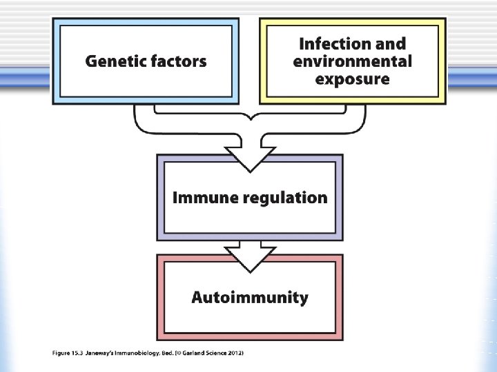
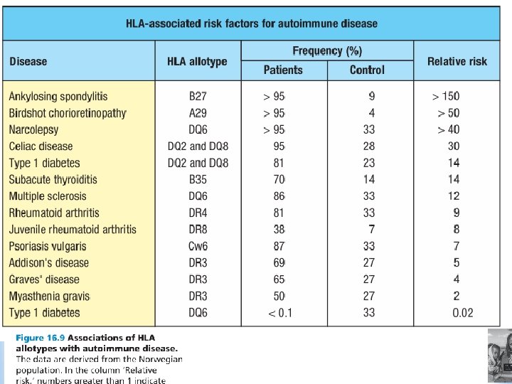
Autoimmunity is the result of genetic susceptibility and environmental trigger? Identical twin studies: If one twin develop an autoimmune disease, 10 -50% of the other twin develop the disease Some HLA alleles occur at higher frequency in autoimmune disease patients than in the general population Multiple polymorphisms (variants) in non. HLA genes associated with autoimmune disease susceptibility: CTLA 4, PTPN 22, CD 25 (IL-2 receptor α chain), IL-23 R Single-gene defects cause autoimmunity: FAS/Fas. L (ALPS), AIRE (APS), FOXP 3 (IPEX), complement genes (lupus-like disease)
Susceptibility loci for autoimmune diseases. The chromosomal loci associated with some autoimmune diseases are shown. The location of candidate genes of immunologic interest are indicated as ovals on the left of the chromosomes. These ovals are color coded to indicate the diseases to which the genes are linked. SLE, systemic lupus erythematosus; AITD, autoimmune thyroid disease; RA, rheumatoid arthritis; T 1 D, type 1 diabetes. PTPN 22, protein tyrosine phosphatase (Yamada R and K Ymamoto. Recent findings on genes associated with inflammatory diseases. Mutation Research 573: 136 -151, )
p arm centromere q arm Genetic loci implicated as risk factors for the development of SLE, MS, RA, and IDDM in humans. CTLA 4 is located at 2 q 33. CTLA 4 gene polymorphism associated with type 1 diabetes, autoimmune thyroiditis, Graves’ disease
Treg Figure 13‐ 4. Mechanisms of selftolerance. Tolerance relies on the talent of self-recognition that B cells and T cells probably acquire during maturation. Lymphocytes unable to recognize self or lacking the willingness to tolerate self are deleted or rendered anergic. These mechanisms are sequestration, lack of anergy, antigen presentation, clonal energy, clonal deletion, and suppressor cells. In sequestration (1) some tissue proteins (eg, myelin basic protein, lens of the eye) are anatomically isolated from exposure to lymphocytes; anatomic barriers (eg, the blood–brain barrier) preclude contact with T cells. In lack of antigen presentation (2), some tissues contain cells lacking the capability of antigenpresenting cells (APCs)—cells unable to express major histocompatibility complex (MHC). Neurons, for example, cannot express MHC. In clonal anergy (3), T-cell activation requires a secondary costimulatory signal received by the CD 28 complex. If a T cell confronts an antigen on a cell that lacks a functional B 7 protein, the T cell becomes inactivated or tolerant. In clonal deletion (4), during maturation, T cells must demonstrate recognition of self and tolerance of self. This takes place in the thymus (Th). T-cell lines failing to pass these tests are deleted or inactivated. Regulatory T cells (5) (Treg) and other cells have been proposed to induce self-tolerance.
sequestration Immunologically privileged sites (brain, etc)
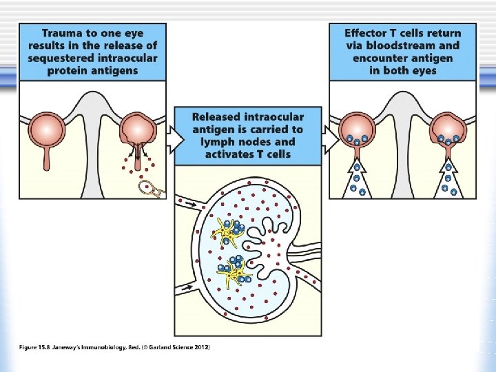
Release of Sequestered Antigens Ø Some self-antigens are sequestered in specialized tissues and cannot be expressed in the thymus or bone marrow. Ø These are not seen by the developing immune system – will not induce self-tolerance. Ø Exposure of T cells to these normally sequestered/tissuespecific self-antigens in the periphery results in their activation.
Examples of Sequestered Antigens Ø Central nervous system (CNS) antigens, such as myelin oligodendrocyte glycoprotein (MOG), associated with multiple sclerosis (MS) Ø Lens and corneal proteins of the eye following infection or trauma
• Tissue grafts placed in these sites do not elicit immune responses • No conventional lymphatics • Surrounded by tissue barriers, e. g. blood-brain barrier
Immunopathology of autoimmune diseases Ø Effector mechanisms Ø Damage by cytotoxic Ig. G antibody (type II), immune complex (type III) or T cellmediated inflammation (type IV) Ø Induction (sensitization) of autoimmune responses Ø Molecular mimicry Ø Epitope spreading
Immune pathology of autoimmune diseases Ø Effector mechanisms Ø Damage by cytotoxic Ig. G antibody (type II), immune complex (type III) or T cellmediated inflammation (type IV) Ø Induction (sensitization) of autoimmune responses Ø Molecular mimicry Ø Epitope spreading
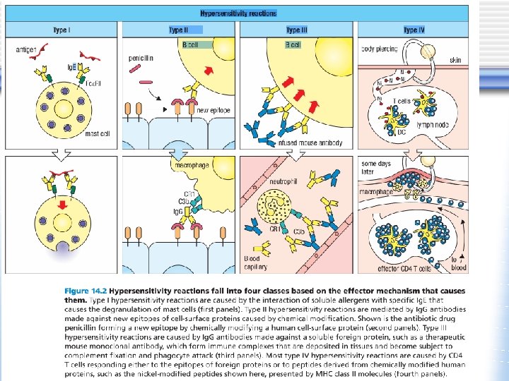
Table 18 -1. Classification of Immunological Diseases Type of hypersensitivity Pathologic immune mechanisms Mechanisms of tissue injury and disease Immediate Ig. E antibody hypersensitivity: Type I Antibody mediated: Ig. M, Ig. G antibodies Type II against cell surface or extracellular matrix antigens Mast cells and their mediators (vasoactive amines, lipid mediators, cytokines) Immune complex mediated: Type III Complement- and Fc receptor-mediated recruitment and activation of leukocytes T cell mediated: Type IV Immune complexes of circulating antigens and Ig. M or Ig. G antibodies 1. CD 4+ T cells (delayedtype hypersensitivity) 2. CD 8+ CTLs (T cellmediated cytolysis) Opsonization and phagocytosis of cells Complement- and Fc receptor-mediated recruitment and activation of leukocytes (neutrophils, macrophages) Abnormalities in cellular functions, e. g. , hormone receptor signaling 1. Macrophage activation, cytokinemediated inflammation 2. Direct target cell killing, cytokinemediated inflammation Hypersensitivity reaction • Allergy; reaction against environmental antigens (allergen), food pollen, house dust • Autoimmunity; reaction against self-antigens (autoantigen)
Type III Types of antibodymediated diseases. Antibodies may bind specifically to tissue antigens (A), or they may be deposited as immune complexes that are formed in the circulation (B). In both cases, the deposited antibodies induce inflammation, leading to tissue injury.
Effector mechanisms of antibody-mediated disease. A. Antibodies opsonize cells and may activate complement, generating complement products that also opsonize cells, leading to phagocytosis of the cells through phagocyte Fc receptors or C 3 receptors. B. Antibodies recruit leukocytes by binding to Fc receptors or by activating complement and thereby releasing by-products that are chemotactic for leukocytes. C. Antibodies specific for cell surface receptors for hormones or neurotransmitters may stimulate the activity of the receptors even in the absence of the hormone (left panel) or may inhibit binding of the neurotransmitter to its receptor (right panel). TSH, thyroidstimulating hormone.
12 -5 Antibody-mediated autoimmune diseases • Autoantibody binds to targets, leading to damage by Fc receptor+ macrophage and/or complement lysis (type II hypersensitivity-type) w Autoimmune hemolytic anemia (antibody against red blood cells) w Autoimmune thrombocytic purpura (antibody against platelets) w Goodpasture’s syndrome (antibody against collagen type IV in the kidney and lung basement membrane) • Immune complex formation and deposition, activating phagocytes and causing damage (type III hypersensitivity-type) w Systemic lupus erythematosus • Direct effect of autoantibodies on the autoantigen affecting its important function w Pemphigus vulgaris w Myasthenia gravis http: //missinglink. ucsf. edu/lm/dermatologyglossary/pemphigus_vulgaris. html
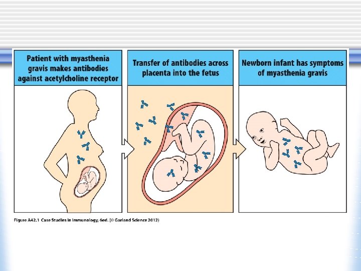
Myasthenia gravis (MG) • Autoimmune disease of the postsynaptic neuromuscular junction • 200 cases per million • Weakness and fatigability • Acetylcholine receptor antibodies in 85% • Thymic hyperplasia in 65% • Thymoma in 15% • Treatment: cholinesterase inhibitors for symptom control, immunosuppressive therapy, thymectomy, plasma exchange
Myasthenia gravis (MG) • MG: Latin. Gravis =grave. Greek. mys, muscle, + astheneia, weakness] severe muscle weakness • Acetylcholine (ACh) receptor in the neuromuscular junction receives Ach from motor neurons to induce muscle contraction • In MG, autoantibody reacts with the ACh receptor • Autoantibody induces the internalization of receptors and can also block the binding of Ach, and block receptor function and prevent muscle contraction
Figure 13‐ 24. Neuromuscular junction in myasthenia gravis. A, The normal neuromuscular junction releases acetylcholine from presynaptic vesicles that bind to acetylcholine receptors at the apices of postsynaptic junctional folds. Bound acetylcholine receptors allow sodium influx, which causes depolarization of postsynaptic membranes. Acetylcholine is degraded by acetylcholinesterase, located deep within the postsynaptic crypts. B, Acetylcholine receptor antibodies bind to acetylcholine receptors, causing accelerated endocytosis and degradation of receptors, blockade of acetylcholine binding sites, and complement-mediated damage to the postsynaptic membrane. The density of acetylcholine receptors is reduced, and the remaining receptors are blocked by the presence of bound antibody. Postsynaptic junctional folds are flattened, damaged, and disorganized by this complement-mediated attack. The reduced population of receptor disallows sufficient membrane depolarization to originate a propagating muscle membrane action potential.
Systemic circulation Thymus Cortex Medulla Cortical thymic epithelial cell Medullary thymic epithelial cell (m. TEC) AIRE MHC CCL 19 CCL 21 autoantigen TCR CD 4 T tissue specific autoantigen RANKL, CD 40 L Mediated m. TEC proliferation CD 8 CCR 7 expression RANKL, CD 40 L expression DP Cell death Positive selection CD 4 SP S 1 P 1 expression. CD 8 T CD 8 SP Systemic circulation Negative selection Cell death or n. Treg generation Dendritic cell Foxp 3 n. Treg Myasthenia gravis: CHRNA 1 gene (encoding Ach receptor α subunit) polymorphism? AIRE modulates CHRNA 1 expression level? Treg abnormality?
Graves’ disease Basedow’s disease • Autoimmune disease in which autoantibodies react with the receptor for thyroid-stimulating hormone (TSH) and activate it • Hyperthyroidism: thyroid cells continually secrete thyroid hormones • Hyperactivity, sweating, fatigue, weight loss with increased appetite, tachycardia, atrial fibrillation (President George HW Bush), exophthalmos(eye protrusion, Barbara Bush) (“Millie” the Bushes' dog has lupus) • Graves' disease occurs in up to 2% of women • Typically occurs between 20 and 50 years of age, but it also occurs in the elderly http: //www. accessmedicine. com/content. aspx? a. ID=2877433 Harrison’s Internal Medicine
Graves’ Ophthalmopathy (GO) • In addition to hyperthyroidism, over 25– 50% of individuals with Graves’ disease have clinical involvement of the eyes known as thyroidassociated ophthalmopathy (TAO) or GO Prabhakar, B. S. et al. Endocr Rev 2003; 24: 802 -835 Current Perspective on the Pathogenesis of Graves’ Disease and Ophthalmopathy FIG. 2. Patient with severe GO Copyright © 2003 The Endocrine Society • 3– 5% of patients suffer from intense pain and inflammation with double vision or even loss of vision • GO can be explained by an increase in the volume of both the orbital fatty connective tissues and the extraocular muscle bodies
Gail Devers, Olympian Masako Natsume, Actress Missy Elliott, rapper Barbara Bush, first lady
• • Thyroid hormones regulate basal metabolism and body temperature Thyroid hormones T 4 and T 3 feed back to inhibit hypothalamic production of thyrotropin-releasing hormone (TRH) and pituitary production of thyroid-stimulating hormone (TSH). TSH stimulates thyroid gland production of T 4 and T 3. Right. Thyroid follicles are formed by thyroid epithelial cells surrounding proteinaceous colloid, which contains thyroglobulin. Follicular cells, which are polarized, synthesize thyroglobulin and carry out thyroid hormone biosynthesis. TSH-R, thyroid-stimulating hormone receptor; Tg, thyroglobulin; NIS, sodium-iodide symporter; TPO, thyroid peroxidase; DIT, diiodotyrosine; MIT, monoiodotyrosine.
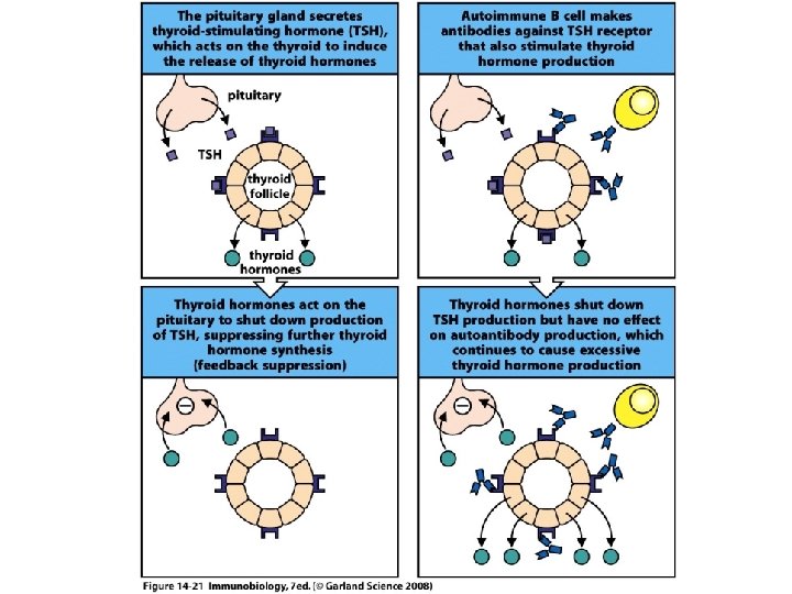
Hyperthyroidism in Graves’ disease • Thyroid stimulating hormone (TSH) from the pituitary gland acts on thyroid cells through the TSH receptor to stimulate the release of thyroid hormones • Thyroid hormones negatively regulate TSH production • In Graves’ disease, TSH receptor antibodies act like TSH to stimulate cells to secrete thyroid hormones • This secretion is unregulated, since the antibodies are always present
Autoimmune thyroiditis (Hashimoto’s thyroiditis) • Reported by Dr. Hakaru Hashimoto • Autoimmune hypothyroidism • Tiredness, weakness, feeling cold, weight gain with poor appetite • CD 4+, CD 8+ T cells and B cells infiltrate in the thyroid • CD 8+ cytotoxic T cells mediated thyroid cell destruction • Local cytokine production, TNF, IL 1, IFN-γ • Autoantibody • HLA-DR 3, 4, 5, CTLA-4 Puffy eyes, thickened, pale skin
Pathologic features of antibody-mediated glomerulonephritis. A. Glomerulonephritis induced by an antibody against the glomerular basement membrane (Goodpasture's syndrome): the light micrograph shows glomerular inflammation and severe damage, and immunofluorescence shows smooth (linear) deposits of antibody along the basement membrane. normal glomerulus B. Glomerulonephritis induced by the deposition of immune complexes (SLE): the light micrograph shows neutrophilic inflammation, and the immunofluorescence and electron micrograph show coarse (granular) deposits of antigen-antibody complexes along the basement membrane.
T Cell-Mediated Diseases • T cells may cause disease. . . w in response to persistent antigens § Delayed-type hypersensitivity w in response to self antigens § Autoimmunity
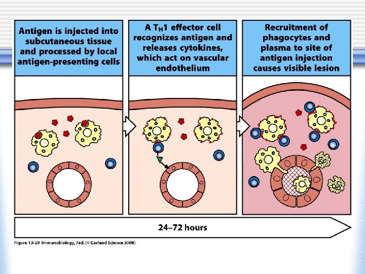
APC, antigenpresenting cell Mechanisms of T cell-mediated diseases. A. In delayed-type hypersensitivity reactions, CD 4+ T cells (and sometimes CD 8+ cells) respond to tissue antigens by secreting cytokines that stimulate inflammation and activate phagocytes, leading to tissue injury. B. In some diseases, CD 8+ CTLs directly kill tissue cells
A) Perivascular mononuclear cell (lymphocyte and macrophage) infiltrates in the dermis. B) B) Immunohistochemistry demonstrate CD 4+ T cells DTH reaction is manifested by induration with redness and swelling at the site of the challenge, which peaks at 48 hours
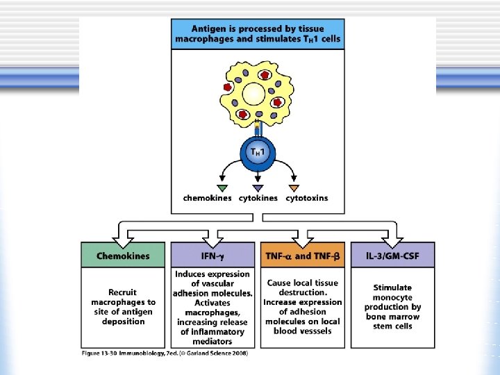
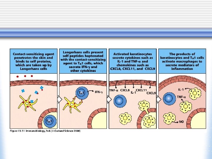
13 -0 Overview: Causes and nature of hypersensitivity reactions • Type IV hypersensitivity is cell-mediated, usually by Th 1 cells, less often by cytotoxic T cells • Known as delayed-type hypersensitivity (DTH), because local exposure to antigen must be followed by migration of antigen specific T cells into the site • In many cases, the sensitizing antigen is a small molecule (hapten, such as poison ivy, nickel) covalently attached to proteins (haptenation) • Most DTH is elicited by haptenated proteins, although some antigens may bind directly to MHC molecules
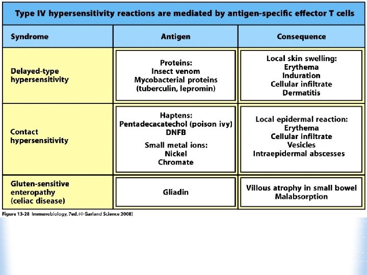
13 -5 Delayed-type hypersensitivity reactions • In most cases the proteins that become haptenated to produce DTH are extracellular, which bind to MHC class II • Lipids-soluble antigens can cross the cell membrane and haptenate cytoplasmic proteins, bind to MHC class I molecule • Contact dermatitis w Poison ivy, nickel (in jewelry) • Celiac disease
Delayed-type Hypersensitivity The sensitization (induction) phase occurs during the first exposure to an antigen. It results in the clonal expansion of antigen-specific T cells as well as the maturation of both effector and memory cells. The effector phase involves the localization of effector T cells to the site of antigen deposition and their subsequent production of specific cytokines that direct the actions of other cell types.
Poison Ivy Reaction Antigenic compound in urushiol is a small hydrophobic molecule, pentadecacatechol
Urushiol Urushi (Japanese) = lacquer Blistering skin lesions on hand of patient with poison ivy contact dermatitis
Autoimmune Diseases due to Type IV Hypersensitivity
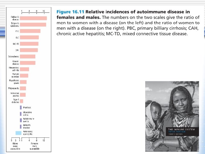
Abbas et al: Cellular and Molecular Immunology Updated 6 E Self-assessment Which one of the following statement concerning autoimmune disease is true? A) Autoimmunity manifests as organ-specific, not systemic, disease B) Infectious microorganisms are frequently present in autoimmune lesions. C) Effector mechanisms in autoimmunity include circulating autoantibodies, immune complexes, and autoreactive T lymphocytes. D) Among the genes associated with autoimmunity, associations are particularly prevalent with class I MHC genes. E) Many autoimmune diseases show higher incidence in males than in females. Explanation: Various effector mechanisms are responsible for tissue injury in different autoimmune diseases. These include circulating autoantibodies, immune complexes, and autoreactive T lymphocytes. Autoimmune diseases may be either systemic (i. e. , systemic lupus erythematosus) or organ specific (i. e. , type 1 diabetes mellitus, multiple sclerosis). Among the genes associated with autoimmunity, the strongest associations are with MHC genes and usually with class II MHC genes (ankylosing spondylitis is an exception). In most cases of autoimmunity, infectious microorganisms are neither present in lesions nor detectable in patients when autoimmunity develops; this suggests that lesions in autoimmunity result not from the infectious agent directly but from host immune responses that may be triggered by microbes. Finally, many autoimmune diseases show higher incidence in females than in males, although the reasons for this are not well understood.