
4 th Universal Definition of MI CDI Implications


- Slides: 22
4 th Universal Definition of MI CDI Implications ACDIS Radio – August 29, 2018 James S. Kennedy, MD, CCS, CDIP President and Chief Medical Officer CDIMD – Physician Champions Smyrna, Tennessee jkennedy@cdimd. com – (615) 479 -7021 1
Disclosures • This presentation is designed to provide accurate and authoritative information in regard to the subject matter covered. The information includes both reporting and interpretation of materials in various publications, as well as interpretation of policies of various organizations. This information is subject to individual interpretation and to changes over time. – VP-MA Health Solutions, dba CDIMD, HCPro, ACDIS, the individual speakers, and all affiliated entities do not warrant that the written or oral opinions expressed in this lecture apply to every situation. Prior to implementing any of the suggestions discussed at this meeting, the attendee is advised to seek counsel from his or her compliance officer or their legal counsel. – CDIMD, HCPro, ACDIS, the individual speakers, and all affiliated entities support accurate coding of every clinical circumstance based upon physician documentation, recognize the role and responsibility of treating physicians to utilize language they deem appropriate to their circumstances, and support compliance to all local, state, and federal laws. 2
Learning Objectives • Discuss the new 4 th Universal Definition of Acute Myocardial Infarction and its impact on CDI and coding practices • Advocate solutions that promote clinical validity and coding compliance 3
4 th Universal Definition Now Available http: //www. tinyurl. com/2018 UDMI 4
2018 4 th UDMI New Concepts • Differentiation of myocardial infarction from myocardial injury. • Highlighting peri-procedural myocardial injury after cardiac and noncardiac procedures as discrete from myocardial infarction. • Consideration of electrical remodeling (cardiac memory) in assessing repolarization abnormalities with tachyarrhythmia, pacing, and rate-related conduction disturbances. • Use of cardiovascular magnetic resonance to define the etiology of myocardial injury. • Use of computed tomographic coronary angiography in suspected myocardial infarction. 5
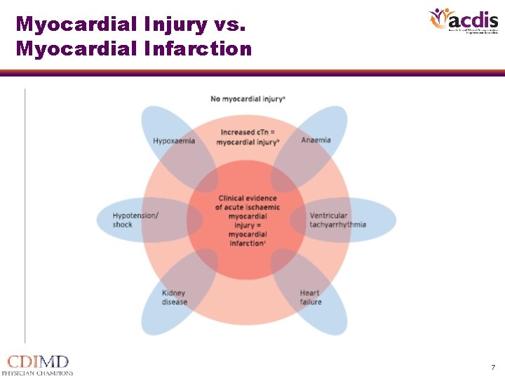
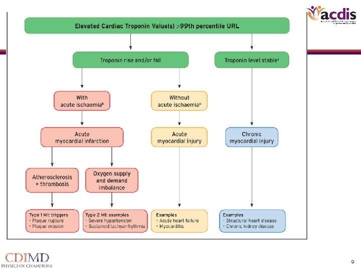
2018 4 th UDMI Definition of Myocardial Injury • The term myocardial injury should be used when there is evidence of elevated cardiac troponin values (c. Tn) with at least one value above the 99 th percentile upper reference limit (URL). The myocardial injury is considered acute if there is a rise and/or fall of c. Tn values. – What many physicians label as “troponin leak”, “troponinemia” – In some circumstances, “demand ischemia” without elaborating as to if it is a myocardial infarction or nonischemic myocardial necrosis is used 6
Myocardial Injury vs. Myocardial Infarction 7
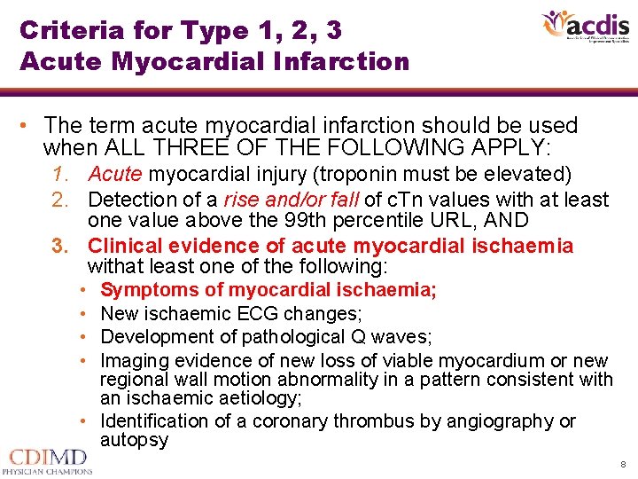
Criteria for Type 1, 2, 3 Acute Myocardial Infarction • The term acute myocardial infarction should be used when ALL THREE OF THE FOLLOWING APPLY: 1. Acute myocardial injury (troponin must be elevated) 2. Detection of a rise and/or fall of c. Tn values with at least one value above the 99 th percentile URL, AND 3. Clinical evidence of acute myocardial ischaemia withat least one of the following: • • Symptoms of myocardial ischaemia; New ischaemic ECG changes; Development of pathological Q waves; Imaging evidence of new loss of viable myocardium or new regional wall motion abnormality in a pattern consistent with an ischaemic aetiology; • Identification of a coronary thrombus by angiography or autopsy 8
9
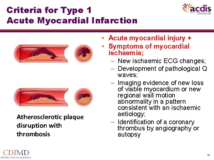
Criteria for Type 1 Acute Myocardial Infarction • Acute myocardial injury + • Symptoms of myocardial ischaemia; – New ischaemic ECG changes; – Development of pathological Q Atherosclerotic plaque disruption with thrombosis waves; – Imaging evidence of new loss of viable myocardium or new regional wall motion abnormality in a pattern consistent with an ischaemic aetiology; – Identification of a coronary thrombus by angiography or autopsy 10
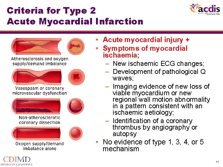
Criteria for Type 2 Acute Myocardial Infarction • Acute myocardial injury + • Symptoms of myocardial ischaemia; – New ischaemic ECG changes; – Development of pathological Q waves; – Imaging evidence of new loss of viable myocardium or new regional wall motion abnormality in a pattern consistent with an ischaemic aetiology; – Identification of a coronary thrombus by angiography or autopsy • No evidence of type 1, 3, 4, or 5 mechanism 11
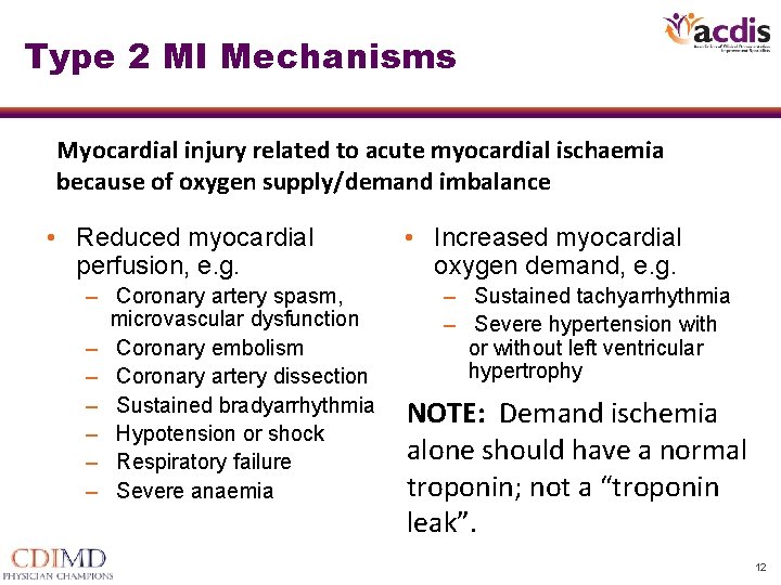
Type 2 MI Mechanisms Myocardial injury related to acute myocardial ischaemia because of oxygen supply/demand imbalance • Reduced myocardial perfusion, e. g. – Coronary artery spasm, microvascular dysfunction – Coronary embolism – Coronary artery dissection – Sustained bradyarrhythmia – Hypotension or shock – Respiratory failure – Severe anaemia • Increased myocardial oxygen demand, e. g. – Sustained tachyarrhythmia – Severe hypertension with or without left ventricular hypertrophy NOTE: Demand ischemia alone should have a normal troponin; not a “troponin leak”. 12
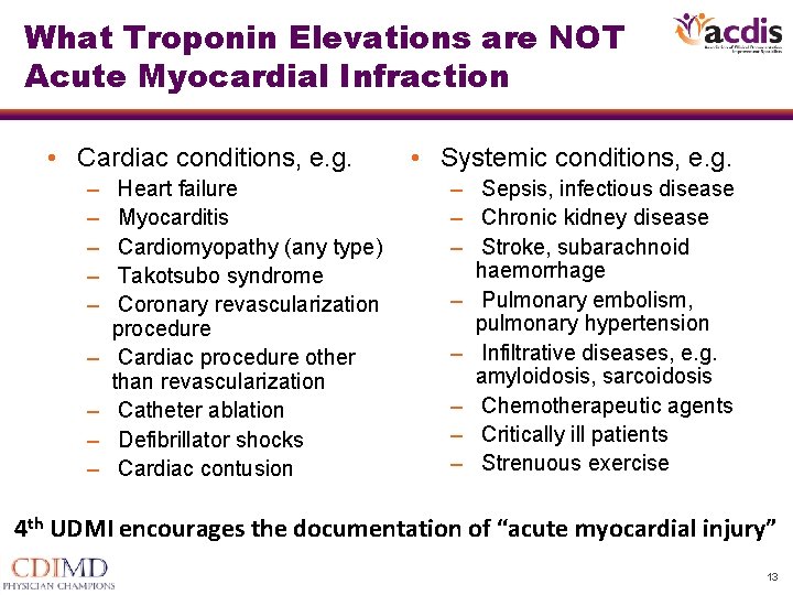
What Troponin Elevations are NOT Acute Myocardial Infraction • Cardiac conditions, e. g. – – – – – Heart failure Myocarditis Cardiomyopathy (any type) Takotsubo syndrome Coronary revascularization procedure Cardiac procedure other than revascularization Catheter ablation Defibrillator shocks Cardiac contusion • Systemic conditions, e. g. – Sepsis, infectious disease – Chronic kidney disease – Stroke, subarachnoid haemorrhage – Pulmonary embolism, pulmonary hypertension – Infiltrative diseases, e. g. amyloidosis, sarcoidosis – Chemotherapeutic agents – Critically ill patients – Strenuous exercise 4 th UDMI encourages the documentation of “acute myocardial injury” 13
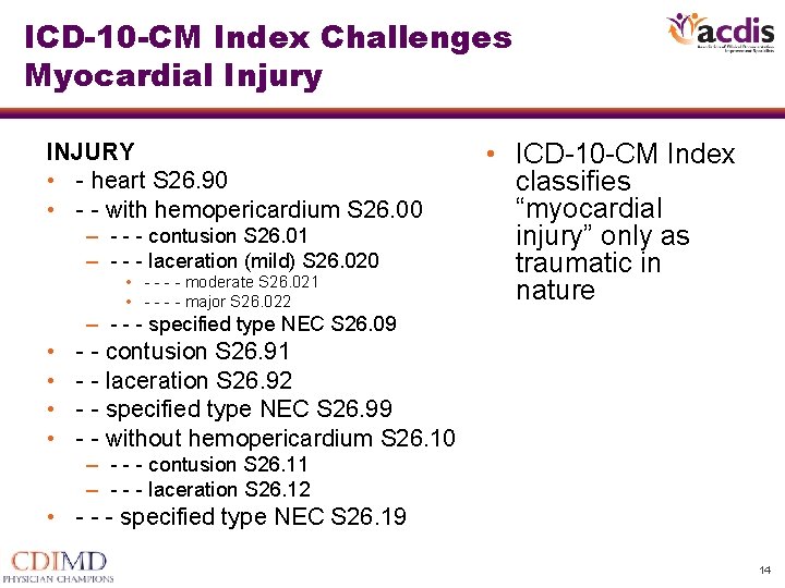
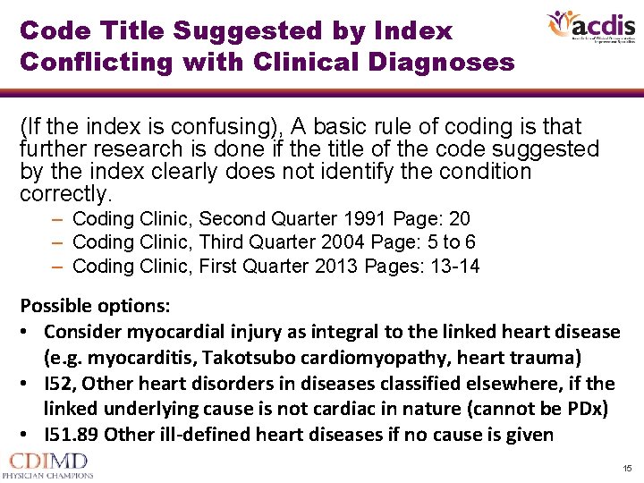
ICD-10 -CM Index Challenges Myocardial Injury INJURY • - heart S 26. 90 • - - with hemopericardium S 26. 00 – - - - contusion S 26. 01 – - - - laceration (mild) S 26. 020 • - - moderate S 26. 021 • - - major S 26. 022 • ICD-10 -CM Index classifies “myocardial injury” only as traumatic in nature – - - - specified type NEC S 26. 09 • • - - contusion S 26. 91 - - laceration S 26. 92 - - specified type NEC S 26. 99 - - without hemopericardium S 26. 10 – - - - contusion S 26. 11 – - - - laceration S 26. 12 • - - - specified type NEC S 26. 19 14
Code Title Suggested by Index Conflicting with Clinical Diagnoses (If the index is confusing), A basic rule of coding is that further research is done if the title of the code suggested by the index clearly does not identify the condition correctly. – Coding Clinic, Second Quarter 1991 Page: 20 – Coding Clinic, Third Quarter 2004 Page: 5 to 6 – Coding Clinic, First Quarter 2013 Pages: 13 -14 Possible options: • Consider myocardial injury as integral to the linked heart disease (e. g. myocarditis, Takotsubo cardiomyopathy, heart trauma) • I 52, Other heart disorders in diseases classified elsewhere, if the linked underlying cause is not cardiac in nature (cannot be PDx) • I 51. 89 Other ill-defined heart diseases if no cause is given 15
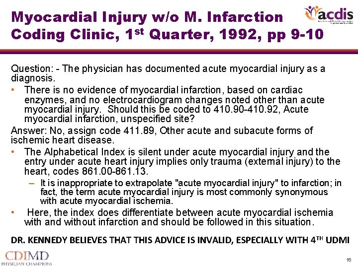
Myocardial Injury w/o M. Infarction Coding Clinic, 1 st Quarter, 1992, pp 9 -10 Question: - The physician has documented acute myocardial injury as a diagnosis. • There is no evidence of myocardial infarction, based on cardiac enzymes, and no electrocardiogram changes noted other than acute myocardial injury. Should this be coded to 410. 90 -410. 92, Acute myocardial infarction, unspecified site? Answer: No, assign code 411. 89, Other acute and subacute forms of ischemic heart disease. • The Alphabetical Index is silent under acute myocardial injury and the entry under acute heart injury implies only trauma (external injury) to the heart, codes 861. 00 -861. 13. – It is inappropriate to extrapolate "acute myocardial injury" to infarction; in fact, the term acute myocardial injury is most commonly synonymous with acute myocardial ischemia. • Here, the index does differentiate between acute myocardial ischemia with and without infarction and should be followed in this situation. DR. KENNEDY BELIEVES THAT THIS ADVICE IS INVALID, ESPECIALLY WITH 4 TH UDMI 16
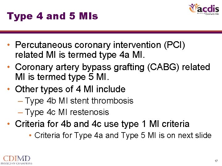
Type 4 and 5 MIs • Percutaneous coronary intervention (PCI) related MI is termed type 4 a MI. • Coronary artery bypass grafting (CABG) related MI is termed type 5 MI. • Other types of 4 MI include – Type 4 b MI stent thrombosis – Type 4 c MI restenosis • Criteria for 4 b and 4 c use type 1 MI criteria • Criteria for Type 4 a and Type 5 MI is on next slide 17
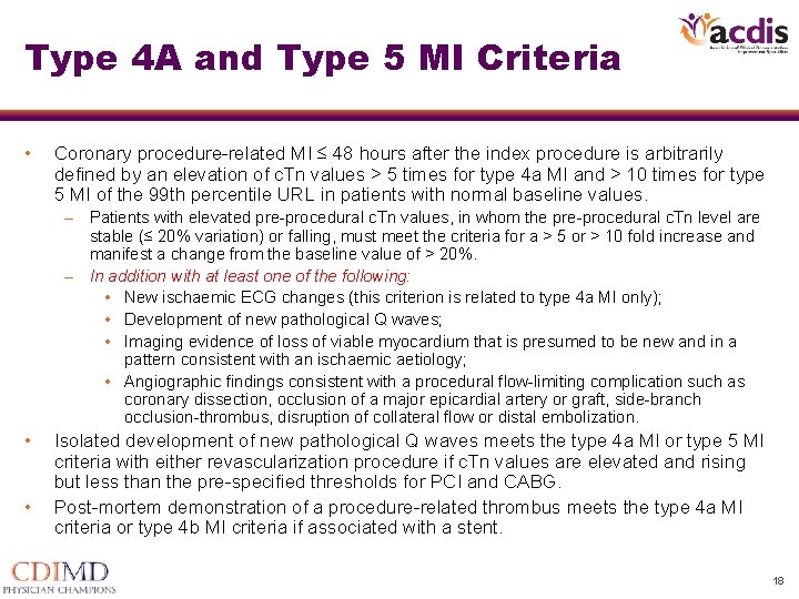
Type 4 A and Type 5 MI Criteria • Coronary procedure-related MI ≤ 48 hours after the index procedure is arbitrarily defined by an elevation of c. Tn values > 5 times for type 4 a MI and > 10 times for type 5 MI of the 99 th percentile URL in patients with normal baseline values. – Patients with elevated pre-procedural c. Tn values, in whom the pre-procedural c. Tn level are stable (≤ 20% variation) or falling, must meet the criteria for a > 5 or > 10 fold increase and manifest a change from the baseline value of > 20%. – In addition with at least one of the following: • New ischaemic ECG changes (this criterion is related to type 4 a MI only); • Development of new pathological Q waves; • Imaging evidence of loss of viable myocardium that is presumed to be new and in a pattern consistent with an ischaemic aetiology; • Angiographic findings consistent with a procedural flow-limiting complication such as coronary dissection, occlusion of a major epicardial artery or graft, side-branch occlusion-thrombus, disruption of collateral flow or distal embolization. • • Isolated development of new pathological Q waves meets the type 4 a MI or type 5 MI criteria with either revascularization procedure if c. Tn values are elevated and rising but less than the pre-specified thresholds for PCI and CABG. Post-mortem demonstration of a procedure-related thrombus meets the type 4 a MI criteria or type 4 b MI criteria if associated with a stent. 18
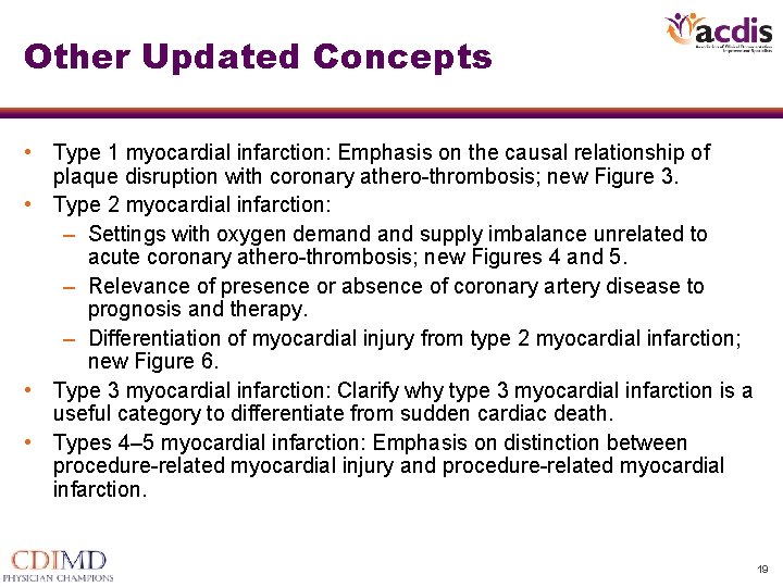
Other Updated Concepts • Type 1 myocardial infarction: Emphasis on the causal relationship of plaque disruption with coronary athero-thrombosis; new Figure 3. • Type 2 myocardial infarction: – Settings with oxygen demand supply imbalance unrelated to acute coronary athero-thrombosis; new Figures 4 and 5. – Relevance of presence or absence of coronary artery disease to prognosis and therapy. – Differentiation of myocardial injury from type 2 myocardial infarction; new Figure 6. • Type 3 myocardial infarction: Clarify why type 3 myocardial infarction is a useful category to differentiate from sudden cardiac death. • Types 4– 5 myocardial infarction: Emphasis on distinction between procedure-related myocardial injury and procedure-related myocardial infarction. 19
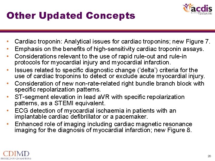
Other Updated Concepts • Cardiac troponin: Analytical issues for cardiac troponins; new Figure 7. • Emphasis on the benefits of high-sensitivity cardiac troponin assays. • Considerations relevant to the use of rapid rule-out and rule-in protocols for myocardial injury and myocardial infarction. • Issues related to specific diagnostic change (’delta’) criteria for the use of cardiac troponins to detect or exclude acute myocardial injury. • Consideration of new non-rate-related right bundle branch block with specific repolarization patterns. • ST-segment elevation in lead a. VR with specific repolarization patterns, as a STEMI equivalent. • ECG detection of myocardial ischaemia in patients with an implantable cardiac defibrillator or a pacemaker. • Enhanced role of imaging including cardiac magnetic resonance imaging for the diagnosis of myocardial infarction; new Figure 8. 20
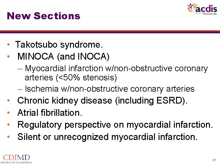
New Sections • Takotsubo syndrome. • MINOCA (and INOCA) – Myocardial infarction w/non-obstructive coronary arteries (<50% stenosis) – Ischemia w/non-obstructive coronary arteries • • Chronic kidney disease (including ESRD). Atrial fibrillation. Regulatory perspective on myocardial infarction. Silent or unrecognized myocardial infarction. 21
Thank you. Questions? James S. Kennedy MD (615) 479 -7021 jkennedy@cdimd. com 22