32 Deuterostome Animals Lecture Presentation by Cindy S

32 Deuterostome Animals Lecture Presentation by Cindy S. Malone, Ph. D, California State University Northridge © 2017 Pearson Education, Inc.

Chapter 32 Roadmap. © 2017 Pearson Education, Inc.

Introduction • The deuterostomes include the largest-bodied and some of the most morphologically complex of all animals • Deuterostomes were initially grouped together because they share important features of embryonic development • The gut develops from posterior to anterior • The coelom develops from outpocketings of mesoderm © 2017 Pearson Education, Inc.

Introduction • Some components of the “deuterostome pattern of development” are not unique to deuterostomes • The developmental distinction between deuterostomes and protostomes has become blurred • Recent molecular phylogenetic studies support grouping the deuterostomes as a monophyletic lineage © 2017 Pearson Education, Inc.

Figure 32. 1 Porifera Ctenophora ANIMALS Cnidaria PROTOSTOMES Rotifera Platyhelminthes Annelida Mollusca Nematoda Tardigrada Onychophora Arthropoda DEUTEROSTOMES Echinodermata Hemichordata Xenoturbellida Chordata © 2017 Pearson Education, Inc.

Introduction • Deuterostomes contain four phyla: 1. Echinoderms • Includes sea stars and sea urchins 2. Hemichordes • Burrowing, deposit- or suspension-feeding acorn worms © 2017 Pearson Education, Inc.

Introduction 3. Xenoturbella • Two recently discovered wormlike species 4. Chordates • Includes the vertebrates (animals with backbones) • Vertebrates comprise hagfish, lampreys, sharks and rays, bony fishes, amphibians, mammals, and reptiles (including birds) © 2017 Pearson Education, Inc.

Introduction • Animals that are not vertebrates are called invertebrates • Over 95% of known animal species are invertebrates • Vertebrates dominate the deuterostomes © 2017 Pearson Education, Inc.

What Is an Echinoderm? • Echinoderms (“spiny-skins”) are named for the spines or spikes observed in many species • All echinoderms are marine animals • To date, about 7000 species of echinoderm have been described • Echinoderms are very abundant, especially in deepwater environments © 2017 Pearson Education, Inc.

The Echinoderm Body Plan • Three main traits are synapomorphies, identifying echinoderms as a monophyletic group 1. Radial symmetry in adults 2. An endoskeleton of calcium carbonate 3. A water vascular system • With tube feet © 2017 Pearson Education, Inc.

The Echinoderm Body Plan • Larvae are bilaterally symmetric, as are all deuterostomes • However, adults have pentaradial symmetry (fivesided radial symmetry) • Pentaradial symmetry originated early in echinoderm evolution © 2017 Pearson Education, Inc.

Figure 32. 2 (a) Echinoderm larvae are bilaterally symmetric. 50 µm (b) Adult echinoderms are radially symmetric. 1 cm © 2017 Pearson Education, Inc.

The Echinoderm Body Plan • Echinoderms have an endoskeleton • A hard protective and supportive structure located inside a thin layer of epidermal tissue • The endoskeleton forms during development through the secretion of calcium carbonate plates inside the skin • In some species, the plates fuse and form a rigid case • In some species, the plates remain independent and flexible • The tissue that connects the plates is reversibly stiff or flexible depending on conditions © 2017 Pearson Education, Inc.

The Echinoderm Body Plan • Echinoderms are also defined by their water vascular system • A series of branching, fluid-filled tubes and chambers that forms a hydrostatic skeleton • An important part of the water vascular system is tube feet • Elongated, fluid-filled appendages, each consisting of • An ampulla on the inside of the body • A tube-like podium projecting on the outside © 2017 Pearson Education, Inc.

Figure 32. 3 (a) Echinoderms have a water vascular system. Opening to exterior Ampulla Tube foot Podium (b) Tube feet project from the underside of the body. Tube feet © 2017 Pearson Education, Inc.

Echinoderms Are Important Consumers • Echinoderms use most methods of feeding, including mass feeding, suspension feeding, and deposit feeding • Tube feet play a key role in obtaining food © 2017 Pearson Education, Inc.

Sea Stars Are Carnivores • Predatory species such as sea stars (Pisaster ochreceus) • Use their tube feet to pry apart bivalve shells • Extrude their stomach through the opening • Secrete digestive enzymes • Absorb the resulting molecules © 2017 Pearson Education, Inc.

Figure 32. 4 a (a) Predator and prey Mytilus californianus Pisaster ochraceus © 2017 Pearson Education, Inc.

Sea Stars Are Carnivores • Ecologist Robert Paine hypothesized that P. ochraceus is an important predator that has a major impact on community structure • Paine measured the number of species present in rocky intertidal communities where P. ochraceus was present and also in experimental plots where P. ochraceus was excluded © 2017 Pearson Education, Inc.

Sea Stars Are Carnivores • When sea stars were removed, the number of species present in the community plummeted • Diverse community overgrown with mussels • M. californianus is a dominant competitor, but its populations had been held in check by predation • P. ochraceus is a “keystone species” • Has a much greater impact on distribution and abundance of surrounding species than its abundance and total biomass would suggest © 2017 Pearson Education, Inc.

Figure 32. 4 b Number of species present (b) Effect of keystone predator on community diversity Community diversity maintained with keystone predator present Community diversity falls with no keystone predator Year © 2017 Pearson Education, Inc.

Sea Urchins Are Herbivores • Sea urchins have a jawlike structure in their mouths —called Aristotle’s lantern • Five calcium carbonate teeth that meet at the center of the mouth • Each tooth is operated by muscles, and can be extended and retracted during feeding • Urchins are important grazers • When their predators (sea otters) are removed, their grazing can destroy kelp forests • Sea otters are a keystone species © 2017 Pearson Education, Inc.

Key Lineages: The Echinoderms • Five major lineages of echinoderms are living today: 1. Crinoidea—feather stars and sea lilies 2. Asteroidea—sea stars 3. Ophiuroidea—brittle stars and basket stars 4. Echinoidea—sea urchins and sand dollars 5. Holothuroidea—sea cucumbers © 2017 Pearson Education, Inc.

Table 32. 1 -1 © 2017 Pearson Education, Inc.

Table 32. 1 -1 © 2017 Pearson Education, Inc.

Table 32. 1 -2 © 2017 Pearson Education, Inc.

Table 32. 1 -2 © 2017 Pearson Education, Inc.

Table 32. 1 -2 © 2017 Pearson Education, Inc.

What Is a Chordate? • All chordates have four morphological features at some stage in their life cycles: 1. Openings into the throat called pharyngeal gill slits 2. A dorsal hollow nerve cord that runs the length of the body, comprised of projections from neurons 3. A stiff and supportive but flexible rod, called the notochord, that runs the length of the body 4. A muscular post-anal tail © 2017 Pearson Education, Inc.

Three Chordate “Subphyla” • The phylum Chordata is made up of three major lineages, or “subphyla”: 1. Cephalochordates 2. Urochordates 3. Vertebrates © 2017 Pearson Education, Inc. Invertebrate chordates

Table 32. 2 -1 © 2017 Pearson Education, Inc.

Table 32. 2 -1 © 2017 Pearson Education, Inc.

Table 32. 2 -2 © 2017 Pearson Education, Inc.

Table 32. 2 -2 © 2017 Pearson Education, Inc.

The Cephalochordates • Cephalochordates are also called lancelets or amphioxus • They are small, mobile suspension feeders that resemble fish • Adults burrow in sand in their ocean-bottom habitats • Their dorsal hollow nerve cord runs parallel to a notochord • The notochord stiffens the body • Muscle contractions on either side result in fishlike movement © 2017 Pearson Education, Inc.

Figure 32. 5 a (a) Cephalochordates Pharyngeal slits or pouches Dorsal hollow nerve cord Notochord Muscular, post-anal tail Water flow © 2017 Pearson Education, Inc. Adult

The Urochordates • Urochordates are also called tunicates • Both larvae and adults have pharyngeal gill slits that function in feeding and gas exchange • The notochord, dorsal hollow nerve cord, and tail occur only in the larvae or in sexually mature forms of motile species © 2017 Pearson Education, Inc.

Figure 32. 5 b (b) Urochordates Pharyngeal slits or pouches Dorsal hollow nerve cord Notochord Muscular, post-anal tail Water flow Larva © 2017 Pearson Education, Inc. Adult

The Vertebrates • Vertebrates include hagfish, lampreys, sharks and rays, bony fishes, amphibians, mammals, and reptiles (including birds) • The dorsal hollow nerve cord is elaborated into the spinal cord • A bundle of nerve cells that runs from the brain to the body’s posterior • The pharyngeal pouches present in embryos develop into gills in aquatic species, but not in terrestrial species © 2017 Pearson Education, Inc.

The Vertebrates • A notochord develops in all vertebrate embryos • However, the vertebral column, rather than the notochord, functions in body support and movement • The notochord helps organize the body plan early in development by secreting proteins that induce somite formation • Somites are segmented blocks of tissue that later differentiate into vertebrae, ribs, and skeletal muscles © 2017 Pearson Education, Inc.

Figure 32. 5 c (c) Vertebrates Pharyngeal slits or pouches Dorsal hollow nerve cord Notochord Muscular, post-anal tail Cross section of embryo Embryo Pharyngeal pouches become gill slits in aquatic vertebrates © 2017 Pearson Education, Inc.

What Is a Vertebrate? • The vertebrates are a monophyletic group distinguished by two synapomorphies: 1. A column of cartilaginous or bony structures called vertebrae, which form along the dorsal side of most species • Important because it protects the spinal cord 2. A cranium—a bony, cartilaginous, or fibrous case that encloses the brain • Important because it protects the brain and sensory organs © 2017 Pearson Education, Inc.

What Is a Vertebrate? • Together, the vertebrae and cranium protect the central nervous system and key sensory structures • The coordinated movements of vertebrates are possible in part because vertebrates have complex brains © 2017 Pearson Education, Inc.

What Is a Vertebrate? • Vertebrate brains are divided into three distinct regions: 1. A forebrain, housing the sense of smell 2. A midbrain, associated with vision 3. A hindbrain, responsible for balance and hearing © 2017 Pearson Education, Inc.

What Is a Vertebrate? • All living vertebrates have three brain regions, but the structure and functions of the regions have evolved over time • In the jawed vertebrates, or gnathostomes, the hindbrain consists of enlarged regions called the cerebellum and medulla oblongata © 2017 Pearson Education, Inc.

What Is a Vertebrate? • Part of the forebrain became elaborated into a large structure called the cerebrum, especially in birds and mammals • The evolution of a large, three-part brain, protected by a hard cranium, was a key innovation in vertebrate evolution © 2017 Pearson Education, Inc.

Figure 32. 6 Ray-finned fishes Amphibians Turtles Snakes, lizards Crocodiles, alligators Birds Tetrapods Sharks, rays, skates Hagfishes, lampreys © 2017 Pearson Education, Inc. Mammals

What Key Innovations Occurred during the Evolution of Vertebrates? • Synthesis of diverse sources of new data: 1. New fossil evidence continues to provide major insights 2. Phylogenetic analysis now incorporates fossil evidence and also a treasure trove of new molecular data 3. Evidence from developmental biology is enabling scientists to test hypothesized relationships between structures in different vertebrate lineages © 2017 Pearson Education, Inc.

What Key Innovations Occurred during the Evolution of Vertebrates? • Three general themes of vertebrate evolution: 1. Most vertebrates are extinct 2. Some traits evolved more than once • Convergent evolution has occurred in multiple lineages 3. Traits are sometimes lost • Evolution is not a progression from simple to complex and is not limited to addition of new traits © 2017 Pearson Education, Inc.

Figure 32. 7 Loss of pharyngeal slits Echinodermata (Echinoderms) Protostomes Hemichordata (Acorn worms) Outgroups to Chordata Deuterostomes CHORDATA Xenoturbellida Loss of pharyngeal slits (Xenoturbella) CHORDATA Cephalochordata (Lancelets) Pharyngeal slits Urochordata (Tunicates) CHORDATA Dorsal hollow nerve cord Notochord Muscular, post-anal tail VERTEBRATA Loss of vertebrae Myxinoidea (Hagfishes) Petromyzontoidea (Lampreys) GNATHOSTOMATA Cranium, vertebrae Paired sense organs Chondrichthyes (Sharks, rays, skates) Actinopterygii (Ray-finned fishes) Jaws Paired appendages SARCOPTERYGII Actinistia (Coelacanths) Lungs Internal bone (endoskeleton) Dipnoi (Lungfishes) Lobed fins AMPHIBIA TETRAPODA Anura (Frogs, toads) Urodela (Salamanders) Limbs Lactation, fur Mammalia (Mammals) REPTILIA Amniotic egg Scales with hard keratin Lepidosauria (Lizards, snakes) Testudinia (Turtles) Crocodilia (Alligators, crocodiles) Aves (Birds) © 2017 Pearson Education, Inc. AMNIOTA

Urochordates: Outgroup to Vertebrates • According to the most recent molecular data, the urochordates are the closest living relatives of the vertebrates • Not the cephalochordates, as previously proposed • Urochordates are a sister group to the vertebrates © 2017 Pearson Education, Inc.

Table 32. 3 © 2017 Pearson Education, Inc.

Table 32. 3 © 2017 Pearson Education, Inc.

Fossil Evidence for Early Vertebrates • Earliest vertebrate fossils are in Chengjiang formation of China and Burgess Shale in Canada • Dated to Cambrian explosion • Earliest members lived in the ocean about 520 mya • Streamlined, fishlike bodies, gills, a notochord, postanal tail, and paired eyes • Cranium made of cartilage and possibly a series of cartilaginous reinforcements to the notochord © 2017 Pearson Education, Inc.

Fossil Evidence for Early Vertebrates • The specialized neural crest cells and some other cells responsible for brain, cranium, and sensory cell formation are synapomorphies for vertebrates • But, genes responsible for differentiation of these cells have been co-opted and modified from genes present in invertebrate chordate ancestors • Tinkering of the regulatory networks for neural crest may have been central to key innovations in evolution of vertebrate head © 2017 Pearson Education, Inc.

The Hagfish Hypothesis for Early Vertebrates • Hagfishes and lampreys have a notochord, gills, and a muscular post-anal tail • Plus a three-part brain, paired eyes, and cartilaginous cranium • Lampreys have cartilaginous reinforcements (vertebrae) on their notochord, but hagfishes do not © 2017 Pearson Education, Inc.

The Hagfish Hypothesis for Early Vertebrates • The phylogenetic relationships among the hagfishes, lampreys, and gnathostomes have been debated • Hagfishes and lampreys used to be grouped as class Cyclostomata, based on unique mouthparts and absence of jaws • In 1970 s, hagfishes and lampreys in two lineages: • Hagfishes—basal lineage of vertebrates • Lampreys—sister group to gnathostomes • Molecular phylogenies support Cyclostomata grouping © 2017 Pearson Education, Inc.

Table 32. 4 -1 © 2017 Pearson Education, Inc.

Table 32. 4 -1 © 2017 Pearson Education, Inc.

Table 32. 4 -2 © 2017 Pearson Education, Inc.

Table 32. 4 -2 © 2017 Pearson Education, Inc.

Gnathostomes: Origin of the Vertebrate Jaw • Jawed fishes are a grade that includes four major living lineages: • Cartilaginous fishes • Ray-finned fishes • Coelacanths • Lungfishes • A grade is a sequence of lineages that are paraphyletic (that include some, but not all, of the descendants of a common ancestor) © 2017 Pearson Education, Inc.

Gnathostomes: Origin of the Vertebrate Jaw • The fishes are a grade • The jawed fishes are a grade • The bony fishes are a grade • The lobe-finned fishes are a grade © 2017 Pearson Education, Inc.

Fossil Evidence for the Origin of the Jaw • Cartilaginous fishes (sharks and rays) have jaws made of reinforced cartilage rather than bone • Fossil evidence disproved shark-like origin of jawed fishes, pointing instead to jawed fishes that show up early in the Silurian (~430 mya) • Rather than being shark-like, these armored fishes, including placoderms, had heads covered with bony shields © 2017 Pearson Education, Inc.

Fossil Evidence for the Origin of the Jaw • After appearance of jawbones, teeth appear in the fossil record • Evolution of jaws was significant because it improved the ability of fishes to capture prey and also to bite • No longer limited to suspension or deposit feeding • Other key traits: • Paired fins • Internal fertilization © 2017 Pearson Education, Inc.

The Gill-Arch Hypothesis for the Origin of the Jaw • One hypothesis for the origin of the jaw proposes that • Natural selection acted on developmental regulatory genes that determine gill arch morphology • Gill arches are curved regions of tissue between the gills © 2017 Pearson Education, Inc.

The Gill-Arch Hypothesis for the Origin of the Jaw • One hypothesis for the origin of the jaw proposes that • Natural selection acted on developmental regulatory genes that determine gill arch morphology • Gill arches are curved regions of tissue between the gills • Mutation and natural selection increased the size of the most anterior arch and modified its orientation slightly, producing the first working jaw © 2017 Pearson Education, Inc.

Figure 32. 8 (a) Jawless vertebrate Gill arches Mouth (b) Intermediate form (basal gnathostomes) Gill arches Jaw (c) Later gnathostome © 2017 Pearson Education, Inc. Gill arches Jaw

The Gill-Arch Hypothesis for the Origin of the Jaw • Evidence for the gill-arch hypothesis includes • Both gill arches and jaws consist of bars of bony or cartilaginous tissue that hinge and bend forward • During development, the same population of cells gives rise to the muscles that move jaws and the muscles that move gill arches • Jaws and gill arches are derived from specialized cells in embryo called neural crest cells • Expression of developmental regulatory genes (Hox and Dlx) are similar in jaws and gill arches © 2017 Pearson Education, Inc.

The Gill-Arch Hypothesis for the Origin of the Jaw • This hypothesis is not yet supported by fossil data • Discovery of new fossil species of armored fishes will provide insights on origin of jaw • Once jaws originated, changes in size and shape enabled a rapid diversification of feeding strategies, from suction feeding to biting © 2017 Pearson Education, Inc.

The Gill-Arch Hypothesis for the Origin of the Jaw • Other modifications to the jaw also occurred: • In most ray-finned fishes, jaw is protrusible (can be extended to bite at food) • Several lineages of ray-finned fishes have a second specialized jaw called a pharyngeal (“throat”) jaw • Modified gill arches located in back of throat • Makes food processing efficient © 2017 Pearson Education, Inc.

Origin of the Bony Endoskeleton • In some lineages of fishes from late in Silurian, the cartilaginous endoskeleton began to be stiffened by deposition of bone • Unlike the dermal bone that had evolved earlier and was derived from neural crest cells (ectodermal in origin), the bony endoskeleton was mesodermally derived • Functioned to support swimming movements © 2017 Pearson Education, Inc.

Figure 32. 7 Loss of pharyngeal slits Echinodermata (Echinoderms) Protostomes Hemichordata (Acorn worms) Outgroups to Chordata Deuterostomes CHORDATA Xenoturbellida Loss of pharyngeal slits (Xenoturbella) CHORDATA Cephalochordata (Lancelets) Pharyngeal slits Urochordata (Tunicates) CHORDATA Dorsal hollow nerve cord Notochord Muscular, post-anal tail VERTEBRATA Loss of vertebrae Myxinoidea (Hagfishes) Petromyzontoidea (Lampreys) GNATHOSTOMATA Cranium, vertebrae Paired sense organs Chondrichthyes (Sharks, rays, skates) Actinopterygii (Ray-finned fishes) Jaws Paired appendages SARCOPTERYGII Actinistia (Coelacanths) Lungs Internal bone (endoskeleton) Dipnoi (Lungfishes) Lobed fins AMPHIBIA TETRAPODA Anura (Frogs, toads) Urodela (Salamanders) Limbs Lactation, fur Mammalia (Mammals) REPTILIA Amniotic egg Scales with hard keratin Lepidosauria (Lizards, snakes) Testudinia (Turtles) Crocodilia (Alligators, crocodiles) Aves (Birds) © 2017 Pearson Education, Inc. AMNIOTA

Origin of the Bony Endoskeleton • Three living lineages of bony fishes: 1. Ray-finned fishes 2. Coelacanths Lobe-finned fishes 3. Lungfishes • Bodies are covered with interlocking scales that provide a stiff but flexible covering • Many have a gas-filled swim bladder • Allowed ray-finned fishes to maintain neutral buoyancy and avoid sinking © 2017 Pearson Education, Inc.

Tetrapods: Origin of the Limb • Major event in evolution of vertebrates was transition to living on land • First vertebrates that had limbs and were capable of moving on land date to ~365 mya (late Devonian) • First tetrapods—animals with four limbs © 2017 Pearson Education, Inc.

Tetrapods: Origin of the Limb • Transition from water to land occurred once in evolution of vertebrates, giving rise to three major lineages of living tetrapods: 1. Amphibians 2. Mammals 3. Reptiles © 2017 Pearson Education, Inc.

Limbs-from-Fins Hypothesis • Most living species of lungfishes inhabit shallow, oxygen-poor water • Breathe with lungs, supplementing O 2 taken in by gills • Each fin or limb has a single bony element closest to the body that articulates with two bones arranged side by side, next to a series of elements farthest from the body • The structures are similar, and because no other animal groups have limb bones in this arrangement, the evidence for homology is strong © 2017 Pearson Education, Inc.

Limbs-from-Fins Hypothesis • Several regulatory proteins involved in development of fish fins and upper parts of mammal limbs are homologous • Proteins produced by several different Hox genes are found at the same times during development and in the same locations in fins and limbs • Once tetrapod limb evolved, adaptive radiation resulted in diverse structures used for running, gliding, crawling, burrowing, swimming, and flying © 2017 Pearson Education, Inc.

Figure 32. 9 Eusthenopteron (~385 mya) Lobe-like fin Panderichthys (~380 mya) Tiktaalik (~375 mya) Acanthostega (~365 mya) Tulerpeton (~362 mya) Limb that can support walking on land © 2017 Pearson Education, Inc.

Table 32. 5 © 2017 Pearson Education, Inc.

Table 32. 5 © 2017 Pearson Education, Inc.

Table 32. 5 © 2017 Pearson Education, Inc.

Amphibians • The first tetrapods to live on land were amphibians (“both-sides-living”) • Adults of most amphibians feed on land but lay eggs in water • Most undergo metamorphosis from aquatic larva to terrestrial or semiterrestrial adult • Gas exchange occurs across their moist mucus covered skin © 2017 Pearson Education, Inc.

Amphibians • Living amphibians represent a monophyletic group, Amphibia • Frogs and toads • Salamanders • Snake-like caecilians © 2017 Pearson Education, Inc.

Amniotes: Origin of the Amniotic Egg • Amniota is a lineage that includes all tetrapods other than amphibians • Amniotes named for synapomorphy—the amniotic egg • Protective covering that reduces the rate of drying • Reptiles (including birds) and the few mammals that lay eggs produce an amniotic egg and lay them outside of water © 2017 Pearson Education, Inc.

The Amniotic Egg • The three inner membranes surround • The embryo itself (amnion) • The yolk provided by the mother (the yolk sac) • The waste from the embryo (allantois) • Albumen cushions developing embryo and provides nutrients • Membranes provide mechanical support and increase surface area for gas exchange • Surrounded by shell © 2017 Pearson Education, Inc.

Figure 32. 10 Embryo Albumen provides water and mechanical support © 2017 Pearson Education, Inc. Yolk sac contains nutrients

Mammals: Origin of Lactation and Fur • Mammals are a monophyletic group of amniotes named for mammary glands, which produce milk • Milk nourishes developing young (lactation) • Only vertebrates with cheek muscles and lips • Make suckling milk possible • Endotherms (“inside-heated”)—maintain high body temperatures by oxidizing large amounts of food and generating large amounts of heat • Body covered with layers of hair or fur made of keratin © 2017 Pearson Education, Inc.

Mammals: Origin of Lactation and Fur • Three major lineages of mammals alive today: 1. Egg-laying monotremes 2. Pouch-bearing marsupials 3. Placental (eutherians) • Produce placenta within uterus or oviduct during pregnancy © 2017 Pearson Education, Inc.

Table 32. 6 © 2017 Pearson Education, Inc.

Table 32. 6 © 2017 Pearson Education, Inc.

Table 32. 6 © 2017 Pearson Education, Inc.

Mammals: Origin of Lactation and Fur • The placenta is an organ combining maternal and embryonic tissues • Rich in blood vessels that facilitate flow of O 2 and nutrients from mother to developing embryo and also remove nitrogenous wastes and CO 2 from embryo • Embryo contributes to placenta—allantois and chorion • The diffusion of gases, nutrients, and wastes • After development period (gestation), embryo emerges from mother’s body © 2017 Pearson Education, Inc.

Figure 32. 11 Yolk sac Embryo © 2017 Pearson Education, Inc.

Mammals: Origin of Lactation and Fur • Evolutionary advantages of viviparity and placenta? 1. Offspring develop at a more constant, favorable temperature 2. Offspring are protected 3. Offspring are portable—mothers are not tied to a nest • All adaptive traits have trade-offs • Placenta is energetically expensive to produce, and bearing live young is energetically costly © 2017 Pearson Education, Inc.

Mammals: Origin of Lactation and Fur • Earliest mammals in fossil record appear ~195 mya • Mammals were widespread and ecologically diverse by ~165 mya (Jurassic period) • Mammals began to diversify when dinosaurs and other reptiles were dominant large herbivores and predators in terrestrial and aquatic environments © 2017 Pearson Education, Inc.

Mammals: Origin of Lactation and Fur • Many lineages died out during end-Cretaceous mass extinction 66 mya • Lineages that survived underwent an adaptive radiation • Mammalian orders familiar today • Filled ecological roles once dominated by dinosaurs and ocean-dwelling, extinct reptiles (ichthyosaurs and plesiosaurs) © 2017 Pearson Education, Inc.

Reptiles: Origin of Scales and Feathers Made of Keratin • Reptiles are a monophyletic group that represents second major living lineage of amniotes besides mammals. • Four major lineages: 1. Lizards and snakes 2. Turtles 3. Crocodiles and alligators 4. Birds © 2017 Pearson Education, Inc.

Reptiles: Origin of Scales and Feathers Made of Keratin • Adaptations for life on land: • Skin is watertight by a layer of scales made of keratin • Breathe air through well-developed lungs • Lay shelled, amniotic eggs • Many living reptiles are ectotherms (“outside-heated”) • Do not use internally generated heat to regulate their body temperature • Bask in sunlight, seek shade, and other behaviors to keep body temperature at appropriate level © 2017 Pearson Education, Inc.

Table 32. 7 -1 © 2017 Pearson Education, Inc.

Table 32. 7 -1 © 2017 Pearson Education, Inc.

Table 32. 7 -2 © 2017 Pearson Education, Inc.

Table 32. 7 -2 © 2017 Pearson Education, Inc.

Reptiles: Origin of Scales and Feathers Made of Keratin • The fossil record for reptiles includes dinosaurs, ichthyosaurs, plesiosaurs, pterosaurs, and other extinct lineages that flourished from ~250 mya until end-Cretaceous mass extinction © 2017 Pearson Education, Inc.

Reptiles: Origin of Scales and Feathers Made of Keratin • Phylogenetic evidence supports inclusion of birds within monophyletic group called reptiles • Birds descended from one of several lineages of dinosaurs that had feathers • Outgrowths of the skin composed of keratin • Provide insulation, used for display, and furnish the lift and steering required for flight • Birds are endothermic (evolved independently in birds and mammals) © 2017 Pearson Education, Inc.

Reptiles: Origin of Scales and Feathers Made of Keratin • Wings and flight evolved independently in three lineages of tetrapod amniotes 1. Pterosaurs 2. Birds 3. Bats • How did flight evolve? © 2017 Pearson Education, Inc.

Reptiles: Origin of Scales and Feathers Made of Keratin • Did birds evolve from dinosaurs? • Fossil species show that birds are part of monophyletic group called dinosaurs • How did feathers evolve? • Fossils suggest feathers evolved in steps culminating in complex, branched structures familiar today • But, feathers occurred in multiple, nonflying dinosaur lineages • Feathers originally may have been used for insulation and/or mating displays © 2017 Pearson Education, Inc.

Reptiles: Origin of Scales and Feathers Made of Keratin • Did birds begin flying from the ground up or from the trees down? • Did flight evolve with running species that began to jump and glide with the aid of feathers to provide lift? • Did flight evolve from tree-dwelling species that used feathers to glide from tree to tree or tree to ground? • Alternate hypothesis—feathers may have helped early birds to run rapidly up steep slopes to high perches © 2017 Pearson Education, Inc.

Figure 32. 12 © 2017 Pearson Education, Inc.

Reptiles: Origin of Scales and Feathers Made of Keratin • Once feathers evolved in dinosaurs, a series of adaptations made powered, flapping flight increasingly efficient • Reduced in size compared to many of their theropod ancestors, enabling them to diversify into new niches • Light for their size, thanks to a reduced number of bones compared to their ancestors • Heads of birds have retained the proportions of the juveniles of their theropod ancestors © 2017 Pearson Education, Inc.

Reptiles: Origin of Scales and Feathers Made of Keratin • Most dinosaurs have a flat sternum (“breastbone”), but bird sternum has a projection (keel) that provides a large surface area to which flight muscles attach • Birds are capable of sustained activity year-round because they are endotherms © 2017 Pearson Education, Inc.

Figure 32. 13 Reduced snout and enlarged brain and eyes Elongated “keel” on sternum for attachment of flight muscles Hollow bones strengthened by “struts” Produce heat in their tissues (endothermic) © 2017 Pearson Education, Inc.

Parental Care • Numerous vertebrate lineages evolved a variety of methods of parental care • Physiological, morphological, or behavioral investment that improves the likelihood of offspring to survive • Fanning aquatic eggs with oxygen-rich water • Guarding eggs and/or new young from predators • Keeping eggs and young moist (amphibians) or keeping them warm and dry (reptiles and mammals) • Supplying young with food • Teaching survival skills to young © 2017 Pearson Education, Inc.

Figure 32. 14 © 2017 Pearson Education, Inc.

Parental Care • Mammals and birds provide particularly extensive parental care • Energetically expensive to provide, but can improve the animals’ fitness by increasing the likelihood that their offspring will survive and reproduce • Evolution of extensive parental care is hypothesized to be a major reason for evolutionary success of mammals and birds © 2017 Pearson Education, Inc.

The Primates and Hominins • Humans occupy a tiny twig on the tree of life, but their origins have been studied extensively © 2017 Pearson Education, Inc.

The Primates • The primate lineage consists of two main groups: 1. Prosimians (“before-monkeys”) 2. Anthropoids (“human-like”) © 2017 Pearson Education, Inc.

The Primates • Prosimians include • Lemurs from Madagascar • Tarsiers, pottos, and lorises from Africa and south Asia • Most prosimians are small-bodied, arboreal, and nocturnal © 2017 Pearson Education, Inc.

Figure 32. 15 a (a) Prosimians © 2017 Pearson Education, Inc. Lemur catta

The Primates • Anthropoids include • New World monkeys from Central and South America • Old World monkeys from Africa and Asia • Gibbons from southeast Asia • The Hominidae or great apes—orangutans, gorillas, chimpanzees, and humans © 2017 Pearson Education, Inc.

Figure 32. 15 b (b) Anthropoids © 2017 Pearson Education, Inc. Gorilla gorilla

Figure 32. 16 Marsupials EUTHERIANS Sloths, armadillos Elephants, manatees Deer, dogs, cats Rodents, rabbits PRIMATES PROSIMIANS Eyes in front of face, grasping hand Larger brain New World monkeys Old World monkeys Gibbons Orangutan Fist walking Knuckle walking Eastern gorilla Bipedalism, long legs Human Bonobo Chimpanzee © 2017 Pearson Education, Inc. ANTHROPOIDEA Western gorilla HOMINIDAE (GREAT APES) Long arms, short legs, no tail

What Makes a Primate? • Primates have many defining characteristics • Hands and feet that are efficient at grasping • Flattened nails instead of claws on the fingers and toes • Relatively large brains • Color vision • Complex social behavior • Extensive parental care of offspring • Forward-facing eyes © 2017 Pearson Education, Inc.

What Makes a Great Ape? • Great apes are also called hominids and are relatively large bodied with long arms, short legs, and no tail • Hominids have distinct ways of walking • Orangutans live primarily in the trees, but fist-walk when on the ground • Gorillas, bonobos, and chimpanzees knuckle-walk • Humans are the only living great ape that is fully bipedal (“two-footed”), walking upright on two legs © 2017 Pearson Education, Inc.

What Makes a Great Ape? • Bipedalism is the synapomorphy that defines the hominins • Hominins are the monophyletic group comprising Homo sapiens and more than 20 extinct, bipedal relatives © 2017 Pearson Education, Inc.

Fossil Humans • DNA sequence data show that humans are most closely related to the common chimpanzees and bonobos, followed by the gorillas • The fossil record indicates that the common ancestor of chimps and humans lived in Africa 6 to 7 mya © 2017 Pearson Education, Inc.

Fossil Humans • Naming hominin species and interpreting their characteristics are intensely controversial topics • Most researchers agree that hominins can be organized into four general groups: 1. Gracile australopithecines 2. Robust australopithecines 3. Early Homo 4. Recent Homo • These lineages appeared after the oldest known hominin, Ardipithecus ramidus © 2017 Pearson Education, Inc.

Table 32. 8 © 2017 Pearson Education, Inc.

Figure 32. 17 Ardipithecus A. ramidus Australopithecus A. afarensis A. africanus A. deyiremeda A. garhi A. sediba Paranthropus P. aethiopicus P. boisei P. robustus Early Homo H. habilis H. rudolfensis H. erectus Recent Homo H. heidelbergensis Denisovans H. neanderthalensis H. floresiensis H. sapiens Present Time (millions of years ago) © 2017 Pearson Education, Inc.

Gracile Australopithecines • Australopithecus is composed of species of small hominins called gracile (“slender”) australopithecines • Several lines of evidence support the hypothesis that the gracile australopithecines were bipedal © 2017 Pearson Education, Inc.

Figure 32. 18 a (a) Gracile australopithecines (Australopithecus africanus) Vertical posture © 2017 Pearson Education, Inc.

Robust Australopithecines • Paranthropus is composed of three known species of bipedal robust australopithecines • All three species had • Massive cheek teeth and jaws • Very large cheekbones • A sagittal crest—a flange of bone at the top of the skull • This lineage is hypothesized to be a monophyletic group that became extinct © 2017 Pearson Education, Inc.

Figure 32. 18 b (b) Robust australopithecines (Paranthropus robustus) Sagittal crest Massive cheek teeth © 2017 Pearson Education, Inc. Massive cheekbones

Early Homo • All species in the genus Homo are called humans • Compared to the earlier hominins, early Homo species have • Flatter and narrower faces • Smaller jaws and teeth • Larger braincases • Many researchers argue that extensive toolmaking was a diagnostic trait of early Homo © 2017 Pearson Education, Inc.
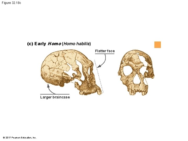
Figure 32. 18 c (c) Early Homo (Homo habilis) Flatter face Larger braincase © 2017 Pearson Education, Inc.
Recent Homo • Recent Homo date from 1. 2 mya to the present • Compared to early Homo species, recent Homo species have • Flatter faces • Smaller teeth • Larger braincases © 2017 Pearson Education, Inc.
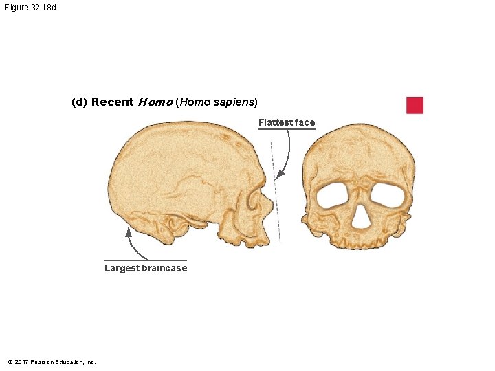
Figure 32. 18 d (d) Recent Homo (Homo sapiens) Flattest face Largest braincase © 2017 Pearson Education, Inc.
Recent Homo • Recent Homo includes our species, Homo sapiens (including Cro-Magnons), and the Neanderthals • There is evidence that both populations created art and buried their dead in an organized manner © 2017 Pearson Education, Inc.
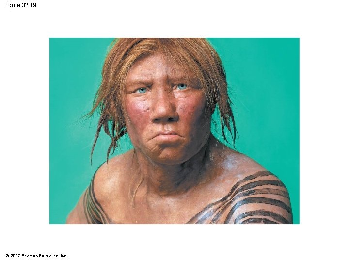
Figure 32. 19 © 2017 Pearson Education, Inc.
Recent Homo • Homo floresiensis discovered in 2003 on the island of Flores in Indonesia • H. floresiensis consisted of individuals who were about a meter tall; their braincases were smaller than those of gracile australopithecines • Fossil finds suggest that the species inhabited the island from about 100, 000 to 12, 000 years before present © 2017 Pearson Education, Inc.
What Can Be Deduced from the Fossil Record? • The fossil evidence indicates • The shared, derived character that defines the hominins is bipedalism • The hominin family tree has many branches. That is, there was not a linear progression from one species to another • Multiple species of hominins lived on Earth at the same time • Compared with other hominins, species in the genus Homo have extremely large brains relative to overall body size © 2017 Pearson Education, Inc.

What Can Be Deduced from the Fossil Record? • Several lines of evidence support the hypothesis that natural selection for the ability to reason and communicate • Was triggered by increased tool and language use • Resulted in humans with large brains © 2017 Pearson Education, Inc.
The Out-of-Africa Hypothesis • The leading hypothesis for the evolution of Homo sapiens is called the out-of-Africa hypothesis, which contends that • H. sapiens evolved its distinctive traits in Africa and then dispersed throughout the world • H. sapiens evolved independently of earlier European and Asian species of Homo © 2017 Pearson Education, Inc.
Fossil Evidence • First H. sapiens fossils appear in East African rocks that date to about 195, 000 years ago • For some 130, 000 years thereafter, the fossil record indicates that our species occupied Africa while H. neanderthalensis resided in Europe and the Middle East • Some evidence suggests that H. erectus may still have been present in Asia at that time © 2017 Pearson Education, Inc.
Fossil Evidence • In rocks dated between 60, 000 and 30, 000 years ago, H. sapiens fossils found throughout Europe, Asia, Africa, and Australia • H. erectus had disappeared by this time • H. neanderthalensis went extinct after coexisting with H. sapiens in Europe for thousands of years • Fossil evidence provides support for the African origin of H. sapiens and subsequent migration • Cannot resolve the question of whether H. sapiens ever interbred with Neanderthals © 2017 Pearson Education, Inc.
Molecular Evidence • Phylogenetic trees based on genetic comparisons of diverse human populations living today agree with pattern in fossil record • First lineages to branch off led to descendant populations that live in Africa today • Later branches gave rise to lineages residing today in central Asia, Europe, East Asia, Polynesia, and the Americas © 2017 Pearson Education, Inc.
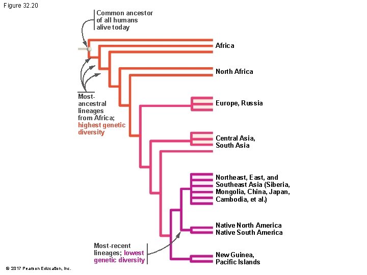
Figure 32. 20 Common ancestor of all humans alive today Africa North Africa Mostancestral lineages from Africa; highest genetic diversity Europe, Russia Central Asia, South Asia Northeast, East, and Southeast Asia (Siberia, Mongolia, China, Japan, Cambodia, et al. ) Native North America Native South America Most-recent lineages; lowest genetic diversity © 2017 Pearson Education, Inc. New Guinea, Pacific Islands
Molecular Evidence • Did H. sapiens leave Africa in one wave? • Sequence data from modern Aboriginal Australians support the hypothesis that the human migration out of Africa occurred in more than one wave— • First wave moved east and south to Australia • Second wave moved to Europe and the rest of mainland Asia © 2017 Pearson Education, Inc.
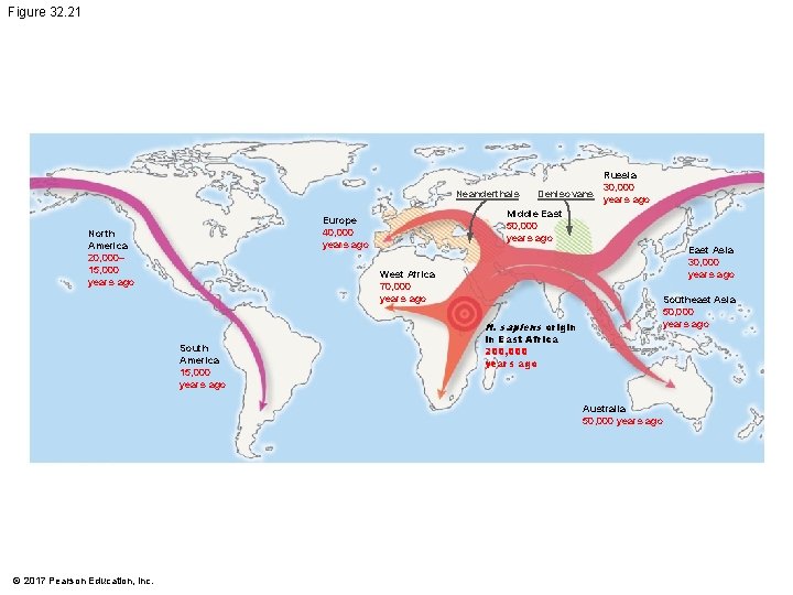
Figure 32. 21 Russia 30, 000 Neanderthals Denisovans years ago Middle East 50, 000 years ago Europe 40, 000 years ago North America 20, 000– 15, 000 years ago East Asia 30, 000 years ago West Africa 70, 000 years ago H. sapiens origin South America 15, 000 years ago Southeast Asia 50, 000 years ago in East Africa 200, 000 years ago Australia 50, 000 years ago © 2017 Pearson Education, Inc.
Molecular Evidence • What about interbreeding? • Two lines of evidence support hypothesis that H. sapiens interbred with other species as they migrated out of Africa 1. From 1% to 4% of the genome of indigenous Europeans and Asians—but not Africans—is derived from Neanderthals • H. sapiens interbred with Neanderthals in the Middle East after H. sapiens left Africa, but before migrating through Europe and Asia © 2017 Pearson Education, Inc.
Molecular Evidence • What about interbreeding? 2. Modern humans interbred with a recently discovered cousin of Neanderthals (Denisovans in central Asia during their first migration out of Africa • About 5% of the genome of Aboriginal Australians is derived from Denisovans • The overall data support an out-of-Africa hypothesis with “leakage” © 2017 Pearson Education, Inc.
Have Humans Stopped Evolving? • All four processes of evolution—mutation, genetic drift, gene flow, and natural selection—are measurable in human populations today and are expected to continue • Coevolution with pathogens • Effects of C-section on evolution of head size? • Spread of lactose tolerance mutation © 2017 Pearson Education, Inc.
- Slides: 152