3 Protein structures Why are we interested Function


 Some with NMR (13%) Very little")
, so we can measure the distance")


- Slides: 22
3. Protein structures
Why are we interested? Function is related to the structure Understand biological processes (DNA, RNS, enzymes, hormones, receptors) Diseases Drug design, protein – drug interactions
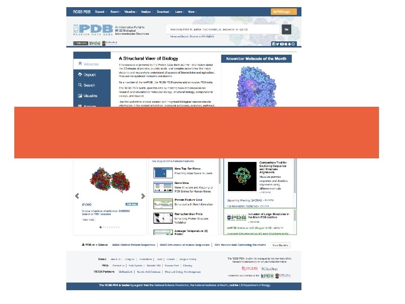
Protein structures How to find a specific structure? Database Wordwide Protein Data Bank (ww. PDB) 3 entries: RSCB PDB, PDB Europe, PDB Japan Easy to use • Search by name, ID • Direct links
Most structures are solved by x-ray crystallography (86%) Some with NMR (13%) Very little by electron microscopy (1%)
X-ray - Short wavelength (ca. 1. 5 Å), so we can measure the distance between atoms - The output is an electron density - Crystallization artefacts - Not physiological - Hydrogens are not visible
NMR - Solution - Usually a crowd of structures that fulfill the constrains - Only small molecules ( > 35 k. Da) - Less accurate - Can be used on flexible proteins
PDB ID In the DPB each atomic coordinate file has a unique ID which contains 4 specific characters First is always a number, a followed by 3 characters that can be either letters or numbers. F. E. : • 1 mbn - The very first structure from 1973, the myoglobin • 1 tna - Form 1975 yeast phenylalanine transfer RNS, the first RNS structure • 1 bna - The first DNA double helix solved in 1980 with X-ray, (confirmation of the Watson & Crick modell from 1953). • 2 hhd - human hemoglobin, (deoxy form) • 9 ins - insulin
The. pdb file format HEADER TITLE. . . EXPDTA AUTHOR. . . REMARK. . . SEQRES. . . ATOM ATOM. . . HETATM. . . EXTRACELLULAR MATRIX 22 -JAN-98 1 A 3 I X-RAY CRYSTALLOGRAPHIC DETERMINATION OF A COLLAGEN-LIKE 2 PEPTIDE WITH THE REPEATING SEQUENCE (PRO-GLY) X-RAY DIFFRACTION R. Z. KRAMER, L. VITAGLIANO, J. BELLA, R. BERISIO, L. MAZZARELLA, 2 B. BRODSKY, A. ZAGARI, H. M. BERMAN 350 BIOMOLECULE: 1 350 APPLY THE FOLLOWING TO CHAINS: A, B, C 350 BIOMT 1 1 1. 000000 0. 000000 350 BIOMT 2 1 0. 000000 1. 000000 0. 000000 1 A 1 B 1 C 1 2 3 4 5 130 131 132 9 6 6 N CA C O CB 0. 00000 PRO PRO GLY PRO PRO GLY PRO PRO PRO C ACY OXT ACY A A A 1 1 1 8. 316 7. 608 8. 487 9. 466 6. 460 21. 206 20. 729 20. 707 21. 457 21. 723 21. 530 20. 336 19. 092 19. 005 20. 211 1. 00 17. 44 22. 26 N C C O C 401 401 3. 682 2. 807 4. 306 22. 541 23. 097 23. 101 11. 236 10. 553 12. 291 1. 00 21. 19 C O O
The. pdb file format
Model The model obtained with NMR or X-ray is not a direct one. Through the measurement we derive the model from a collection of data (NMR spectrum or diffraction pattern).
Resolturion • Indicates how much can we trust in a position of specific atom Low: <3. 0 A Average: 1. 8 -3. 0 A Good: 1. 0 – 1. 8 A Atomic: >1. 0 A The resolution varies through the structure!
Resolution
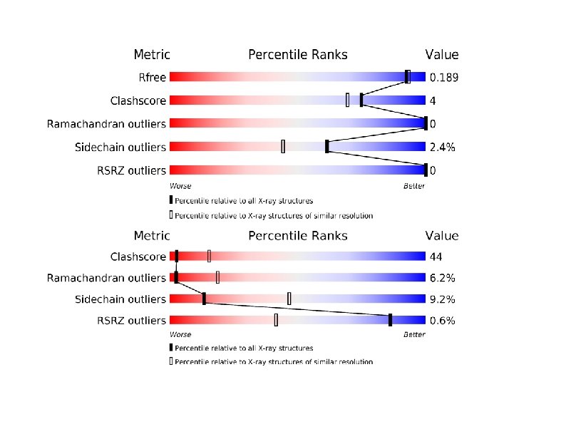
How good is a structure? How much is the resolution of the collected data? (In case of X-ray) R-factor/Free. R-factor (X-ray) How well does the data fit to the experimental mesurements? There is no standard for NMR structures Ramachandran Plot Geometry and stereo-chemistry How similar are these to good known structures
Asymetric Unit PISA
Visualization of structures 1. PDB file 3. Computer 2. Program for visualization F. E. : Rasmol, Pymol, Chimera, VMD, Jmol, Swiss PDB viewer 4. Picture of a molecule
Why Chimera? Free liscence Well maintained Intuitive Not just for visualization Subjective http: //www. cgl. ucsf. edu/chimera/
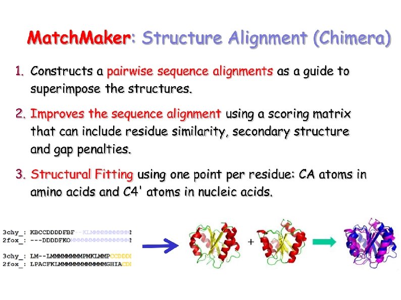
How similar are two structures? Superposition: Minimizing the distance of positions
RMSD
Structural alignment What are the equivalent positions? - If the sequences are alike we can use that - In case of low sequence identity we can use the structure RMSD depends on the equivalent positions Linker regions may cause problems