3 D echografie Elke Sleurs Vrouwenkliniek UZ Gent


: Het oppervlak zal zich tonen")


 Ultrasound Obstet Gynecol 2007; 30")
")


")


 maternale ademhalingsbewegingen foetale extrasystolen")


")
- Slides: 70
3 D echografie Elke Sleurs Vrouwenkliniek UZ Gent Dienst. Gentverloskunde - prenatale geneeskunde © 2008 Universitair Ziekenhuis 1
3 D echografie Inleiding • Overzicht • ~ 50 jaar echografie i/d verloskunde - gynaecologie Van 2 D naar 3 D - 4 D echografie: techniek • Verschillende modaliteiten - toepassingen • Samenvatting - Kritische noot • 2
Inleiding 3
Inleiding: hoe het allemaal begon? • Gebruik van “ ultrageluid” in de geneeskunde: begin 20 ste eeuw: als therapeutisch middel: interne geneeskunde: maagulcera neurochirurgie: vernietigen basale ganglia bij Parkinson fysische geneeskunde en revalidatie: reumatoïde arthritis 4
Inleiding: hoe het allemaal begon? Jaren 1940: diagnostisch middel H. Gohr - Th. Wedekind, 1940: ” Der Ultraschall in der Medizin” geen overtuigende gegevens Karl Theo Dussik: wordt beschouwd als eerste arts die ultrageluid gebruikte in de diagnostiek 5
Inleiding: hoe het allemaal begon? applicatie in verloskunde en gynaecologie late jaren 1950 - 1960 Professor Ian Donald - Glasgow A- en B- mode cephalometry - placenta localisatie 6
Inleiding: hoe het allemaal begon? boom vanaf de jaren 1966 Europa - Verenigde Staten - Japan oa. Alfred Kratochwil - Stuart Campbell -. . . oa. begrippen CRL; thoracic circumference; abdominal cricumference b-scan image of the maternal abdomen showing abdominal circumference and placenta using a compound contact scanner without gray-scale in the late 1960 s. 7
Inleiding: verdere ontwikkelingen toevoegen van “gray - scale” technologie B-scan image with gray scale of a similar section of the maternal abdomen showing abdominal circumference and placenta using the Nuclear Enterprise® NE 4102 in the late 1970 s A gray scale Octoson® image of the abdominal circumference and placenta in the late 1970 s. The Octoson® produced superior images as compared to articulated arm scanners but loosed out on mobility and flexibility. A gray scale longitudinal scan of a section of the fetal trunk and placenta made with the very popular Picker® 80 L static scanner in the early 1980 s. Despite the very good images that could be obtained with these machines, they were soon replaced by the new real-time scanners. 8
Inleiding: verdere ontwikkelingen real time scanners: real evolution The large hand-held circular rotating transducer (Combison 100) from Kretz. Technik® and the resultant sector image. The transducer is connected to the main console by a flexible cable. convexe sondes Toshiba's advertisement of the SAL 77 A in mid 1985, which used for the first time a convex-array probe. 9
Inleiding: verdere ontwikkelingen doppler: late-comer From the paper "Clinical Applications of a Transcutaneous Ultrasonic Flow Detector" by Robert Rushmer, Donald baker, Wayne Johnson and D. Eugene Strandness in 1967 in the JAMA. van flow velocity waveforms tot color flow mapping, power doppler en doppler tissue imaging 10
Inleiding: verdere ontwikkelingen Veranderingen in beeldkwaliteit van 1985, 1990 tot 1995 respectievelijk. Verbeteringen in “spatial and contrast resolution, background noise reduction, dynamic range, and near and far field visualization”. Meer significante verbeteringen kwamen midden jaren ‘ 90. 11
Van 2 D tot 3 D echografie 12
Van 2 D tot 3 D echografie: basis principe In een 2 D echografisch onderzoek probeert de onderzoeker zelf een driedimensioneel beeld in zijn geest te hebben door het roteren en het inclineren van de probe. Bij 3 D echografie zal de software een aangeduid gebied in ± 250 Bscan frames snijden en die opslaan in het geheugen. 13
Van 2 D tot 3 D echografie: basis principe Automatische volumescan 14
Van 2 D tot 3 D echografie: basis principe Multi Planar Plane Translation 15
Van 2 D tot 3 D echografie: basis principe Intersection of orthogonal planes A B C C Schematic representation of multiplanar analysis of ultrasound volumes, with corresponding multiplanar analysis of the fetal face. A is the plane parallel to the acquisition or ‘start’ scan; B and C are the reconstructed orthogonal planes. The C-plane is also commonly referred to as the coronal plane. The arrows indicate the points that represent the intersection of the three planes. Ultrasound Obstet Gynecol 2007; 30
3 D echografie: multiplanar view After acquisition of the volume data set one has a multiplanar image of the different planes. The volumes box gives an idea of the volume data set. 17
3 D echografie: volume rendering Surface mode: surface light > face, limbs, organs Transparant mode: maximum > skeleton minimum > organs/ liquid > bloodvessels X-ray > brain Power doppler mode 18
3 D echografie: volume rendering De 3 D - renderbox bepaalt de ROI voor de 3 D - berekening en bepaalt de kijkrichting door het volumeblok. De aanpassing van de renderbox gebeurt met behulp van de 3 orthogonale vlakken A, B en C. De groene lijn van de renderbox in het A- en B-vlak bepaalt de richting en de grens van de analyse. 19
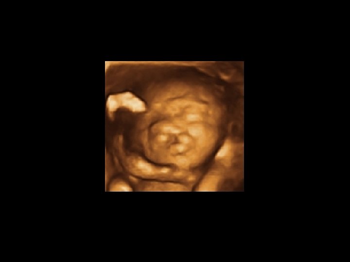
3 D echografie: render modes Surface modus Surface (texture): Het oppervlak zal zich tonen in de "texture" mode: de grijswaarden van het 3 D - beeld zijn identiek met de grijswaarden van de originele 2 D - scan. Light Mode: structuren dichterbij worden lichter structuren verder verwijderd worden donkerder 20
3 D echografie: render modes Surface modus Smooth Surface: Het oppervlak toont zich "smoothed" in "texture" mode. Gradient Light Mode: Structuren met “surface normals (gradients)” georiënteerd naar de onderzoeker tonen zich helder. Structuren minder georiënteerd naar de onderzoeker tonen zich donkerder. 21
3 D echografie: render modes Transparant modus Maximum Mode: De maximum grijswaarden van de ROI worden getoond. > skelet en echodense structuren X-Ray Mode: Presentatie van alle grijswaarden in de ROI. > weefselstructuren ( tumoren of gelijkaardige structuren) 22
3 D echografie: render modes Transparant modus Minimum mode: De minimum grijswaarden van de ROI worden getoond > vaten en holle organen Opmerking: De software berekent altijd twee render modes tergelijkertijd waardoor men twee verschillende modussen naar gewenst percentage kan mengen. 23
3 D echografie: threshold ”Threshold Low”: structuren, die het oppervlak omringen, kunnen geëlimineerd worden als de grijswaarden veel lager zijn dan de grijswaarde van het eigenlijke oppervlak maw. threshold wordt dus gebruikt om signaalruis te elimineren Zelfde volume met verschillende threshold parameters 24
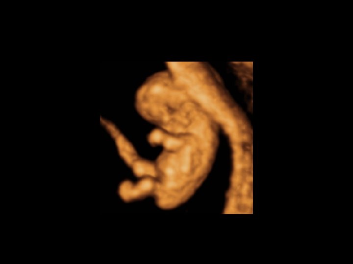
Reversed face @ E. Roets
Flipped face @ E. Roets
VCI - Volume Contrast Imaging 36
VCI - Volume Contrast Imaging Basis principe van “Volume Contrast Imaging”: een volume acquisitie combineren met surface en maximum gradient rendering, met als doel geluidsartefacten te verminderen. Fetus in B-mode and VCI 37
layer 1 Anatomical structures Noise and speckles layer 2 Standard multiplanar Improved signal to noise ratio Door het superponeren van verschillende lagen van het weefsel wordt omliggend ruis gereduceerd en worden de anatomische structuren beter en contrastrijker gevisualiseerd. VCI kan ofwel gebruikt worden als postprocessing techniek voor statische volumes (static VCI) of als een modaliteit voor rendering in 4 D mode (VCI in the coronal plane, VCI-C). Static VCI 3 mm thickness
VCI in coronal plane ( VCI-C) Ultrasound Obstet Gynecol 2007; 30
VCI in coronal plane ( VCI-C)
STIC - Spatio-Temporal Image Correlation 41
STIC - Spatio-Temporal Image Correlation • Foetale echocardiografie is nog steeds één van de moeilijkste delen van de foetale echografie • Moeilijkheden: complexe anatomie relatief klein oppervlak “snel kloppend” hart afwezigheid van foetaal ECG • • STIC = eerste commerciële 4 D - technologie door Kretz US Doel: automatische volume acquisitie met mogelijkheid van gating 42
STIC - Spatio-Temporal Image Correlation • Onderzoeker bepaalt de „sweep“ hoek ( 15°- 45°) de acquisitietijd ( 7, 5 - 10 - 12, 5 - 15 sec) het volume • Computer: herkent automatisch het hartritme beoordeelt hartcyclus reconstrueert een real-time volume 43
STIC: techniek • automatische volume acquisitie: sonde voert één enkele, trage 3 D sweep uit gezien de kleine ROI is de frame rate zeer hoog ( ± 150/ s) en bevat het volume een hoog aantal 2 D frames Bv: bij een acquisitie van 10 s en een sweep van 25° worden er 1500 B-mode beelden in het volume geheugen opgeslagen gedurende deze 10 s slaat het foetale hart 20 - 25 x wat betekent dat het onderzoek 20 - 25 beelden produceert die een systolische piek tonen Raw data volume showing a beating fetal heart during a slow 3 D sweep. This information is used to calculate the fetal heart rate. 44
STIC: techniek • • Vervolgens bewerkt de sofware de gegevens, detecteert die systolische pieken en berekent het hartritme. Nu gaat het systeem de Bmode frames reorganiseren in een nieuwe orde. 45
STIC: techniek Dit resulteert in 40 volumes, die elk op zich representatief zijn voor een “snapshot” van het hart gedurende een gehele hartcyclus. Nu zijn er 40 opeenvolgende volumes die één complete hartcyclus vertegenwoordigen. 46
STIC: beperkingen • • foetale bewegingen en ademhalingsbewegingen ( hikken) maternale ademhalingsbewegingen foetale extrasystolen acoustische schaduw 47
STIC: mogelijkheden STIC Gray scale Display of Volume information Multiplanar mode Surface Mode Minimum Mode Inversion Mode By Chaoui - VISUS course
STIC in Color-Doppler, Power-Doppler or B-Flow Display of Volume information Multiplanar mode Glass Body Mode Color/Power Glass body Diastole/Systole By Chaoui - VISUS course
TUI - Tomographic Ultrasound Imaging 53
TUI - Tomographic Ultrasound Imaging • TUI of Multi-Slice View: “ 3 D image processing” technologie die een op CT- en NMR gelijkende technologie incorporeert in het echografie toestel • De volumes worden voorgesteld in honderden beelden welke off-line kunnen geroteerd worden met post-processing mogelijkheden • statisch als dynamisch • combinatie met de andere 3 D/4 D modaliteiten 54
• verwerven van een adequaat 2 D-beeld • vervolgens wordt een 3 D-volume gemaakt • tijdens de sweep: transducer onbeweeglijk • acquisitie hoek en kwaliteit instellen • volumes worden opgeslagen 23/10/2010 VVOG Jaarcongres 55
23/10/2010 VVOG Jaarcongres 56
VOCAL 59
VOCAL = Virtual Organ Computer aided Ana. Lysis • Computer Aided Sonography Tool: • berekening van contouren en volumes van complexe anatomische structuren • 2 manieren: • de contour volgen met een vinger op het paneel > de software zal het juiste tracé vinden • Semi - automatisch: traceer de contour met 2 orthogonale vlakken > de software zal de contour vinden in het overblijvende vlak By Kratochwil - VISUS course 60
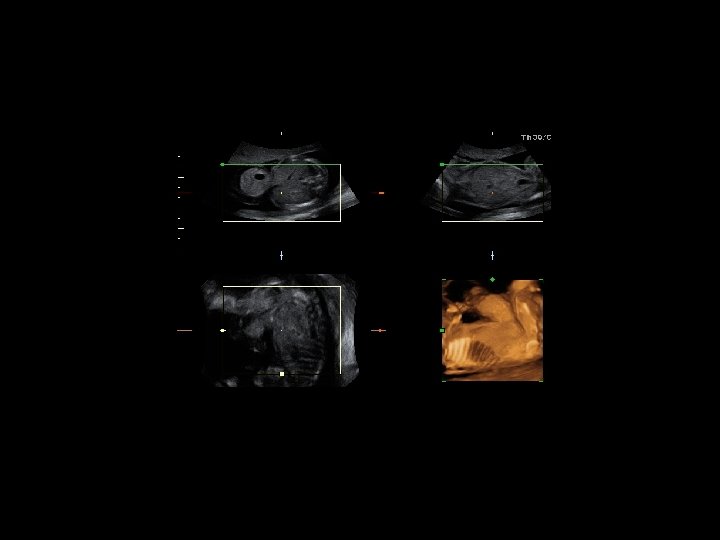
VOCAL Ultrasound images of the fetal stomach at 20 w gestation showing volume measurement by VOCAL. A 30° rotational angle was used and the organ contours were manually traced (Panel A). Panels B and C show the trace in the transverse and coronal sections. Panel D displays a three-dimensional image of the fetal stomach. The volume is expressed in cubic centimeters. 61 Ultrasound Obstet Gynecol 2008; 31
Samenvatting 62
Samenvatting • 3 D - 4 D: veel mogelijkheden 63
Samenvatting - Kritische noot G. Pilu: “ 3 D ultrasound imaging is being used increasingly in obstetric examinations. It remains to be demonstrated whether this tool will have a measurable impact on the prenatal diagnosis of fetal anomalies. However, there is a general consensus that some benefits exist, at least in terms of facilitating fetal examination and data storage. At present, 2 D ultrasound examination remains the basis of fetal diagnosis for many reasons. . 3 D ultrasound imaging does not overcome the physical limitations of ultrasound and sections reconstructed from ultrasound volumes are far less accurate than those obtained by direct insonation; images should therefore always be interpreted with caution. . However the value of ultrasound volumes in terms of data storage, offline analysis and training should not be understated. ” 64
Samenvatting - Kritische noot • 3 D - 4 D: veel mogelijkheden doch daardoor ook duurder aankoop toestel: betaalbaarheid? gevaar commercialisering > “geneeskunde” 65
Samenvatting - Kritische noot • 3 D - 4 D: veel mogelijkheden doch daardoor ook duurder aankoop toestel: betaalbaarheid? gevaar commercialisering > “geneeskunde” tijdsdruk post - processing: fantastische tool: maar wanneer? 66
Samenvatting - Kritische noot • 3 D - 4 D: veel mogelijkheden doch daardoor ook duurder aankoop toestel: betaalbaarheid? gevaar commercialisering > “geneeskunde” tijdsdruk post - processing: fantastische tool: maar wanneer? vragen ivm veiligheid 67
Kritische noot 68
3 D echografie Dank voor jullie aandacht. 69
VCI in coronal plane ( VCI-C)