
3 1 1 1 The structure and functions
- Slides: 46
3. 1. 1. 1 – The structure and functions of the musculo-skeletal system Learning objectives To be able to describe the functions of the skeleton. To be able to recognise and label a skeleton. To understand the different joint classifications. To be able to describe and analyse the different types of joint movements and how they are used during sporting movements. To be able to label the voluntary muscles in our body To explain the term ‘antagonist pair’ and provide examples
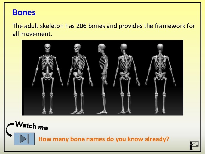
Bones The adult skeleton has 206 bones and provides the framework for all movement. Watch me How many bone names do you know already?
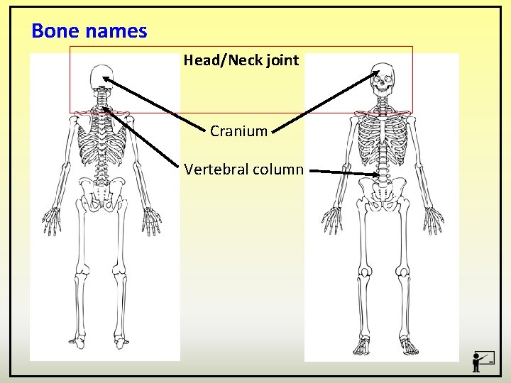
Bone names Head/Neck joint Cranium Vertebral column
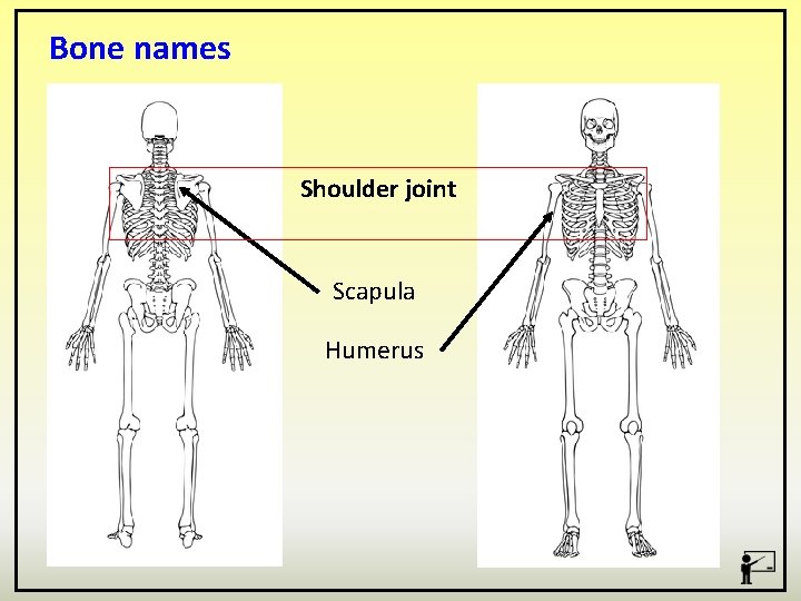
Bone names Shoulder joint Scapula Humerus
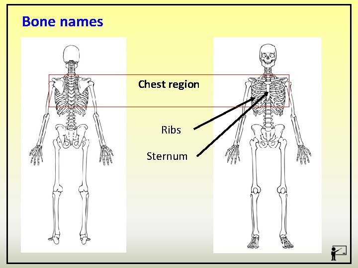
Bone names Chest region Ribs Sternum
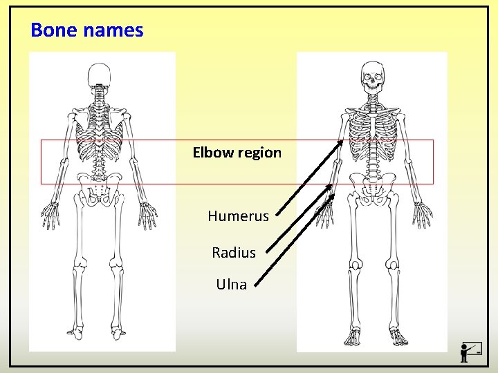
Bone names Elbow region Humerus Radius Ulna
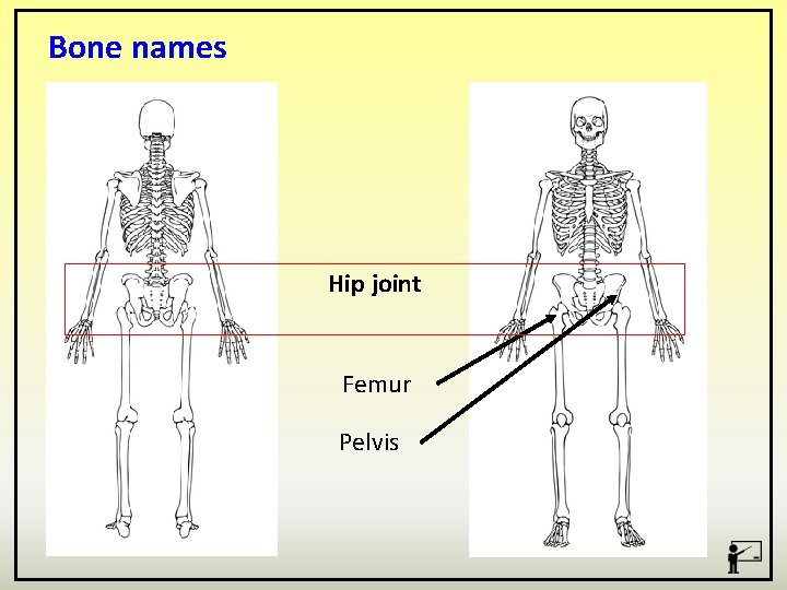
Bone names Hip joint Femur Pelvis
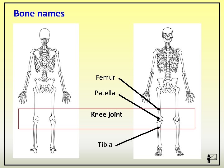
Bone names Femur Patella Knee joint Tibia
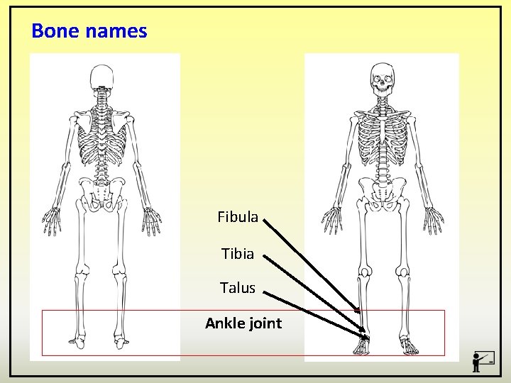
Bone names Fibula Tibia Talus Ankle joint
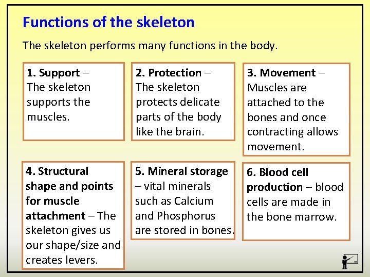
Functions of the skeleton The skeleton performs many functions in the body. 1. Support – The skeleton supports the muscles. 2. Protection – The skeleton protects delicate parts of the body like the brain. 3. Movement – Muscles are attached to the bones and once contracting allows movement. 4. Structural shape and points for muscle attachment – The skeleton gives us our shape/size and creates levers. 5. Mineral storage – vital minerals such as Calcium and Phosphorus are stored in bones. 6. Blood cell production – blood cells are made in the bone marrow.
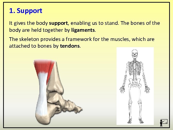
1. Support It gives the body support, enabling us to stand. The bones of the body are held together by ligaments. The skeleton provides a framework for the muscles, which are attached to bones by tendons.
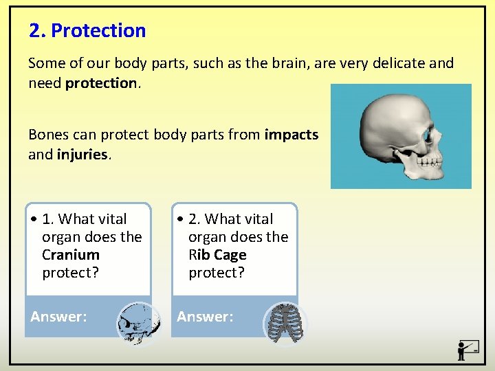
2. Protection Some of our body parts, such as the brain, are very delicate and need protection. Bones can protect body parts from impacts and injuries. • 1. What vital organ does the Cranium protect? • 2. What vital organ does the Rib Cage protect? Answer:
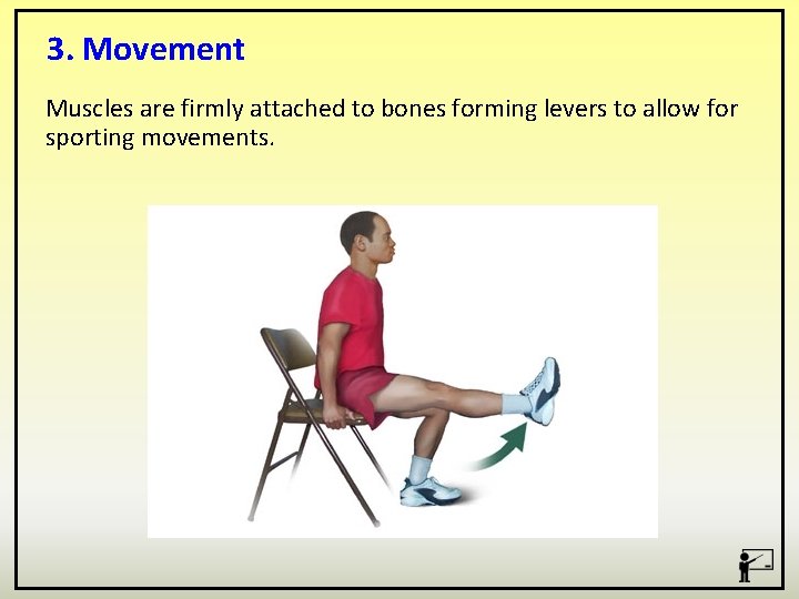
3. Movement Muscles are firmly attached to bones forming levers to allow for sporting movements.
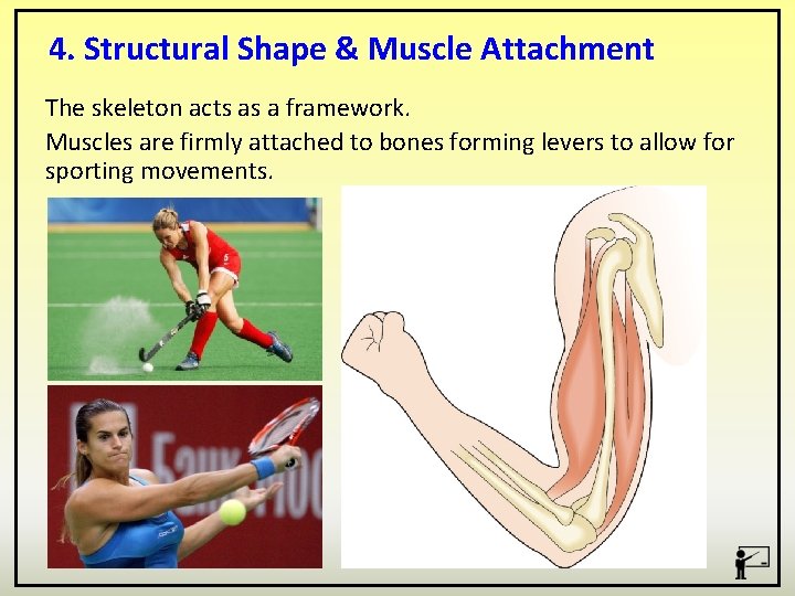
4. Structural Shape & Muscle Attachment The skeleton acts as a framework. Muscles are firmly attached to bones forming levers to allow for sporting movements.
5. Mineral Storage The minerals in your bones serve two main functions. Minerals transform spongy bone matrix into a rigid structure and in turn increase density and strength. Your bones also function as a mineral storage depot, releasing dissolved calcium, phosphorus and magnesium into your bloodstream if needed.
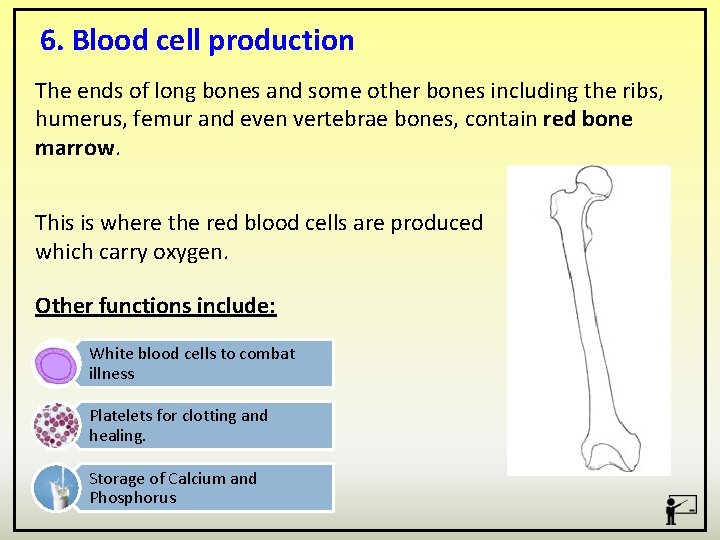
6. Blood cell production The ends of long bones and some other bones including the ribs, humerus, femur and even vertebrae bones, contain red bone marrow. This is where the red blood cells are produced which carry oxygen. Other functions include: White blood cells to combat illness Platelets for clotting and healing. Storage of Calcium and Phosphorus
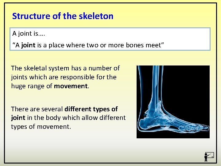
Structure of the skeleton A joint is…. “A joint is a place where two or more bones meet” The skeletal system has a number of joints which are responsible for the huge range of movement. There are several different types of joint in the body which allow different types of movement.
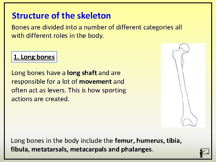
Structure of the skeleton Bones are divided into a number of different categories all with different roles in the body. 1. Long bones have a long shaft and are responsible for a lot of movement and often act as levers. This is how sporting actions are created. Long bones in the body include the femur, humerus, tibia, fibula, metatarsals, metacarpals and phalanges.
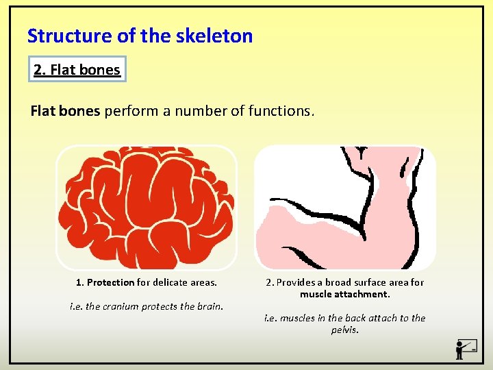
Structure of the skeleton 2. Flat bones perform a number of functions. 1. Protection for delicate areas. i. e. the cranium protects the brain. 2. Provides a broad surface area for muscle attachment. i. e. muscles in the back attach to the pelvis.
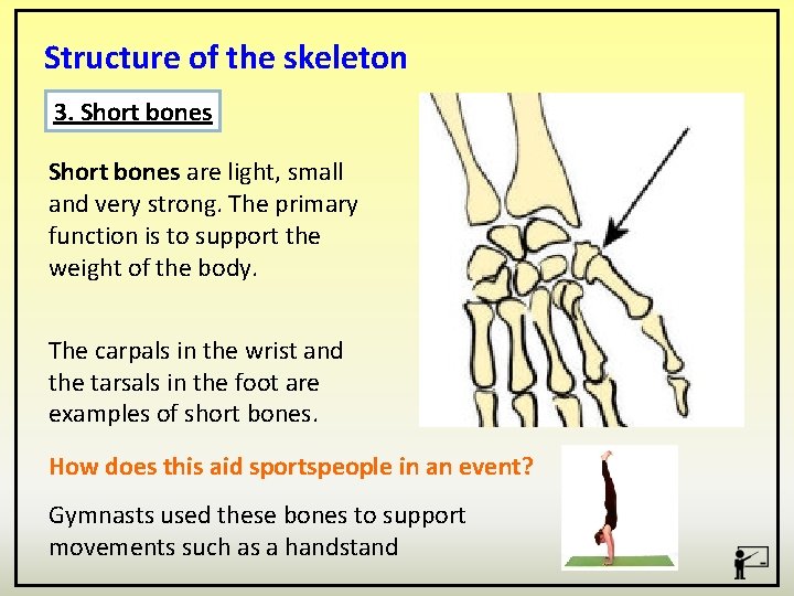
Structure of the skeleton 3. Short bones are light, small and very strong. The primary function is to support the weight of the body. The carpals in the wrist and the tarsals in the foot are examples of short bones. How does this aid sportspeople in an event? Gymnasts used these bones to support movements such as a handstand
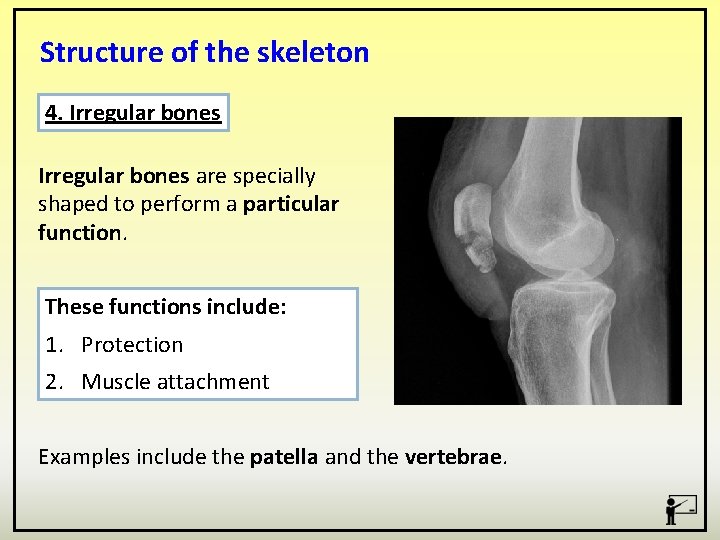
Structure of the skeleton 4. Irregular bones are specially shaped to perform a particular function. These functions include: 1. Protection 2. Muscle attachment Examples include the patella and the vertebrae.
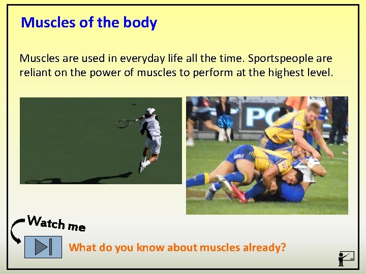
Muscles of the body Muscles are used in everyday life all the time. Sportspeople are reliant on the power of muscles to perform at the highest level. Watch me What do you know about muscles already?
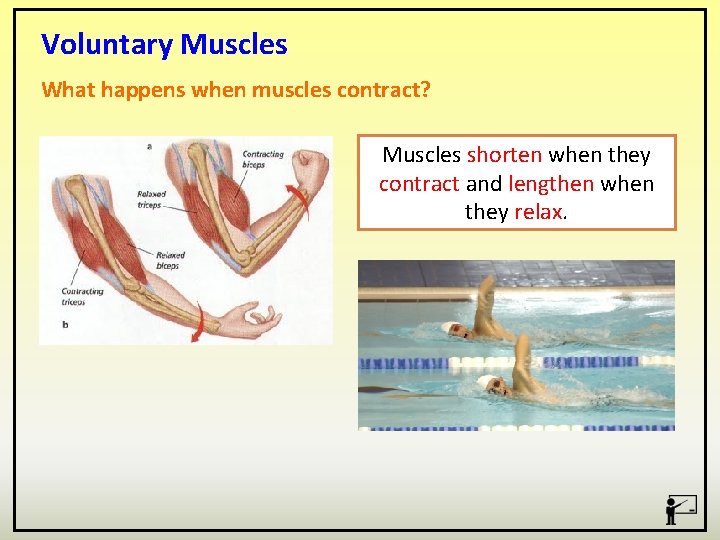
Voluntary Muscles What happens when muscles contract? Muscles shorten when they contract and lengthen when they relax.
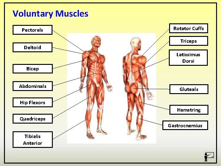
Voluntary Muscles Pectorals Deltoid Rotator Cuffs Triceps Latissimus Dorsi Bicep Abdominals Gluteals Hip Flexors Hamstring Quadriceps Tibialis Anterior Gastrocnemius
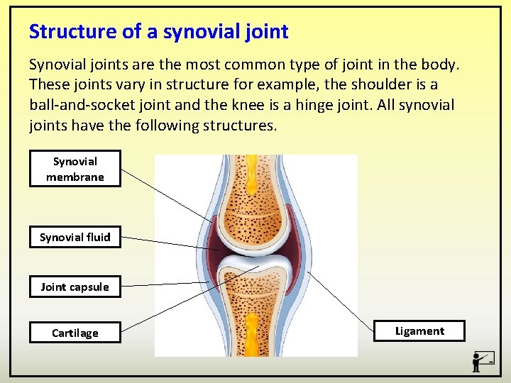
Structure of a synovial joint Synovial joints are the most common type of joint in the body. These joints vary in structure for example, the shoulder is a ball-and-socket joint and the knee is a hinge joint. All synovial joints have the following structures. Synovial membrane Synovial fluid Joint capsule Cartilage Ligament
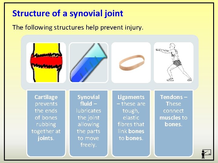
Structure of a synovial joint The following structures help prevent injury. Cartilage prevents the ends of bones rubbing together at joints. Synovial fluid – lubricates the joint allowing the parts to move freely. Ligaments – these are tough, elastic fibres that link bones to bones. Tendons – These connect muscles to bones.
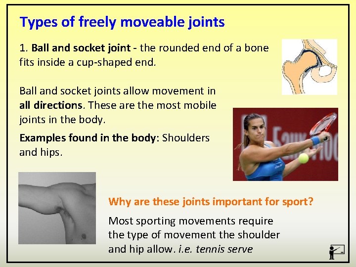
Types of freely moveable joints 1. Ball and socket joint - the rounded end of a bone fits inside a cup-shaped end. Ball and socket joints allow movement in all directions. These are the most mobile joints in the body. Examples found in the body: Shoulders and hips. Why are these joints important for sport? Most sporting movements require the type of movement the shoulder and hip allow. i. e. tennis serve
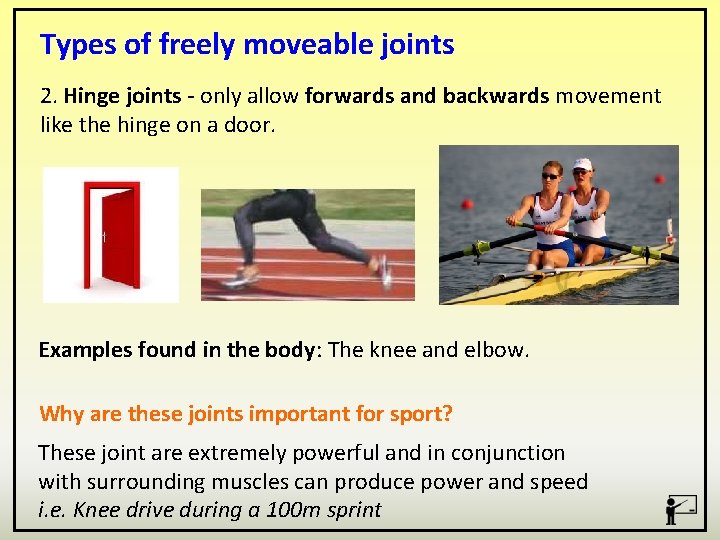
Types of freely moveable joints 2. Hinge joints - only allow forwards and backwards movement like the hinge on a door. Examples found in the body: The knee and elbow. Why are these joints important for sport? These joint are extremely powerful and in conjunction with surrounding muscles can produce power and speed i. e. Knee drive during a 100 m sprint
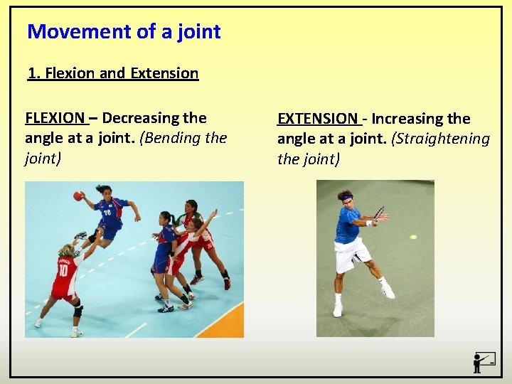
Movement of a joint 1. Flexion and Extension FLEXION – Decreasing the angle at a joint. (Bending the joint) EXTENSION - Increasing the angle at a joint. (Straightening the joint)
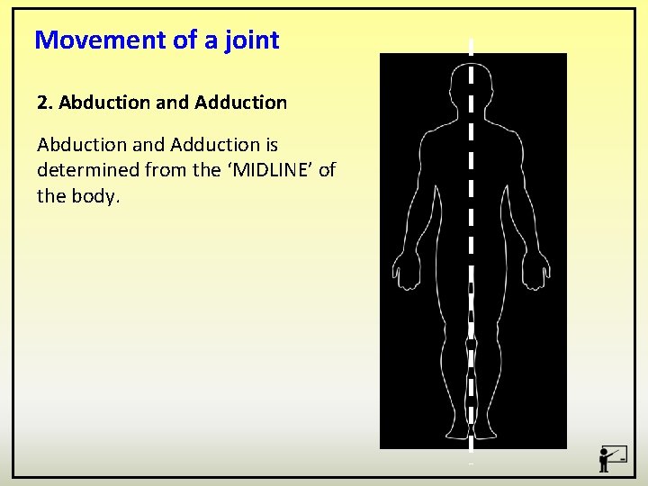
Movement of a joint 2. Abduction and Adduction is determined from the ‘MIDLINE’ of the body.
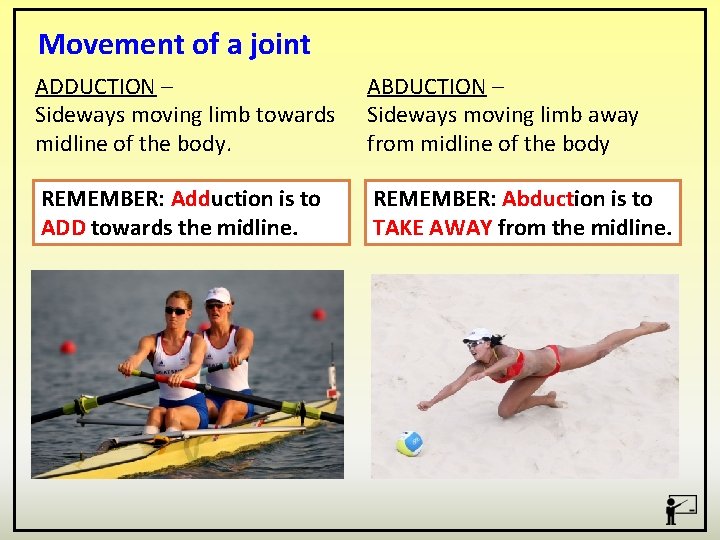
Movement of a joint ADDUCTION – Sideways moving limb towards midline of the body. ABDUCTION – Sideways moving limb away from midline of the body REMEMBER: Adduction is to ADD towards the midline. REMEMBER: Abduction is to TAKE AWAY from the midline.
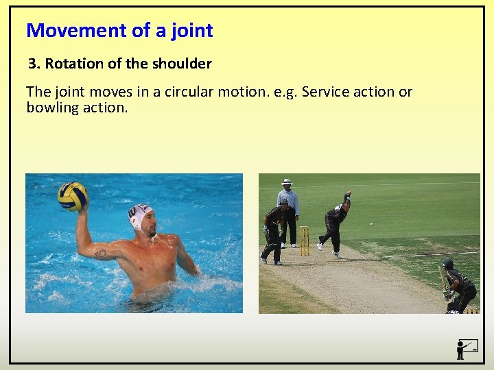
Movement of a joint 3. Rotation of the shoulder The joint moves in a circular motion. e. g. Service action or bowling action.
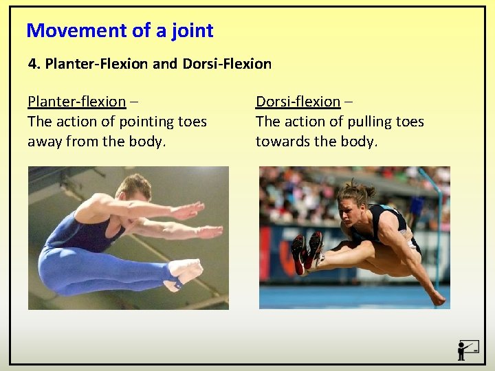
Movement of a joint 4. Planter-Flexion and Dorsi-Flexion Planter-flexion – The action of pointing toes away from the body. Dorsi-flexion – The action of pulling toes towards the body.
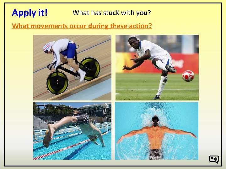
Apply it! What has stuck with you? What movements occur during these action?
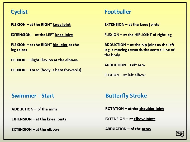
Cyclist Footballer FLEXION – at the RIGHT knee joint EXTENSION – at the knee joints EXTENSION - at the LEFT knee joint FLEXION – at the HIP JOINT of right leg FLEXION – at the RIGHT hip joint as the leg raises ADDUCTION – at the hip joint as the left leg is moving towards the central line of the body FLEXION – Slight Flexion at the elbows FLEXION – Torso (body is bent forwards) ADDUCTION – Left arm FLEXION – at left elbow Swimmer - Start Butterfly Stroke ADDUCTION – of the arms ROTATION – at the shoulder joint EXTENSION – at the knee joints EXTENSION – at elbow joints EXTENSION – at the elbows ABDUCTION – of the arms
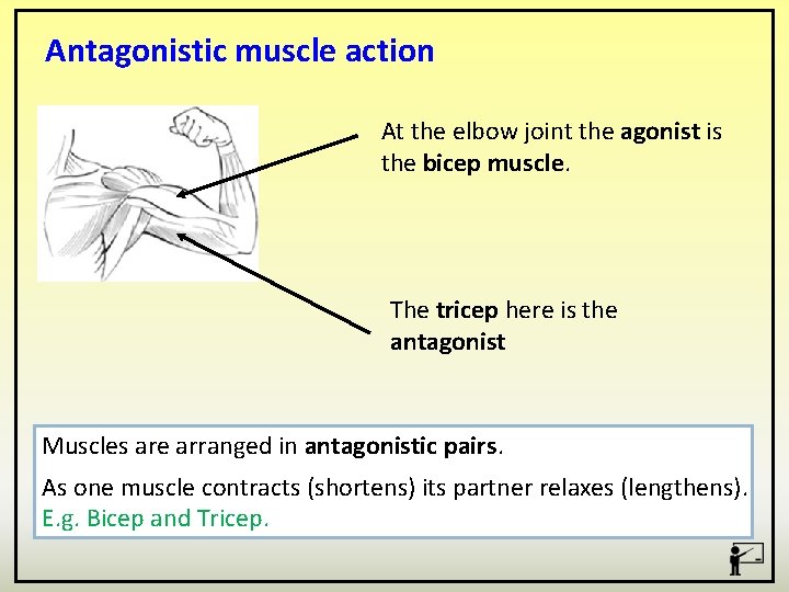
Antagonistic muscle action At the elbow joint the agonist is the bicep muscle. The tricep here is the antagonist Muscles are arranged in antagonistic pairs. As one muscle contracts (shortens) its partner relaxes (lengthens). E. g. Bicep and Tricep.
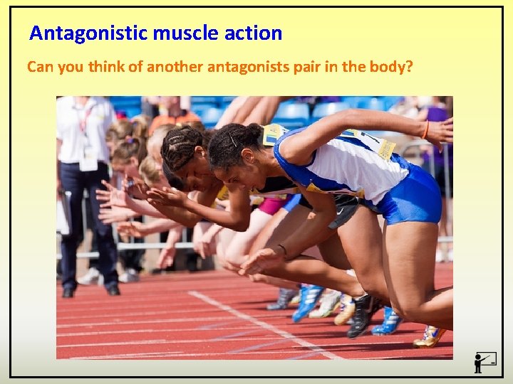
Antagonistic muscle action Can you think of another antagonists pair in the body?
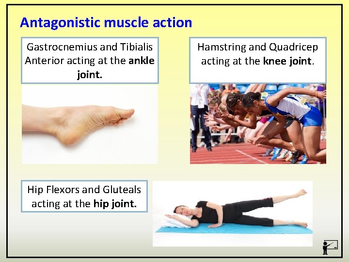
Antagonistic muscle action Gastrocnemius and Tibialis Anterior acting at the ankle joint. Hip Flexors and Gluteals acting at the hip joint. Hamstring and Quadricep acting at the knee joint.
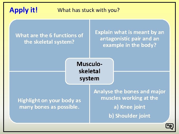
Apply it! What has stuck with you? What are the 6 functions of the skeletal system? Explain what is meant by an antagonistic pair and an example in the body? Musculoskeletal system Highlight on your body as many bones as possible. Analyse the bones and major muscles working at the a) Knee joint b) Shoulder joint
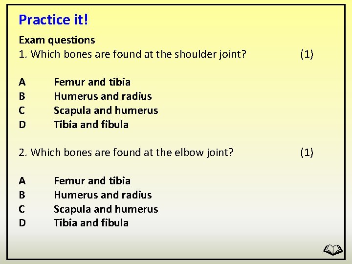
Practice it! Exam questions 1. Which bones are found at the shoulder joint? Femur and tibia Humerus and radius Scapula and humerus Tibia and fibula 2. Which bones are found at the elbow joint? (1) A B C D Femur and tibia Humerus and radius Scapula and humerus Tibia and fibula (1)
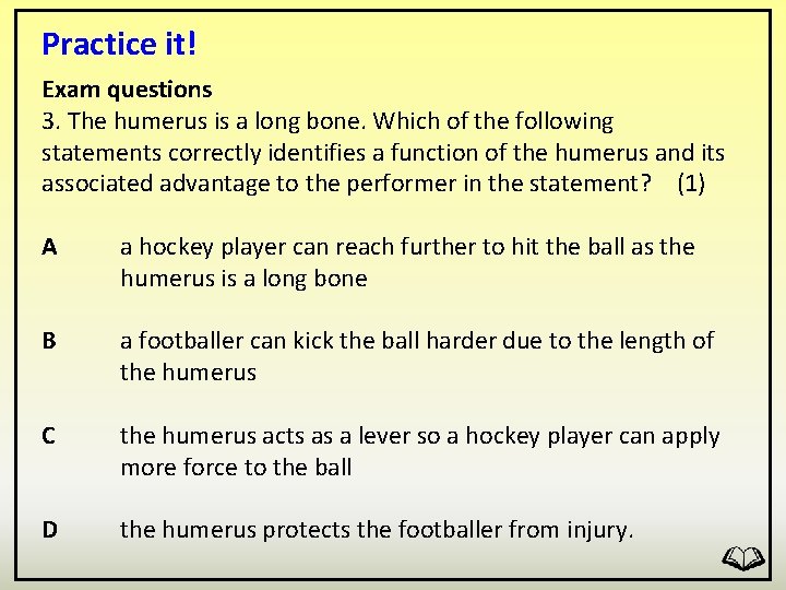
Practice it! Exam questions 3. The humerus is a long bone. Which of the following statements correctly identifies a function of the humerus and its associated advantage to the performer in the statement? (1) A a hockey player can reach further to hit the ball as the humerus is a long bone B a footballer can kick the ball harder due to the length of the humerus C the humerus acts as a lever so a hockey player can apply more force to the ball D the humerus protects the footballer from injury.
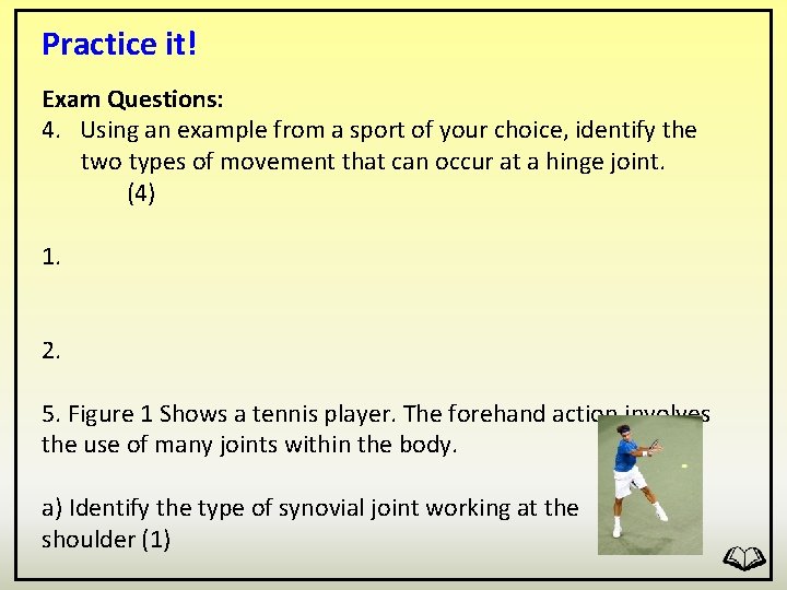
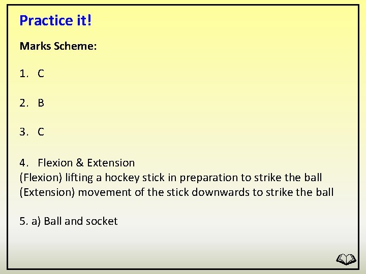
Practice it! Exam Questions: 4. Using an example from a sport of your choice, identify the two types of movement that can occur at a hinge joint. (4) 1. 2. 5. Figure 1 Shows a tennis player. The forehand action involves the use of many joints within the body. a) Identify the type of synovial joint working at the shoulder (1)
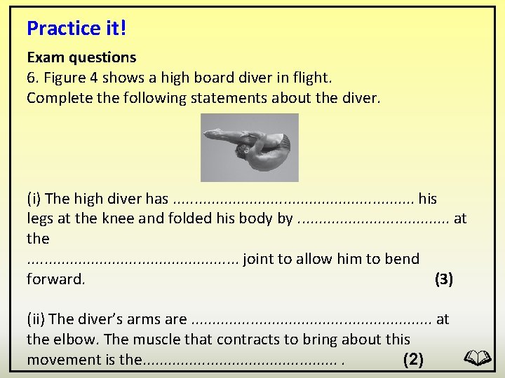
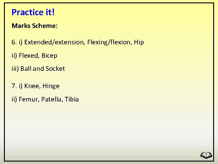
Practice it! Exam questions 6. Figure 4 shows a high board diver in flight. Complete the following statements about the diver. (i) The high diver has. . . . his legs at the knee and folded his body by. . . . . at the. . . joint to allow him to bend forward. (3) (ii) The diver’s arms are. . . . at the elbow. The muscle that contracts to bring about this movement is the. . . (2)
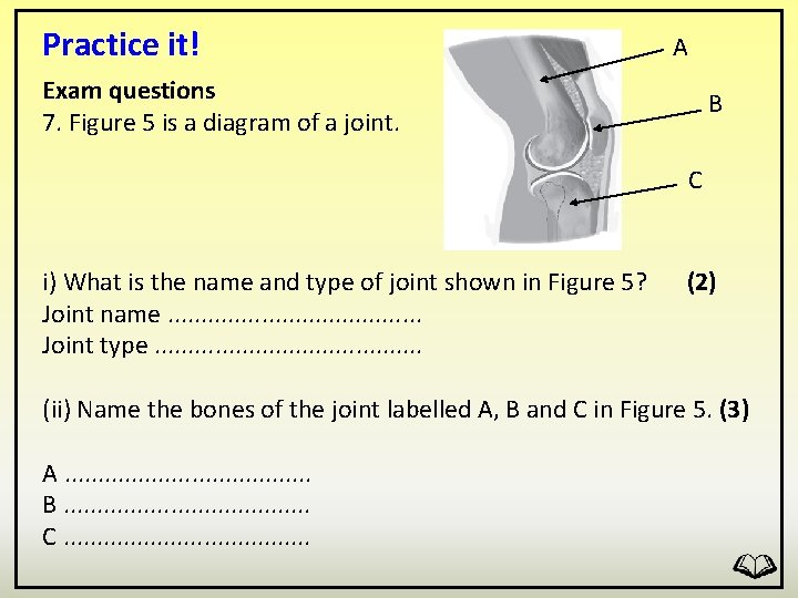
Practice it! A Exam questions 7. Figure 5 is a diagram of a joint. B C i) What is the name and type of joint shown in Figure 5? Joint name. . . . . Joint type. . . . . (2) (ii) Name the bones of the joint labelled A, B and C in Figure 5. (3) A. . . . . B. . . . . C. . . . .
Practice it! Marks Scheme: 1. C 2. B 3. C 4. Flexion & Extension (Flexion) lifting a hockey stick in preparation to strike the ball (Extension) movement of the stick downwards to strike the ball 5. a) Ball and socket
Practice it! Marks Scheme: 6. i) Extended/extension, Flexing/flexion, Hip ii) Flexed, Bicep iii) Ball and Socket 7. i) Knee, Hinge ii) Femur, Patella, Tibia