
26 RNA Metabolism 2017 W H Freeman and
26| RNA Metabolism © 2017 W. H. Freeman and Company
CHAPTER 26 RNA Metabolism Learning goals: • Transcription: DNA-dependent synthesis of RNA • Capping and splicing: RNA processing • RNA-dependent synthesis of RNA and DNA
Overview of RNA Function • Ribonucleic acids play three well-understood roles in living cells: – Messenger RNAs encode the amino acid sequences of all the polypeptides found in the cell. – Transfer RNAs match their anticodon to the m. RNA while carrying a specific amino acid used for protein synthesis. – Ribosomal RNAs are constituents of the large and small ribosomal subunits. • Ribonucleic acids play several less-understood functions in eukaryotic cells: – Micro. RNAs regulate the expression of genes, possibly via binding to specific nucleotide sequences. – Ribozymes are catalytic RNA molecules that act as enzymes. • Ribonucleic acids act as genomic material in viruses.
Overview of RNA Metabolism • Transcribed from DNA – Transcription is tightly regulated in order to control the concentration of each protein. • Ribozymes – Being mainly single stranded, many RNA molecules can fold into compact structures with specific functions. – Some RNA molecules can act as catalysts (ribozymes), often using metal ions as cofactors such as the group I introns. • Processing of m. RNAs – Splicing - elimination of introns; joining of exons – Poly-adenylation of the 3’ end – Capping the 5’ end
Transcription in E. Coli • The nucleoside triphosphates add to the 3’ end of the growing RNA strand. • The growing chain is complementary to the template strand in DNA. • The synthesis is catalyzed by enzyme (RNA polymerase) and involves 2 Mg 2+ ions for catalysis. • RNA polymerase unwinds ~17 bp of DNA and covers about 35 bp of DNA.
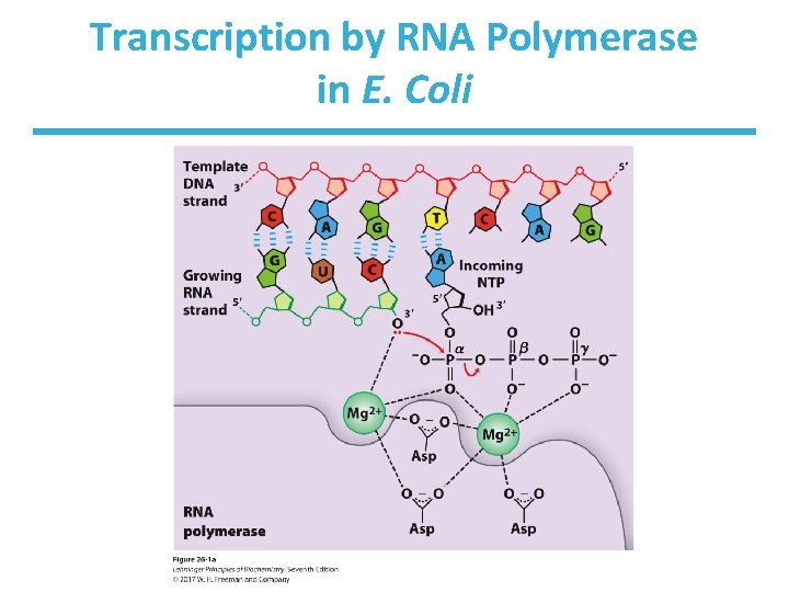
Transcription by RNA Polymerase in E. Coli
Features of Transcription • RNA polymerase binds to a sequence called promoter to begin transcription. – primer not required • The growing end of new RNA temporarily base-pairs with DNA template for ~8 bp and inserts 50 -90 nt/sec. • The DNA duplex unwinds, forming a “bubble” of ~17 bp. • The RNA Pol generates positive supercoils ahead, later relieved by topoisomerases.
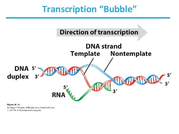
Transcription “Bubble”
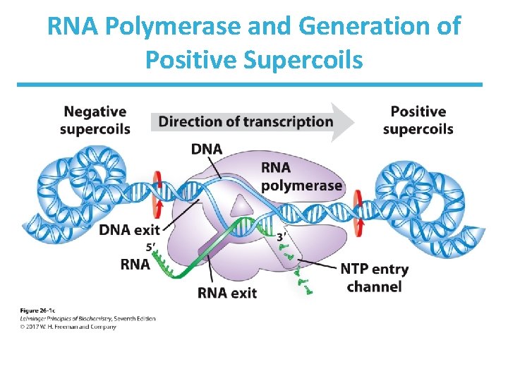
RNA Polymerase and Generation of Positive Supercoils
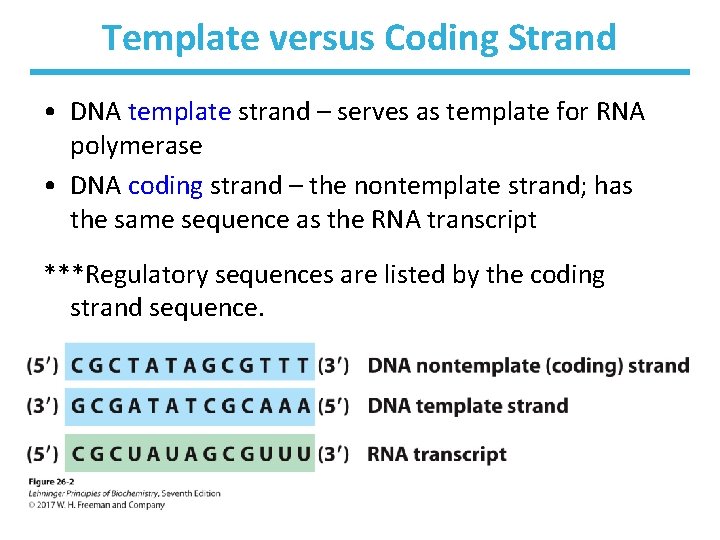
Template versus Coding Strand • DNA template strand – serves as template for RNA polymerase • DNA coding strand – the nontemplate strand; has the same sequence as the RNA transcript ***Regulatory sequences are listed by the coding strand sequence.
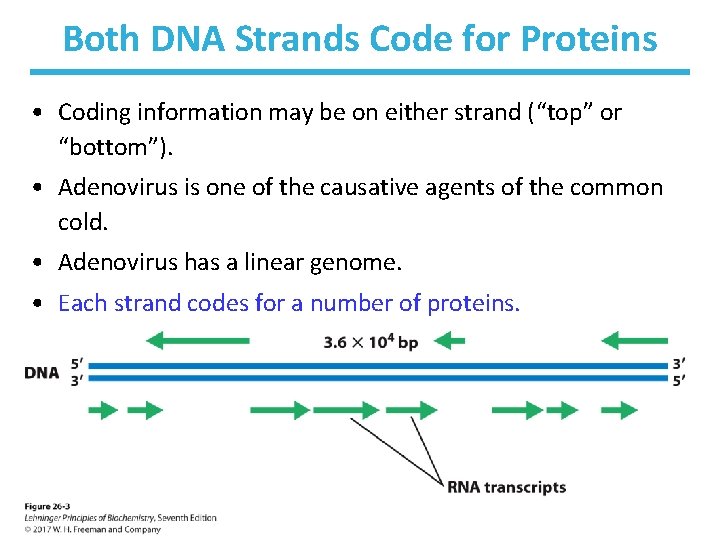
Both DNA Strands Code for Proteins • Coding information may be on either strand (“top” or “bottom”). • Adenovirus is one of the causative agents of the common cold. • Adenovirus has a linear genome. • Each strand codes for a number of proteins.
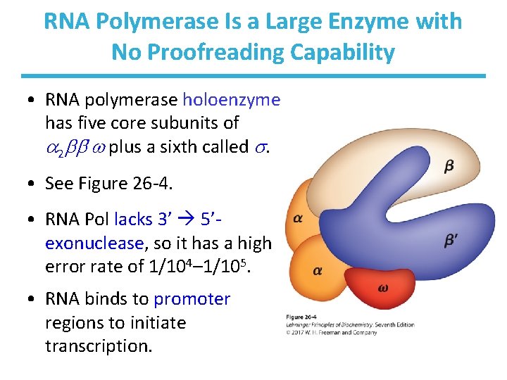
RNA Polymerase Is a Large Enzyme with No Proofreading Capability • RNA polymerase holoenzyme has five core subunits of 2 ’ plus a sixth called . • See Figure 26 -4. • RNA Pol lacks 3’ 5’exonuclease, so it has a high error rate of 1/104– 1/105. • RNA binds to promoter regions to initiate transcription.
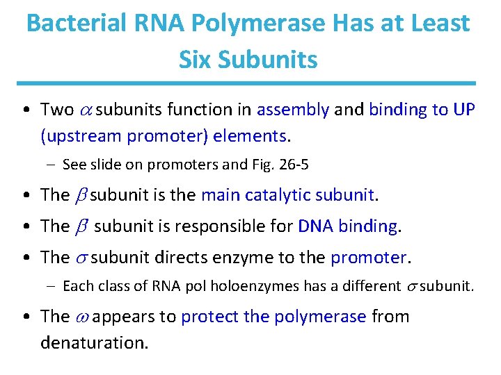
Bacterial RNA Polymerase Has at Least Six Subunits • Two subunits function in assembly and binding to UP (upstream promoter) elements. – See slide on promoters and Fig. 26 -5 • The subunit is the main catalytic subunit. • The ’ subunit is responsible for DNA binding. • The subunit directs enzyme to the promoter. – Each class of RNA pol holoenzymes has a different subunit. • The appears to protect the polymerase from denaturation.
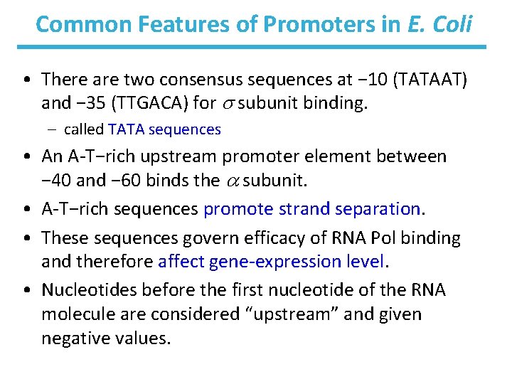
Common Features of Promoters in E. Coli • There are two consensus sequences at − 10 (TATAAT) and − 35 (TTGACA) for subunit binding. – called TATA sequences • An A-T−rich upstream promoter element between − 40 and − 60 binds the subunit. • A-T−rich sequences promote strand separation. • These sequences govern efficacy of RNA Pol binding and therefore affect gene-expression level. • Nucleotides before the first nucleotide of the RNA molecule are considered “upstream” and given negative values.
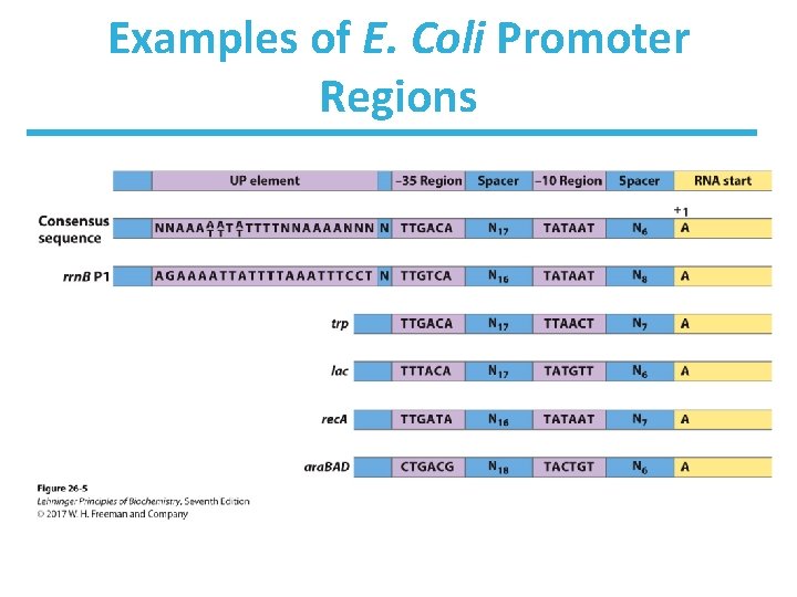
Examples of E. Coli Promoter Regions
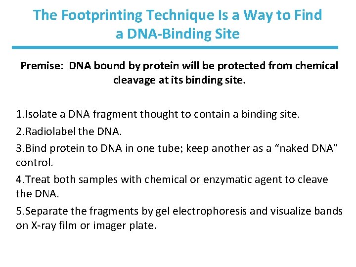
The Footprinting Technique Is a Way to Find a DNA-Binding Site Premise: DNA bound by protein will be protected from chemical cleavage at its binding site. 1. Isolate a DNA fragment thought to contain a binding site. 2. Radiolabel the DNA. 3. Bind protein to DNA in one tube; keep another as a “naked DNA” control. 4. Treat both samples with chemical or enzymatic agent to cleave the DNA. 5. Separate the fragments by gel electrophoresis and visualize bands on X-ray film or imager plate.
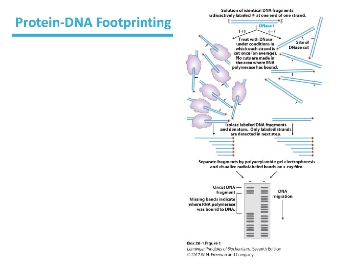
Protein-DNA Footprinting
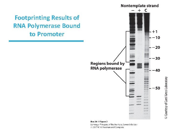
Footprinting Results of RNA Polymerase Bound to Promoter
Initiation of Transcription • RNA Pol binds to promoter with the factor bound. – It creates a closed complex (DNA is not unwound). • An open complex forms. – The region from ~− 10 to ~+2 unwinds. • RNA Pol moves away from the promoter. – is replaced by the protein Nus. A.
Elongation of Transcription • RNA Pol binds to triphosphate nucleosides and generates the RNA transcript. • Nus. G binds to both the RNA polymerase and ribosome linking the two together, directly affecting the rate of transcription in prokaryotic cells.
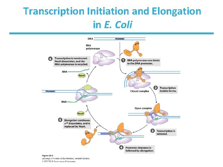
Transcription Initiation and Elongation in E. Coli
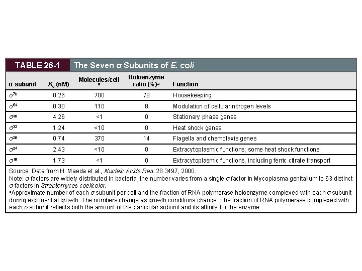
TABLE 26 -1 σ subunit Kd (n. M) The Seven σ Subunits of E. coli Molecules/cell a Holoenzyme ratio (%)a Function σ70 0. 26 700 78 Housekeeping σ54 0. 30 110 8 Modulation of cellular nitrogen levels σ38 4. 26 <1 0 Stationary phase genes σ32 1. 24 <10 0 Heat shock genes σ28 0. 74 370 14 Flagella and chemotaxis genes σ24 2. 43 <10 0 Extracytoplasmic functions; some heat shock functions σ18 1. 73 <1 0 Extracytoplasmic functions, including ferric citrate transport Source: Data from H. Maeda et al. , Nucleic Acids Res. 28: 3497, 2000. Note: σ factors are widely distributed in bacteria; the number varies from a single σ factor in Mycoplasma genitalium to 63 distinct σ factors in Streptomyces coelicolor. a. Approximate number of each σ subunit per cell and the fraction of RNA polymerase holoenzyme complexed with each σ subunit during exponential growth. The numbers change as growth conditions change. The fraction of RNA polymerase complexed with each σ subunit reflects both the amount of the particular subunit and its affinity for the enzyme.
Regulation of Transcription • Transcription is energy-intensive, so it’s logical to regulate gene production here. • Regulation is achieved in many ways. – One way is to regulate the affinity of RNA polymerase for a promoter. • promoter sequence – deviating from the consensus sequence • activator proteins – CRP • repressor proteins – block necessary binding sites
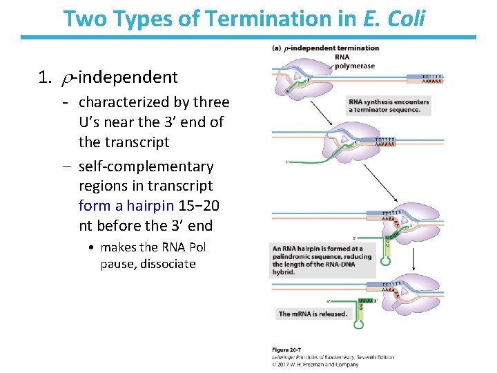
Two Types of Termination in E. Coli 1. -independent - characterized by three U’s near the 3’ end of the transcript – self-complementary regions in transcript form a hairpin 15− 20 nt before the 3’ end • makes the RNA Pol pause, dissociate
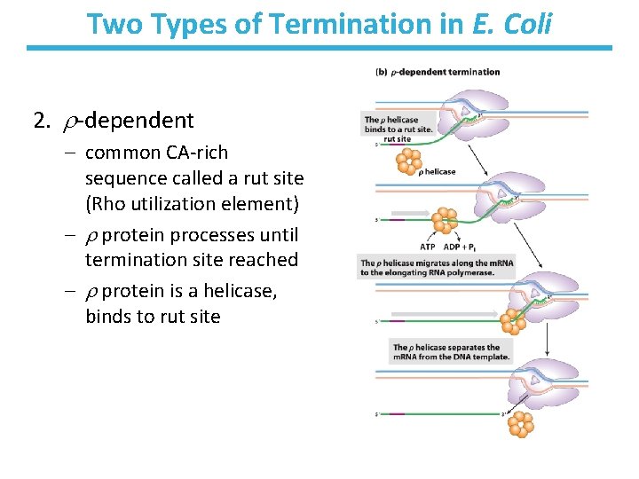
Two Types of Termination in E. Coli 2. -dependent – common CA-rich sequence called a rut site (Rho utilization element) – protein processes until termination site reached – protein is a helicase, binds to rut site
Eukaryotes RNA Polymerases • RNA polymerase I synthesizes pre-ribosomal RNA (precursor for 28 S, 18 S, and 5. 8 r. RNAs). • RNA polymerase II is responsible for synthesis of m. RNA. – very fast (500– 1000 nucleotides/sec) – specifically inhibited by mushroom toxin -amanitin – can recognize thousands of promoters (See Fig. 26 -8) • RNA polymerase III makes t. RNAs and some small RNA products. • Plants appear to have RNA polymerase IV that is responsible for the synthesis of small interfering RNAs. • Mitochondria have their own RNA polymerase.
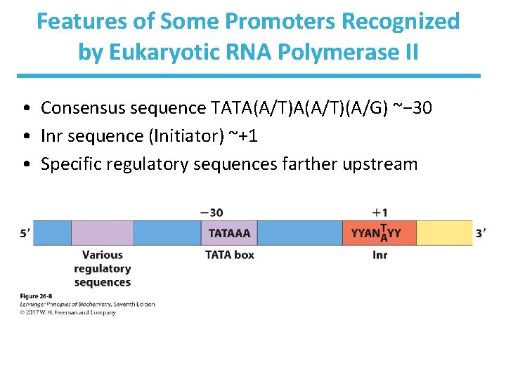
Features of Some Promoters Recognized by Eukaryotic RNA Polymerase II • Consensus sequence TATA(A/T)(A/G) ~− 30 • Inr sequence (Initiator) ~+1 • Specific regulatory sequences farther upstream
Eukaryotic m. RNA Transcription Involves Many Proteins • Relies on protein-protein contacts – many highly conserved transcription factors • RNA Pol II is well-studied. – large complex of 12 subunits • Some subunits have some structural homology to bacterial RNA polymerase. – has carboxy-terminal domain of highly conserved repeats
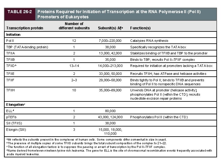
TABLE 26 -2 Proteins Required for Initiation of Transcription at the RNA Polymerase II (Pol II) Promoters of Eukaryotes Number of different subunits Subunit(s) Mra Pol II 12 7, 000– 220, 000 TBP (TATA-binding protein) 1 38, 000 TFIIA 2 13, 000, 42, 000 TFIIB 1 35, 000 TFIIDb 13– 14 14, 000– 213, 000 Required for initiation at promoters lacking a TATA box TFIIE 2 33, 000, 50, 000 Recruits TFIIH; has ATPase and helicase activities TFIIF 2– 3 29, 000– 58, 000 Binds tightly to Pol II; binds to TFIIB and prevents binding of Pol II to nonspecific DNA sequences TFIIH 10 35, 000– 89, 000 Unwinds DNA at promoter (helicase activity); phosphorylates Pol II (within the CTD); recruits nucleotide-excision repair proteins ELLd 1 80, 000 p. TEFb 2 43, 000, 124, 000 SII (TFIIS) 1 38, 000 Elongin (SIII) 3 15, 000, 18, 000, 110, 000 Transcription protein Function(s) Initiation Catalyzes RNA synthesis Specifically recognizes the TATA box Stabilizes binding of TFIIB and TBP to the promoter Binds to TBP; recruits Pol II–TFIIF complex Elongationc a. Mr Phosphorylates Pol II (within the CTD) reflects the subunits present in the complexes of human cells. Some components differ somewhat in size in yeast. presence of multiple copies of some TFIID subunits brings the total subunit composition of the complex to 21– 22. c. The function of all elongation factors is to suppress the pausing or arrest of transcription by the Pol II–TFIIF complex. d. Name derived from eleven-nineteen lysine-rich leukemia. The gene for ELL is the site of chromosomal recombination events frequently associated with acute myeloid leukemia. b. The
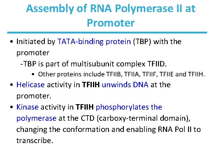
Assembly of RNA Polymerase II at Promoter • Initiated by TATA-binding protein (TBP) with the promoter -TBP is part of multisubunit complex TFIID. • Other proteins include TFIIB, TFIIA, TFIIF, TFIIE and TFIIH. • Helicase activity in TFIIH unwinds DNA at the promoter. • Kinase activity in TFIIH phosphorylates the polymerase at the CTD (carboxy-terminal domain), changing the conformation and enabling RNA Pol II to transcribe.
Elongation and Termination • After 60 -70 nt, TFIIE is released followed by TFIIH. • Elongation factors bound to RNA Pol II enhance processivity and coordinate posttranslational modifications. – Some elongation factors are bound to the phosphorylated CTD. • For termination, Pol II is dephosphorylated. • Regulation is complex (see Chapter 28).
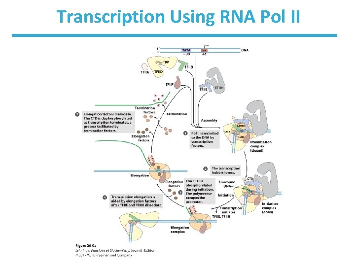
Transcription Using RNA Pol II
TFIIH and Repair • Transcribed genes are more actively repaired than silent genes are. • It may partly be explained that TFIIH also has a role in nucleotide-excision repair (NER). – recruits the NER complex at a lesion • Genetic repair diseases are associated with TFIIH defects: – xeroderma pigmentosum and others
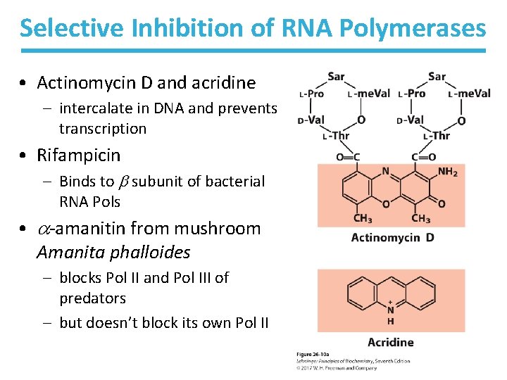
Selective Inhibition of RNA Polymerases • Actinomycin D and acridine – intercalate in DNA and prevents transcription • Rifampicin – Binds to subunit of bacterial RNA Pols • -amanitin from mushroom Amanita phalloides – blocks Pol II and Pol III of predators – but doesn’t block its own Pol II
Processing of m. RNA − Overview • Dozens of proteins coordinate with each other and with proteins involved in RNA transport to ribosomes. • An unprocessed, newly synthesized RNA molecule is referred to as a primary transcript. • Processing includes: – splicing out introns and rejoining any exons for a continuous sequence – adding a 5’-cap – adding a 3’-poly(A) tail – degradation
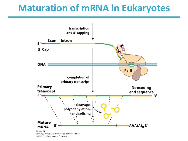
Maturation of m. RNA in Eukaryotes
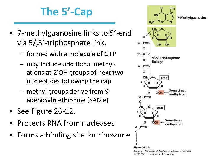
The 5’-Cap • 7 -methylguanosine links to 5’-end via 5/, 5’-triphosphate link. – formed with a molecule of GTP – may include additional methylations at 2’OH groups of next two nucleotides following the cap – methyl groups derive from Sadenosylmethionine (SAMe) • See Figure 26 -12. • Protects RNA from nucleases • Forms a binding site for ribosome
Introns Are Found in Most Genes • Most genes in vertebrates, and some in yeast, a few bacteria have introns. • Exons are usually <1, 000 bp in length. • Introns are 50− 700, 000 bp in length. – 1, 800 bp avg • Some genes have dozens of introns. – The human genome has more than 200, 000 introns spread across ~20, 000 genes.
Four Classes of Introns • Group I and group II introns are self-splicing. – require no additional proteins or ATP – in nuclear, mitochondrial, and chloroplast genomes – differ mainly in the splicing mechanism – found in some bacteria • Spliceosomal introns are spliced by enormous complexes called spliceosomes. – the most common introns – frequent in protein-coding regions of eukaryotic genomes • t. RNA introns are spliced by protein-based enzymes. – Primary transcript is cleaved by endonuclease. – Exons are joined by ATP-dependent ligase.
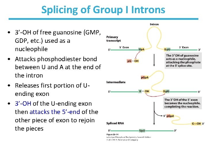
Splicing of Group I Introns • 3’-OH of free guanosine (GMP, GDP, etc. ) used as a nucleophile • Attacks phosphodiester bond between U and A at the end of the intron • Releases first portion of Uending exon • 3’-OH of the U-ending exon then attacks the 5’-end of the other piece of exon to rejoin the pieces
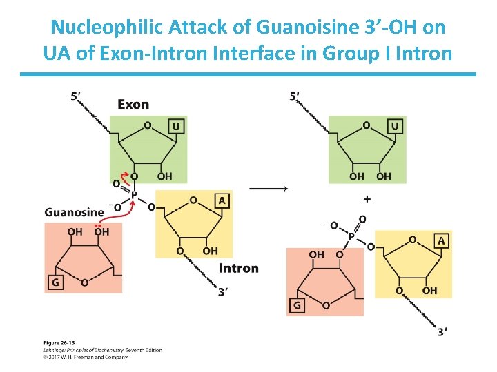
Nucleophilic Attack of Guanoisine 3’-OH on UA of Exon-Intron Interface in Group I Intron
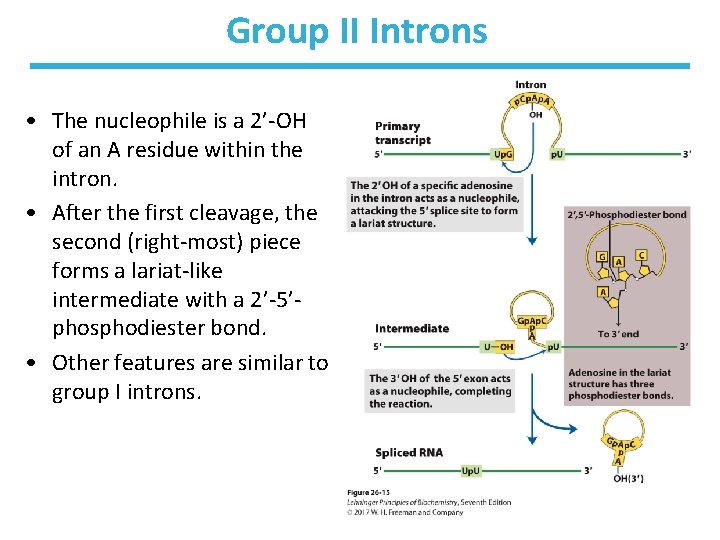
Group II Introns • The nucleophile is a 2’-OH of an A residue within the intron. • After the first cleavage, the second (right-most) piece forms a lariat-like intermediate with a 2’-5’phosphodiester bond. • Other features are similar to group I introns.
Spliceosome Introns Are Removed via a Large Complex Called a Spliceosome • The spliceosome is made up of sn. RNPs (“snurps” for small nuclear ribonuclear proteins). – sn. RNP RNA is called sn. RNA (for small nuclear RNA), which are 100− 200 nt long. • five sn. RNAs known in eukaryotes (U 1, U 2, U 4, U 5, U 6) • GU at 5’-end and AU at 3’-end usually mark sites of splicing.
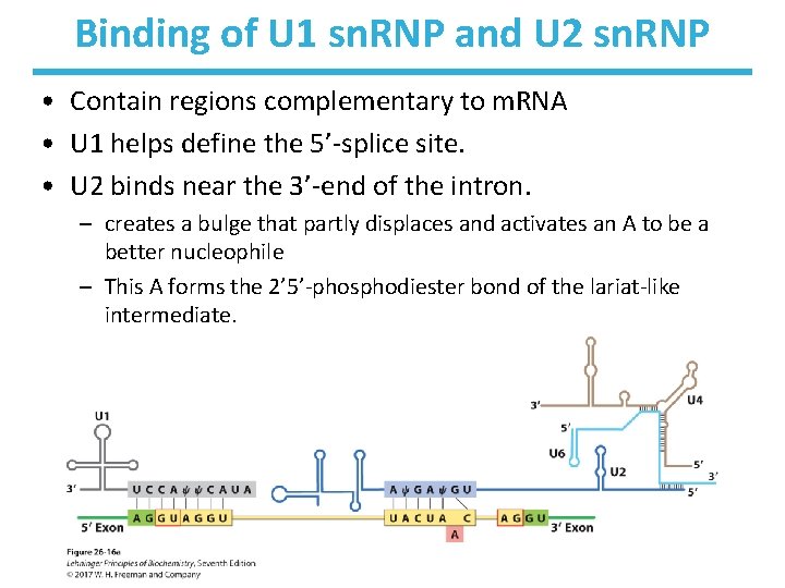
Binding of U 1 sn. RNP and U 2 sn. RNP • Contain regions complementary to m. RNA • U 1 helps define the 5’-splice site. • U 2 binds near the 3’-end of the intron. – creates a bulge that partly displaces and activates an A to be a better nucleophile – This A forms the 2’ 5’-phosphodiester bond of the lariat-like intermediate.
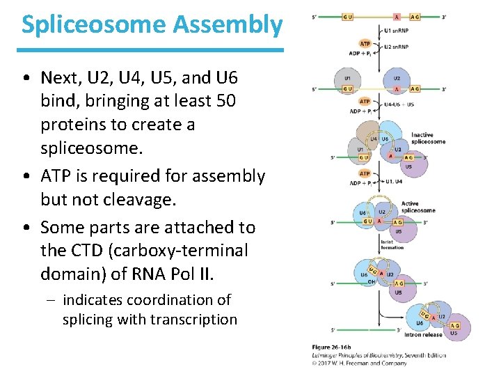
Spliceosome Assembly • Next, U 2, U 4, U 5, and U 6 bind, bringing at least 50 proteins to create a spliceosome. • ATP is required for assembly but not cleavage. • Some parts are attached to the CTD (carboxy-terminal domain) of RNA Pol II. – indicates coordination of splicing with transcription
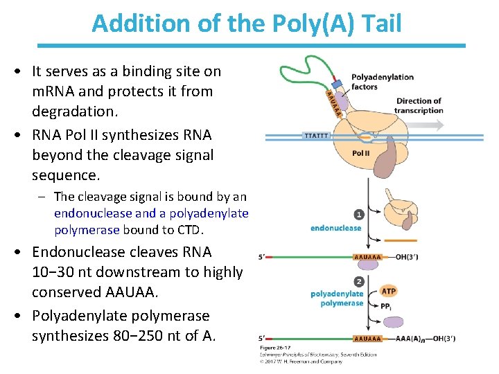
Addition of the Poly(A) Tail • It serves as a binding site on m. RNA and protects it from degradation. • RNA Pol II synthesizes RNA beyond the cleavage signal sequence. – The cleavage signal is bound by an endonuclease and a polyadenylate polymerase bound to CTD. • Endonuclease cleaves RNA 10− 30 nt downstream to highly conserved AAUAA. • Polyadenylate polymerase synthesizes 80− 250 nt of A.
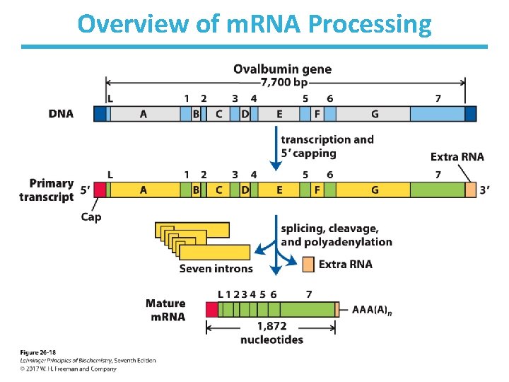
Overview of m. RNA Processing
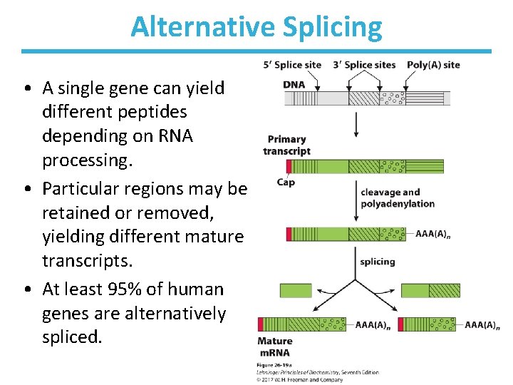
Alternative Splicing • A single gene can yield different peptides depending on RNA processing. • Particular regions may be retained or removed, yielding different mature transcripts. • At least 95% of human genes are alternatively spliced.
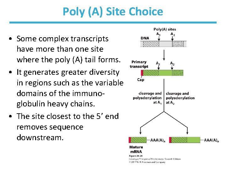
Poly (A) Site Choice • Some complex transcripts have more than one site where the poly (A) tail forms. • It generates greater diversity in regions such as the variable domains of the immunoglobulin heavy chains. • The site closest to the 5’ end removes sequence downstream.
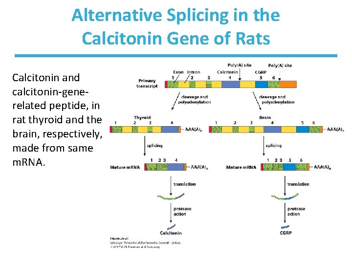
Alternative Splicing in the Calcitonin Gene of Rats Calcitonin and calcitonin-generelated peptide, in rat thyroid and the brain, respectively, made from same m. RNA.
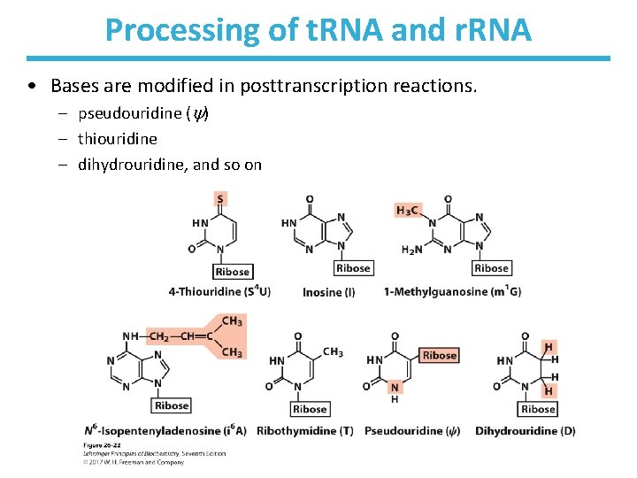
Processing of t. RNA and r. RNA • Bases are modified in posttranscription reactions. – pseudouridine ( ) – thiouridine – dihydrouridine, and so on
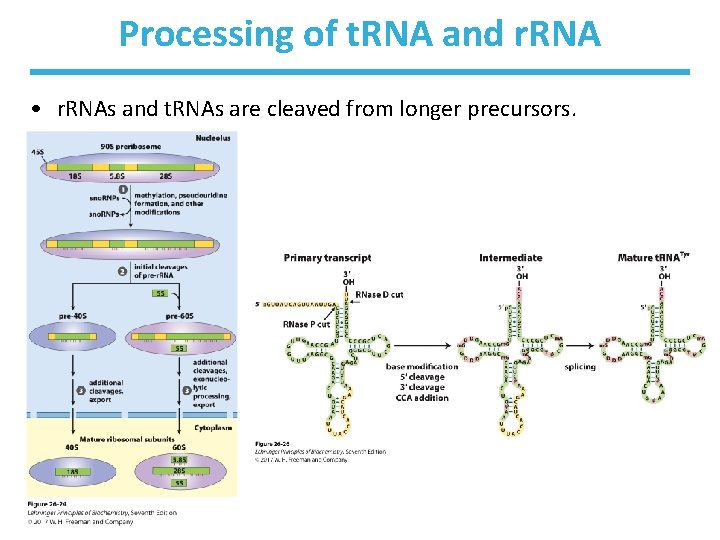
Processing of t. RNA and r. RNA • r. RNAs and t. RNAs are cleaved from longer precursors.
Micro. RNAs Function in Gene Regulation • Micro. RNAs (mi. RNAs): – short noncoding RNAs of ~22 nucleotides – bind to specific regions of m. RNA to alter translation • assist in cleaving the m. RNAs • or block the m. RNA from translation – About 1, 500 human genes encode mi. RNAs and 1 or more affect the expression of MOST proteincoding genes! – Synthesized from larger precursors • processed by two endoribonucleases, Drosha and Dicer
Steps in mi. RNA Processing • Long precursor pri-mi. RNA is made in the nucleus. • Drosha and DGCR 8 cleave pri-mi. RNA to a 70− 80 nt precursor. • Exportin and Ran export this precursor to the cytoplasm. • Dicer cleaves the pre-mi. RNA into ds. RNA. • The complement of mi. RNA is removed by helicase. • mi. RNA is loaded onto a protein complex such as RNAinduced silencing complex (RISC). • RISC binds to target m. RNA.
RISC-mi. RNA Prevents Translation of m. RNA • The mi. RNA sequence in RISC binds to a complement in target m. RNA. • If mi. RNA is an approxiamtely perfect complement, target m. RNA is cleaved. – Thus, the m. RNA is not translated. • If mi. RNA is only a partial complement, translation is blocked.
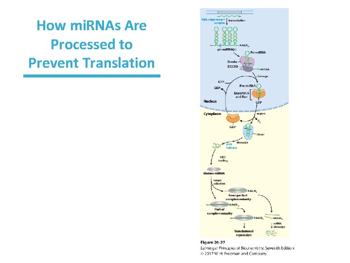
How mi. RNAs Are Processed to Prevent Translation
Ribozymes Are RNA Molecules That Catalytically Cleave RNA • • Cleave themselves or another RNA 3 -D structure integral to function Inactive if denatured Follows Michaelis-Menton kinetics – – – saturable active site measureable KM competitively inhibited nucleophilic attack of sugar OH on phosphate (transesterification) followed by phosphodiester bond hydrolysis
Examples of Well-characterized Ribozymes – Self-splicing group I introns • Example: 26 S r. RNA precursor from protozoan Tetrahymena – Hammerhead ribozyme • cleaves RNA of virusoids (circular RNAs that are replicated by plant viruses) • so named because 2 structure looks like head of hammer – RNase P • cleaves precursors to t. RNAs by recognizing the shape of the pre-t. RNA and the CCA sequence
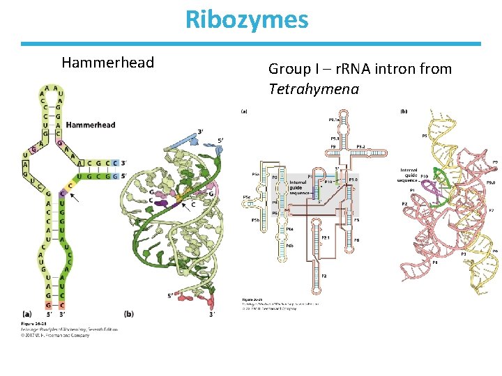
Ribozymes Hammerhead Group I – r. RNA intron from Tetrahymena
Degradation of Cellular m. RNAs • The RNA lifetime is one means of gene regulation. – Each gene product is needed for a unique amount of time. • Half-lives vary from seconds to hours. – – – rate of synthesis : rate of degradation steady state – rates are equal and balanced typical vertebrate m. RNA ~3 hrs ~10 turnovers per cell generation shorter (~1. 5 mins) half-lives for bacterial m. RNAs • Degradation occurs via ribonucleases. • Hairpin structures in bacterial m. RNA can extend half-life. • In eukaryotes, the 5’cap and 3’ poly (A) tail aid in the stability of the m. RNA.
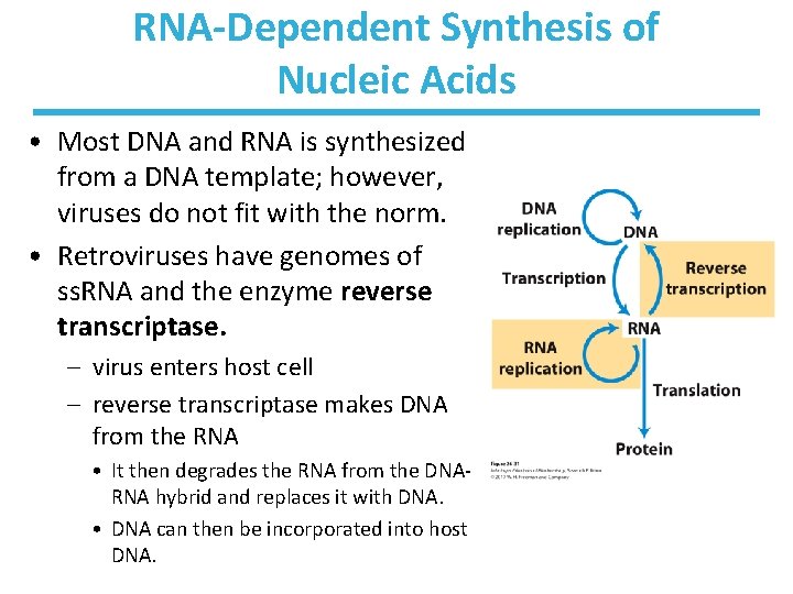
RNA-Dependent Synthesis of Nucleic Acids • Most DNA and RNA is synthesized from a DNA template; however, viruses do not fit with the norm. • Retroviruses have genomes of ss. RNA and the enzyme reverse transcriptase. – virus enters host cell – reverse transcriptase makes DNA from the RNA • It then degrades the RNA from the DNARNA hybrid and replaces it with DNA. • DNA can then be incorporated into host DNA.
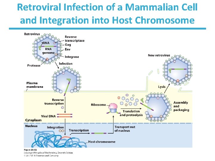
Retroviral Infection of a Mammalian Cell and Integration into Host Chromosome
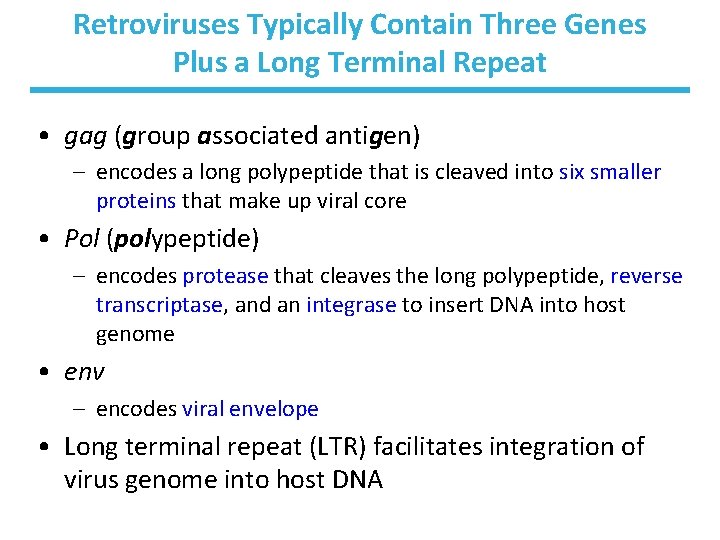
Retroviruses Typically Contain Three Genes Plus a Long Terminal Repeat • gag (group associated antigen) – encodes a long polypeptide that is cleaved into six smaller proteins that make up viral core • Pol (polypeptide) – encodes protease that cleaves the long polypeptide, reverse transcriptase, and an integrase to insert DNA into host genome • env – encodes viral envelope • Long terminal repeat (LTR) facilitates integration of virus genome into host DNA
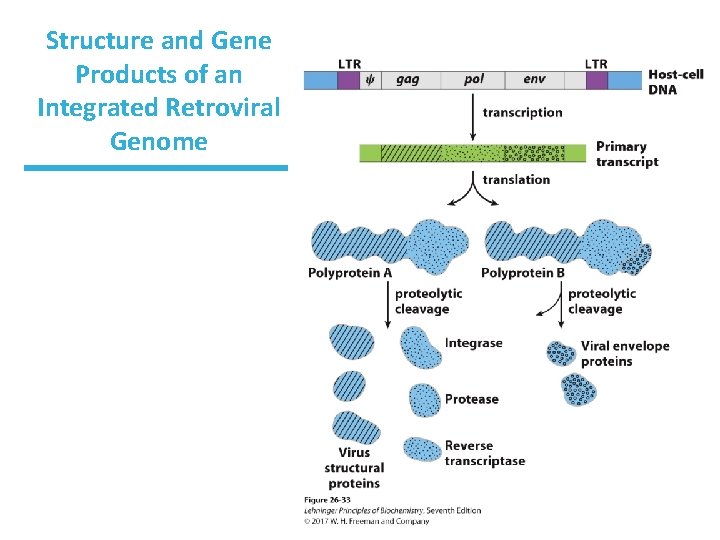
Structure and Gene Products of an Integrated Retroviral Genome

Reverse Transcriptases Catalyze Three Reactions 1. RNA-dependent DNA synthesis 2. RNA degradation 3. DNA-dependent DNA synthesis • contain Zn 2+, like DNA Pol • use a primer of t. RNA • lack 3’ 5’-proofreading, like RNA Pol - make reverse transcriptase error-prone - explains high rate of virus mutation/evolution (1: 20, 000 nt)
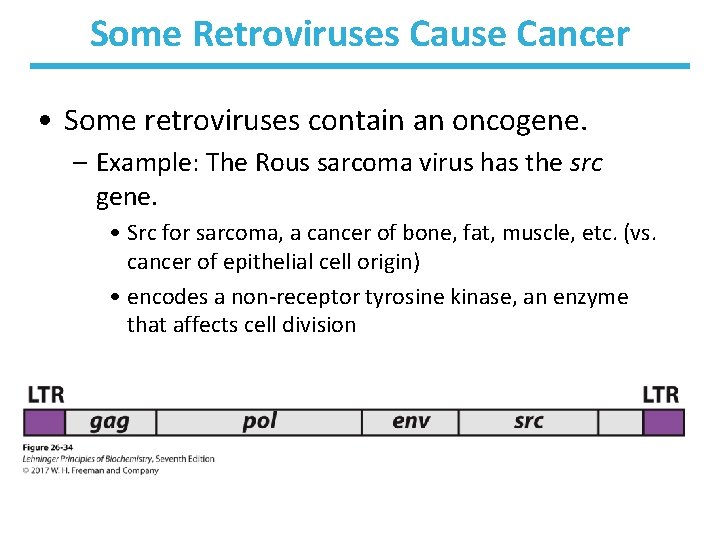
Some Retroviruses Cause Cancer • Some retroviruses contain an oncogene. – Example: The Rous sarcoma virus has the src gene. • Src for sarcoma, a cancer of bone, fat, muscle, etc. (vs. cancer of epithelial cell origin) • encodes a non-receptor tyrosine kinase, an enzyme that affects cell division
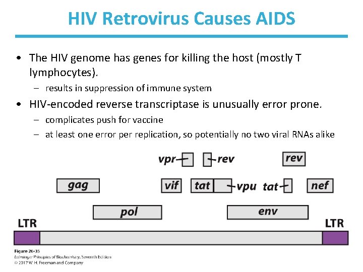
HIV Retrovirus Causes AIDS • The HIV genome has genes for killing the host (mostly T lymphocytes). – results in suppression of immune system • HIV-encoded reverse transcriptase is unusually error prone. – complicates push for vaccine – at least one error per replication, so potentially no two viral RNAs alike
Pharmaceutical Targets for HIV (Antiretroviral Drugs) • Reverse transcriptase inhibitors – nucleotide or nucleoside analogs – drug names ending in “dine” or “sine”: • zidovudine (AZT), didanosine (Videx), and so on • Protease inhibitors – since proteases are used in cleaving proteins for packaging into new viral particles – drug names ending in “avir”: • indinavir, saquinavir, and so on
Retrotransposons in Eukaryotes Have Similarities to Retroviruses • Retrotransposons are mobile genetic elements in eukaryotes. – encode an enzyme with homology to reverse transcriptase of retroviruses – move between positions via RNA intermediates • using their enzyme to make DNA from RNA – unlike bacterial transposons that move directly from DNA to DNA
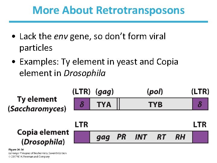
More About Retrotransposons • Lack the env gene, so don’t form viral particles • Examples: Ty element in yeast and Copia element in Drosophila
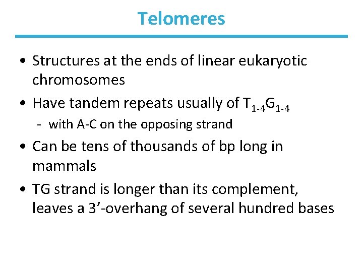
Telomeres • Structures at the ends of linear eukaryotic chromosomes • Have tandem repeats usually of T 1 -4 G 1 -4 - with A-C on the opposing strand • Can be tens of thousands of bp long in mammals • TG strand is longer than its complement, leaves a 3’-overhang of several hundred bases
Telomerase Extends the Ends of Chromosomes • Telomeres are not easily replicated using DNA polymerases. – Beyond an end, there is no template for an RNA primer. – Chromosomes are shortened with each generation. • Telomerase adds telomeric sequences to solve this problem.
The Mechanism of Telomerase • Telomerase has RNA with Cy. Ax repeat to serve as template for synthesis of the Tx. Gy strand of the telomere. • Telomerase binds to the 3’-end of the chromosome and hangs off so that the RNA template extends beyond it. • Telomerase extends the 3’-end, using the RNA of the enzyme as the primer. • The gap on the bottom strand is filled in by DNA polymerases.
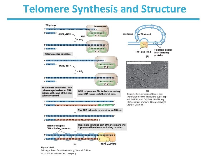
Telomere Synthesis and Structure
Support for the RNA World Hypothesis 1. Synthesis of adenine from cyanide is simple; most enzyme cofactors contain adenosine and it is the most abundant nucleotide constituent. 2. Catalytic RNAs exist and there might have been a self-replicating RNA. 3. Ribozymes can catalyze ester and amide bond formations, SN 2 reactions, metalation of porphyrins, and C-C bond formation. 4. Peptide bond formation requires RNA molecules. 5. Many nonliving entities (RNA viruses, retoviruses, retrotransposons) exist and can parasitize living cells. 6. Is there a ribozyme capable of directing its own synthesis?
Chapter 26: Summary In this chapter, we learned that: • RNA polymerase synthesizes RNA using a strand of DNA as a template and nucleoside triphosphates as substrates • the primary RNA transcript in eukaryotes requires processing before it becomes messenger RNA • the processing involves capping the 5’ end with methylguanosine to stabilize the RNA molecule • the processing involves splicing out introns • some introns have an amazing ability to carry out their own splicing
- Slides: 76