22 1 Text Only in Females slide Only


 1. How many sperms in the")


 Second trimester (second 3 months) Third trimester")
 A. Human chorionic gonadotropin (h.")
 � Increase the most in the 1")
 2. Estrogen (only in females’ slides)")
")


- Slides: 36
ﺍﻟﺒﺎﻗﻲ ، ﺳﻼﻳﺪ ﻓﻘﻂ 22 ﺍﻟﻤﺤﺎﺿﺮﺓ ﺳﻬﻠﺔ ﻭ ﺍﻛﺴﺘﺮﺍ § § § § 1 Text Only in Females’ slide Only in Males’ slides Important Numbers Doctor notes Extra Notes All numbers in this lecture are Important
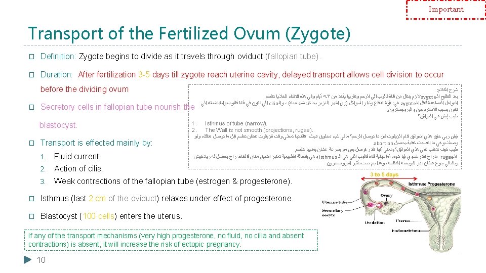
Physiology of Pregnancy By the end of this lecture, students should be able to describe: 1. Describe Fertilization & implantation of the blastocyst into the endometrium. 2. Recognize the development and normal physiology of the placenta. 3. Describe the physiological functions of placental hormones during pregnancy. 4. Explain the physiological responses of mother’s body to pregnancy. All numbers in this lecture are Important ﺍﻟﺒﺎﻗﻲ ، ﺳﻼﻳﺪ ﻓﻘﻂ 22 ﺍﻟﻤﺤﺎﺿﺮﺓ ﺳﻬﻠﺔ ﻭ ﺍﻛﺴﺘﺮﺍ 2
Only in Females’ Slides Revision (Large Group Activity) 1. How many sperms in the ejaculated semen? ü Range of sperms depends on semen. ü Average semen volume is 3 -5 ml and the average of sperms is 100 million. So, if the semen was 5 ml X 100 million = half a billion of sperms. 2. In which stage the ova is after ovulation? 4. Can the ova released from the right ovary reaches the left fallopian tube? The ova can enter the opposite fallopian tube 5. What are the factors that help the ovulated ova to reach the fallopian tube? 1. Fallopian tube contraction. 2. Fimbrial. Metaphase of the 2 nd meiotic division. 3. cilia with fluid 3. What is the parentage of ovulated ova that can reach fallopian tube? 6. What are the factors that help the sperm to travel in the female genital tract? 95 -98% of ova will reach the fallopian tube. Contractions of the uterus and fallopian tubes which stimulated by prostaglandins. 3
Maturation of The Ovum � While still in the ovary, the ovum is in the primary oocyte stage. Shortly before it is released from the ovarian follicle, its nucleus divides by meiosis and a first polar body is expelled from the nucleus of the oocyte. � The primary oocyte then becomes the secondary oocyte. In this process, each of the 23 pairs of chromosomes loses one of its partners, which becomes incorporated in a polar body that is expelled. � This leaves 23 unpaired chromosomes in the secondary oocyte. It is at this time the ovum, which is still in the secondary oocyte stage, is ovulated into the abdominal cavity. Then, almost immediately it enters the fimbriated end of one of the fallopian tubes fertilization. 4
Layers of The Ovum � What are the layers that the sperms needs to reach the ovum? (from outer to inner) 1. Corona radiata. (Before ovulation it is called granulosa cells After ovulation it names becomes (corona radiate) We have ZP 3 receptor on zona pellucida and corona radiata only). 2. Zona pilluceda. 3. Cell membrane. 4. Cortical granules. 5
Fertilization � If the ovum becomes fertilized, a new sequence of events called gestation or pregnancy takes place, and the fertilized ovum eventually develops into a full-term fetus. � during intercourse, half billion sperms are made but only thousands will enter the female reproductive tract (that’s why we need a lot of sperms) 6
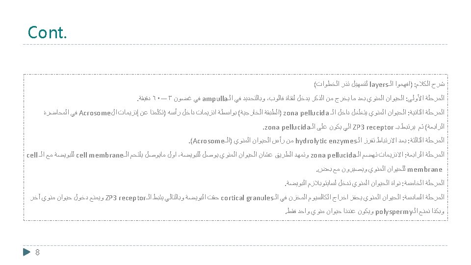
Important Process Fertilization Step 1: After the male ejaculate the semen into the vagina during intercourse, the sperm reach the ampulla of fallopian tube within 30 - 60 minutes (PG & OT actions) Q: what leads to the block of polyspermy? Inhibition of ZP 3 receptor by stimulation of Ca release stored in cortical granules. Step 2: The fertilizing sperm penetrates the corona radiata via membrane-bound enzymes in the plasma membrane of its head and binds to ZP 3 receptors on the zona Step 3: binding of sperm to these receptors triggers the acrosome reaction, in which hydrolytic enzymes in the acrosome are released onto the zona pellucida. Step 4: The acrosomal enzymes Step 6: The sperm stimulates digest the zona pellucida, creating a release of Ca+2 stored in cortical pathway to the plasma membrane of granules in the ovum, which in turn, inactivate ZP 3 receptors, leading to Step 5: The sperm nucleus enters the ovum cytoplasm. the block to polyspermy. When the sperm reaches the ovum, the plasma membrane of the two cells ﺍﻟﺠﺎﻱ ﺑﺎﻟﺴﻼﻳﺪ ﺍﻟﻜﻼﻡ ﺷﺮﺡ 7 the ovum. fuse.
Results of Fertilization 1. Oocyte divides to form mature ovum (female pronucleus 23 unpaired chromosomes) + 2 nd polar body. 2. Head of sperm swells (male pronucleus 23 unpaired chromosomes). 3. Fertilized ovum (zygote) contain 23 paired chromosomes. � The 23 chromosomes of the male and female pronuclei align themselves to re-form a complete complement of 46 chromosomes. � Male sperm either x or y arrive to fertilize the ovum; thus it’s the one determining the embryo’s sex. � . Theory: if a couple wants to have a boy they must do the intercourse in the day of the ovulation cause the y sperm is more faster and if they want a girl do the intercourse 2 days before the ovulation so the y sperm will die by then (sperm could stay in the female maximum 3 days ) and the x sperm could penetrate the ovum. : ﺍﻟﻜﻼﻡ ﺷﺮﺡ )ﻭ (unpaired ﺃﻨﻬﻢ )ﺭﻛﺰﻭﺍ 23 unpaired chromosomes ﻋﻠﻰ ﻳﺤﺘﻮﻱ ﻣﻨﻬﻢ ﻭﺍﺣﺪ ﻛﻞ ﻳﻜﻮﻥ ﺍﻟﻤﻨﻮﻱ ﻭﺍﻟﺤﻴﻮﺍﻥ ﺍﻟﺒﻮﻳﻀﺔ : ﺍﻟﺘﻠﻘﻴﺢ ﻗﺒﻞ . ( ﻟﻠﺒﻮﻳﻀﺔ ﺑﺎﻟﻨﺴﺒﺔ 2 nd polar body. ﻛﺮﻭﻣﻮﺳﻮﻡ ٤٦ = ﺍﻟﻜﺮﻭﻣﻮﺳﻮﻣﺎﺕ ﻣﻦ ﺯﻭﺝ ٢٣ ﻋﻦ ﻋﺒﺎﺭﺓ ﻭﻫﻮ zygote ﺍﻟـ ﻋﻨﺪﻱ ﻳﺘﻜﻮﻥ : ﺍﻟﺘﻠﻘﻴﺢ ﺑﻌﺪ 9
Entry of The Ovum Into The Uterus At day 19 -20 of ovulatory cycle, transportation is done and the zygote entered the uterus. � When ovulation occurs, the ovum, along with a hundred or more attached granulosa cells that constitute the corona radiata, is expelled directly into the peritoneal cavity and must then enter one of the fallopian tubes (also called uterine tubes) to reach the cavity of the uterus. � The fimbriated ends of each fallopian tube fall naturally around the ovaries. The inner surfaces of the fimbriated tentacles are lined with ciliated epithelium, and the cilia are activated by estrogen from the ovaries, which causes the cilia to beat toward the opening or ostium of the involved fallopian tube. One can actually see a slow fluid current flowing toward the ostium. By this means, the ovum enters one of the fallopian tubes. � Although one might suspect that many ova fail to enter the fallopian tubes, conception studies suggest that up to 98 percent of ova succeed in this task. Indeed, in some recorded cases, women with one ovary removed and the opposite fallopian tube removed have had several children with relative ease of conception, thus demonstrating that ova can even enter the opposite fallopian tube. 11
Cleavage � Doctor said: we wont ask about the name of the cells. Remember it just in case. . Following fertilization (1 st thing happening after fertilization) the zygote undergoes several mitotic divisions inside the zona pellucida (overall size does not change). � 1 st cleavage yields a 2 celled embryo, each cell is called a blastomere and is totipotent 1. � Divisions continue rapidly until the 32 cell stage AKA Morula. � Blastocyst (100 cells) enters the uterus. (the zygote will not enter the uterus unless it become Blastocyst which mean 100 cells) : ﺍﻟﻜﻼﻡ ﺷﺮﺡ . ﺍﻹﻧﻘﺴﺎﻡ ﻫﺬﺍ ﻓﻲ ﻳﺰﻳﺪ ﻣﺎ ﺍﻟﺤﺠﻢ ﻟﻜﻦ zona pellucida ﺍﻟـ ﺩﺍﺧﻞ ﺍﻟﻤﺎﻳﺘﻮﺯﻱ ﺍﻻﻧﻘﺴﺎﻡ ﻋﻤﻠﻴﺔ ﺗﺒﺪﺍ ﺍﻟﺘﻠﻘﻴﺢ ﺑﻌﺪ . ﻣﺴﺘﻘﺒﻼ ﺍﻟﺨﻼﻳﺎ ﻣﻦ ﻧﻮﻉ ﺃﻲ ﻳﻌﻄﻴﻨﺎ ﻣﻤﻜﻦ ﺍﻟﻨﻮﻉ ﻭﻫﺬﺍ blstomere ﺍﺳﻤﻬﻢ ﺧﻠﻴﺘﻴﻦ ﻳﻌﻄﻴﻨﺎ ﺍﻧﻘﺴﺎﻡ ﺍﻭﻝ . ﺧﻠﻴﺔ ٣٢ ﻋﻨﺪﻧﺎ ﻳﺼﻴﺮ ﻣﺎ ﺍﻟﻰ ﻣﺘﻰ؟ ﺍﻟﻴﻦ ﺗﻜﻤﻞ ﺭﺍﺡ ﺍﻻﻧﻘﺴﺎﻣﺎﺕ ( )ﻣﻬﻤﻪ blastocyst ﺍﺳﻤﻪ ﻭﻳﺼﻴﺮ ﺧﻠﻴﺔ ١٠٠ ﺍﻟـ ﻭﺻﻞ ﻟﻮ ﺍﻻ ﺍﻟﺮﺣﻢ ﻣﺎﻳﺪﺧﻞ zygote ﺍﻟـ 12 1: Totipotent embryo cells can differentiate into any cell type.
Important Implantation � The blastocyst will plant itself by sending projections from chorionic cells into the endometrium (eating Decidua cells which are filled with glycogen & nutrient). When the implantation occur? Implantation occurs on 5 -7 day after ovulation (day 21). � Steps of implantation: Implantation results from the action of trophoblastic cords from the surface of blastocyst. 1. Digestion of endometrium. 2. Decidua cells 1 : (glycogen, proteins, lipids & minerals) Both estrogen and progesterone are important at this stage: 1. Estrogen for proliferation of the endometrium. 2. Progesterone for the secretory phase (uterine milk) With increasing of the size of the endometrium due to estrogen and progesterone (secretions) it is called (Decidua; endometrium during pregnancy only). : ﺍﻟﻜﻼﻡ ﺷﺮﺡ . implantation ﺍﻟـ ﻋﻤﻠﻴﺔ ﻓﻲ ﺗﺴﺎﻫﻢ ﺍﻟﻲ ﻫﻲ ﻭﻫﺬﻱ trophoblast ﺍﺳﻤﻬﺎ ﺍﻟﺮﺣﻢ ﺩﺧﻠﺖ ﺍﻟﻠﻲ blastocyst ﺍﻟـ ﻣﻦ ﺍﻟﺨﺎﺭﺟﻴﺔ ﺍﻟﻄﺒﻘﺔ . ﻟﻸﻢ ﺍﻟﺘﺎﺑﻌﺔ ﺍﻟﻤﺸﻴﻤﺔ ﻫﻲ ﺍﻟﻲ decidua cells ﺍﻟـ ﻭ endomerium ﺍﻟـ ﻃﺒﻘﺔ ﻫﻀﻢ ﺍﻟﻮﻗﺖ ﻧﻔﺲ ﻭﻓﻲ ﻧﻔﺴﻬﺎ ﻏﺮﺱ ﻓﻲ ﺗﺒﺪﺃ ﺑﺄﻨﻬﺎ ؟ ﺗﺴﺎﻫﻢ ﻛﻴﻒ 13 1: The decidua is the uterine lining (endometrium) during a pregnancy, which forms the maternal part of the placenta.
Embryological Development of Placenta 1. Blastocyst Trophoblastic cords attaching to the uterus (Maternal blood sinuses develop around the trophoblastic cords) blood capillaries grow into the cords from the vascular system of the newly forming embryo. 21 days after fertilization, blood starts to be pumped by fetal heart into the capillaries. 3. When implantation occur Blood sinuses appear supplied with blood from the mother develop around the outsides of the trophoblastic cords. 4. More and more trophoblast projections develop (placental villi). : ﺍﻟﻜﻼﻡ ﺷﺮﺡ . ﺍﻷﺨﺮﻯ ﺍﻟﺠﻬﺔ ﻣﻦ ﺗﻨﻤﻮ ﻟﻠﺠﻨﻴﻦ ﺍﻟﺪﻣﻮﻳﺔ ﺍﻻﻭﻋﻴﺔ ﻭﻛﻤﺎﻥ trophoblastic cord ﺍﻟـ ﺣﻮﻟﻴﻦ ﺗﺘﻜﻮﻥ ﺗﺒﺪﺃ ﺍﻟﺪﻣﻮﻳﺔ ﺍﻻﻭﻋﻴﺔ ،implantation ﺍﻟـ ﺑﻌﺪ . ﺍﻷﻮﻋﻴﺔ ﺩﺍﺧﻞ ﺍﻟﺪﻡ ﺑﻀﺦ ﺍﻟﺠﻨﻴﻦ ﻗﻠﺐ ﻳﺒﺪﺃ ﻳﻮﻡ ٢١ ﺑﻌﺪ . ﺍﻟﺠﻨﻴﻦ ﺍﻟﻰ ﺍﻷﻢ ﻣﻦ nutrients ﺍﻟـ ﺗﺒﺎﺩﻝ ﻋﻠﻰ ﻳﺴﺎﻋﺪ ﻣﻤﺎ surface area ﺍﻟـ ﺗﺰﻳﺪ ﻭﺑﺎﻟﺘﺎﻟﻲ ﺗﺰﻳﺪ (placental villi ﺍﻟـ ﺗﻤﺜﻞ )ﺍﻟﻲ trophoblasts ﺍﻟـ ﻧﺘﻮﺀﺍﺕ 14
Physiologic Anatomy of The Placenta The area with concentrated blood that’s why it is important to prevent this area for rupturing during labor 15 � The final structure of the placenta is shown in picture. � Note that the fetus’s blood flows through two umbilical arteries, then into the capillaries of the villi, and finally back through a single umbilical vein into the fetus. (the umbilical cord consists of two arteries and one vein). � At the same time, the mother’s blood flows from her uterine arteries into large maternal sinuses that surround the villi and then back into the uterine veins of the mother. � the picture also shows the relationship between the fetal blood of each fetal placental villus and the blood of the mother surrounding the outsides of the villus in the fully developed placenta. � The total surface area of all the villi of the mature placenta is only a few square meters - many times less than the area of the pulmonary membrane in the lungs. � Nevertheless, nutrients and other substances pass through this placental membrane mainly by diffusion in much the same manner that diffusion occurs through the alveolar membranes of the lungs and the capillary membranes elsewhere in the body.
Functions of The Placenta 1. Respiration � 2. Nutrition 3. Excretion The first three functions are the major functions of the placenta. � We will discuss each one of these functions in next slides. 16 4. Endocrine 5. Protction
Only in Males’ Slides Respiration ﻟﻠﺠﻨﻴﻦ ﺍﻷﻢ ﻣﻦ ﺍﻷﻮﻛﺴﺠﻴﻦ ﺍﻧﺘﻘﺎﻝ ﺑﻬﺎ ﻳﻘﺼﺪ Placental permeability and membrane diffusion conductance In the early months of pregnancy ü In the early months of pregnancy, the placental membrane is still thick because it is not fully developed. ü The surface area is small because the placenta has not In later pregnancy, The permeability increases because of thinning of the membrane diffusion layers and because the surface area expands many times over. grown. ü So, how does the fetus get the nutrient ? Through the uterine milk (remember: in secretory phase progesterone secrete substances in the endometrium which is called : ﺍﻟﻜﻼﻡ ﺷﺮﺡ UTERINE MILK) ﺻﻐﻴﺮﺓ ﺗﻜﻮﻥ surface area ﻭﺍﻟـ ﺳﻤﻴﻚ ﻳﻜﻮﻥ placental membrane ﺍﻟـ ﺑﺎﻟﺘﺎﻟﻲ ، ﺗﻤﺎﻣﺎ ﺍﻟﻨﻤﻮ ﺍﻛﺘﻤﻠﺖ ﻣﺎ ﻟﺴﺎ ﺗﻜﻮﻥ placenta ﺍﻟـ ﺍﻷﻮﻟﻰ ﺍﻟﺤﻤﻞ ﺷﻬﻮﺭ ﻓﻲ ﺍﻷﻢ ﺑﻴﻦ ﻛﺒﻴﺮ ﺑﺸﻜﻞ ﺍﻷﻮﻛﺴﺠﻴﻦ ﺗﺒﺎﺩﻝ ﻳﻌﻴﻖ ﺭﺍﺡ ﻭﻫﺬﺍ ( ﻧﺨﺘﺮﻗﻬﺎ ﺑﺴﻬﻮﻟﺔ ﺭﻗﻴﻘﺔ ﻛﺎﻧﺖ ﻟﻮ ﺑﻴﻨﻤﺎ ﺍﺧﺘﺮﺍﻗﻬﺎ ﺻﻌﺐ ﺑﺎﻟﺘﺎﻟﻲ ﺳﻤﻴﻜﺔ ﻃﺒﻘﺔ ﻳﻜﻮﻥ ﺷﻴﺀ ﺃﻦ )ﺗﺨﻴﻠﻮﺍ . ﻭﺍﻟﺠﻨﻴﻦ . ﺃﻜﺒﺮ ﻭﺑﻜﻤﻴﺎﺕ ﺃﺴﻬﻞ ﺗﻜﻮﻥ ﺍﻟﺘﺒﺎﺩﻝ ﻋﻤﻠﻴﺔ ﺑﺎﻟﺘﺎﻟﻲ ﻭﺗﺘﻤﺪﺩ ﺗﻮﺳﻊ surface area ﻭﺍﻟـ thin ﻳﺼﻴﺮ membrane ﺍﻟـ ، ﺍﻷﻮﻟﻰ ﺍﻟﺤﻤﻞ ﺷﻬﻮﺭ ﺑﻌﺪ 17
Important Cont. Diffusion of oxygen through the placental membrane: � The mean partial pressure of oxygen (PO 2) of the mother’s blood in the placental sinuses is about 50 mm Hg, and the mean PO 2 in the fetal blood after it becomes oxygenated in the placenta is about 30 mm Hg. � Dissolved O 2 in mother’s blood passes to fetal blood by simple diffusion. � PCO 2 2 -3 mm Hg higher in fetal than maternal blood. � Mean pressure gradient= 50 mm hg – 30 mm hg =20 mm hg. � There are 3 reasons why this low PO 2 is sufficient to deliver O 2 to the fetal tissues: : ﻭﺍﻟﺠﺎﻳﺔ ﻫﺬﻱ ﻟﻠﺴﻼﻳﺪ ﺍﻟﻜﻼﻡ ﺷﺮﺡ ﻋﻨﺪ ﻓﻤﺎ ، ﺍﻟﻐﺎﺯ ﻫﺬﺍ ﺗﺘﺨﻠﺺ ﺭﺋﺔ ﻋﻨﺪﻩ ﻣﺎ ﻷﻦ ﻋﺎﻟﻲ ﻳﻜﻮﻥ ﻟﻠﺠﻨﻴﻦ co 2 ﺍﻟـ . ﺍﻷﻢ ﺇﻻ ﺍﻟﻐﺎﺯ ﻫﺬﺍ ﻣﻦ ﻟﻠﺘﺨﻠﺺ ﻃﺮﻳﻘﺔ ﺍﻟﺠﻨﻴﻦ ﺍﻷﻢ؟ ﻃﺮﻳﻖ ﻋﻦ ﻣﻨﻪ ﺍﻟﺘﺨﻠﺺ ﻳﺘﻢ ﺭﺍﺡ ﻛﻴﻒ ﻣﻦ ﺍﻟﻐﺎﺯ ﻓﻴﻨﺘﻘﻞ concentration gradient ﻳﺴﻮﻱ ﺭﺍﺡ Co 2 ﺍﻟـ ﺍﺭﺗﻔﺎﻉ ﻣﻨﻪ ﺍﻟﺘﺨﻠﺺ ﻭﻳﺘﻢ ﺍﻷﻢ ﻫﻲ ﺍﻟﻠﻲ ﺍﻟﻤﻨﺨﻔﺾ ﺇﻟﻰ ﺍﻟﺠﻨﻴﻦ ﻫﻮ ﺍﻟﻠﻲ ﺍﻟﻌﺎﻟﻲ . (simple diffusion ) ﻛﻴﻒ؟ ﺍﻷﻮﻛﺴﺠﻴﻦ ﻃﻴﺐ diffusion ﻓﻴﺤﺼﻞ ، ﻭﺍﻟﺠﻨﻴﻦ ﺍﻷﻢ ﺑﻴﻦ Po 2 ﺍﻝ ﻓﻲ ﺍﺧﺘﻼﻑ ﻓﻴﻪ ﺍﻟﺸﻴﺀ ﻧﻔﺲ . ﻟﻸﻢ ﺍﻟﺠﻨﻴﻦ ﻣﻦ ﻳﺮﻭﺡ ﻛﺎﻥ Co 2 ﺍﻟـ ﺑﻌﻜﺲ ﻟﻠﺠﻨﻴﻦ ﺍﻷﻢ ﻣﻦ . ﺍﻟﻀﺎﺭ ﻣﻨﻪ ﻭﺗﺎﺧﺬ ﻣﻔﻴﺪ ﻣﺎﻫﻮ ﻛﻞ ﻟﻠﺠﻨﻴﻦ ﺗﻌﻄﻲ ﺍﻷﻢ : ﺑﺎﺧﺘﺼﺎﺭ ﻳﻌﻨﻲ Note that 20 mm Hg seems to be low and not enough for exchange sufficient oxygen to the fetal blood. BUT due to these 3 special reasons it is sufficient more than enough to deliver O 2 and nutrients to the fetus. 1. Hemoglobin of the fetus. 2. Fetal hemoglobin concentration. For each point we know that Hb F has more affinity to bind to 3. The Bohr effect. 18 oxygen than Hb A which is normal human Hb.
Doctor said : There must be a question in the final exam each year about this topic Cont. � 1, 2: Hemoglobin of the fetus & Fetal hemoglobin concentration � What is the type of Hb of the fetus? Hemoglobin F (Hb. F). � The fetal hemoglobin concentration is about 50% greater than that of the mother. So, At the low PO 2 levels in fetal blood (While the PCO 2 in the fetus will be high), the fetal hemoglobin (Hb. F) can carry 20% to 50% more oxygen than maternal hemoglobin (Hb. A) (Hb. F has a higher oxygen carrying capacity than Hb. A). � Follow with the curves: In Maternal curve: when pressure of o 2 is 20 the Oxyhemoglobin become almost 38 and in the same time the oxyhemoglobin of the fetus if 46. � Oxygen-hemoglobin dissociation curves 19 Imp: Conclusion ; whenever the curve is shifted to the right more o 2 will be carried (fetal). While whenever the curve is shifted to the left less o 2 will be carried (mother).
Important Cont. � 3: Bohr effect Recall The Bohr Effect refers to the observation that increases in the carbon dioxide partial pressure of blood or decreases in blood p. H result in a lower affinity of hemoglobin for oxygen. Fetal blood Maternal (mother) blood PCO 2 Lost CO 2 Gains CO 2 PH High p. H (alkaline) Low p. H (acidic) Shifts to the left allowing additional oxygen uptake. Shifts to the right releasing additional oxygen. Shifts of the dissociatio n curves These changes cause the capacity of fetal blood to combine with O 2 to increase, and maternal blood to decrease, which forces more O 2 from the maternal blood while enhancing oxygen uptake by the fetal blood. ü Normally before Exchange the fetal blood has high PCo 2 as a waste product due to his body activity, this will lead to make the PH in the fetal circulation to be more Acidic. ü Shown in the diagram: As a result of the above the CO 2 will diffuse from fetal circulation to the mother passively, this diffusion of Co 2 from the fetal to the mother will change the PH in both sides , when the Co 2 leave the fetal circulation it turns to be Alkaline because it lost (CO 2) which is an acid. 20
Cont. � Important factors facilitate delivery of oxygen to the fetal tissues: High maternal intervillous blood flow (almost double 1. the fetal placental flow). 2. High fetal haemoglobin (16 - 17 g/dl). 3. High fetal cardiac output. 4. The fetal metabolic acidosis which shifts the curve to the right and thus aids delivery of oxygen to the tissues. 21
Important Nutrition ﻟﻠﺠﻨﻴﻦ ﺍﻷﻢ ﻣﻦ ﺍﻟﻐﺬﺍﺀ ﺍﻧﺘﻘﺎﻝ ﺑﻬﺎ ﻳﻘﺼﺪ Nutrition Diffusion of foodstuffs through the placental membrane Glucose Fatty acids Amino acids Electrolyte's Fetus uses mainly glucose for nutrition so, the The placenta actively trophoblast cells in placental K+, Na+ and Cl- villi transport glucose by Fatty acids diffuses due transports all amino carrier molecules; GLUT to high solubility in cell acids with fetal diffuses from (facilitated diffusion). That’s membrane (more concentrations maternal to fetal why pregnant woman desire slowly than glucose). something sweat and if it’s not controlled it could lead to gestational diabetes. 22 exceeding maternal levels. blood.
Excretion � ﻟﻸﻢ ﺍﻟﺠﻨﻴﻦ ﻣﻦ ﺍﻹﺧﺮﺍﺝ ﺑﻬﺎ ﻳﻘﺼﺪ Excretory products of the fetus diffuse through placental membrane to maternal blood to be excreted with waste products of the mother (Urea, uric acid and creatinine). � Higher concentration of excretory products in fetal blood insures continuous diffusion of these substances to the maternal blood. 23
Endocrine In the trimester (first 3 months) Second trimester (second 3 months) Third trimester (Third 3 months) 24 A. The placenta is not yet formed. B. High level of h. CG secreted by the trophoblast of the embryo. C. Progesterone & estrogen are secreted from corpus luteum. A. The placenta is formed & the corpus luteum start to degenerate. B. The placenta will secrete most of h. GC, progesterone & estrogen with slight increase of estrogen and progesterone. C. Estrogen and progesterone are also secreted from the corpus leutum A. The corpus luteum is completely degenerated. B. High estrogen and progesterone with low h. CG, estrogen & Progesterone are only secreted by placenta.
Cont. In Other Word (same of the previous slide) A. Human chorionic gonadotropin (h. CG) Will increases until it reaches a maximum concentration near the end of the first trimester. Then it decrease to low level for the reminder of the pregnancy. ü Progesterone continue to increase until it level off near the end of the pregnancy. B. Progesterone ü Early in the pregnancy, progesterone is produced by the corpus luteum in thee ovary. ü By the second trimester, its production shifts to the placenta. ü Estrogen levels increase slowly throughout the pregnancy, but they increase more quickly as the end of the pregnancy approaches. C. Estrogen ü Early in the pregnancy estrogen is produced only in the ovary. ü by the second trimester, its production shifts to the placenta. 25
Cont. � h. CG level (pregnancy test) � Increase the most in the 1 st trimester then it will decrease, but still it is high comparing with normal women. � Blood h. CG under 5 m. IU/ml: Negative (Not pregnant) � Blood h. CG between 5 -25 m. IU/ml: "Equivocal“, Maybe pregnant maybe not Repeat test in a couple of days. � Blood h. CG over 25 m. IU/ml: You are pregnant! � One single h. CG reading is not enough to make a clear diagnosis. Several h. CG tests days apart give a more accurate assessment of the situation. � The blood h. CG levels should not be used to date a pregnancy since these numbers can vary so widely 26
Cont. Hormone 1. Human Chorionic Gonadotropin (h. CG) 2. Estrogen (only in females’ slides) Nature of the hormone Glycoprotein Steroid hormone Secreted by Syncytial trophoblast cells, then after the formation of Syncytial trophoblast cells towards end of the placenta, h. CG will be secreted from the placenta. pregnancy reaches 30× - Weak androgen (DHEA) released from Derived from maternal & fetal adrenals. 1. Most important function is to maintain corpus luteum (↑estrogen & progesterone) till 13 -17 weeks 1. Enlargement of uterus, breast ducts & external genitalia. of gestation. Functions in the mother 2. Relaxation of pelvic ligaments in 2. Another function is that it act as LH hormone which in preparation to labor. response, exerts interstitial (Leyding) cell-stimulating effect on testes of the male fetus (growth of male sex 27 organs). 3. Activation of the uterus (gap junctions).
Cont. Hormone 3. Progsterone 4. Human Chorionic Somatomamotropin or Human placental lactogen (h. PL) 5. Relaxin Nature of the Steroid hormone Protein hormone Polypeptide Syncytial trophoblast cells towards end of Placenta around 5 th gestational week hormone Secreted by pregnancy reaches 10× Derived from mother 28 2. Placenta Cholesterol - 1. Development of the breast lobules. 1. Breast development (h. PL). 2. Provides nutrition to developing 2. Weak growth hormone‘s action. embryo (uterine secretory phase. 3. Inhibit insulin sensitivity =↓ Functions in the 3. Development of decidual cells. 4. Inhibit the contractility of the uterus. 1. Corpus luteum glucose utilization. 4. Promote release of fatty acids. 1. Relaxation of symphysis pubic ligament (weak). 2. Softens the cervix at delivery.
Sources of Placental Estrogen & Progesterone 29 � Estrogen Production: 1. Cholesterol from maternal blood will diffuse through placenta to fetal blood then it will reach fetal adrenal cortex. 2. Fetal adrenal cortex will use the cholesterol to synthesis DHEA (Androgen). 3. DHEA will diffuse to the placenta and it will be converted to Estrogen. 4. Also maternal adrenal cortex will release DHEA which will be converted to Estrogen in placenta. 5. Finally, Estrogen will go back to maternal blood. � Progesterone Production: 1. Cholesterol from maternal blood will diffuse to placenta. 2. Placenta will use cholesterol to synthesis Progesterone. 3. Finally, Progesterone will diffuse back to maternal blood.
Physiological Adaptation To Pregnancy Changes in maternal endocrine system (Almost everything n the body increases during pregnancy) Anterior pituitary gland enlargement (50%) 1. Release of ACTH, TSH and PL increase. 2. FSH and LH almost totally suppressed due to increase of progesterone & estrogen. Adrenal gland 1. Increase glucocorticoids secretion (mobilize aa). 2. Increase aldosterone (retain fluid). Increase thyroxine production (h. CG) Thyroid gland enlargement 50% (Retain Na & water to increase maternal blood concentration even more blood cells will be produced from bone marrow. To prepare her for losing blood in labor) Parathyroid gland enlargement 30 Increase PTH secretion (maintain normal Ca+2)
Cont. � Changes in Different Organs: � Changes in respiration: 1. Increase in uterine size (50 gm to 1100 gm). 1. Increase in O 2 consumption 20% 2. The breasts double in size. • Increase basal metabolic rate (BMR 3. The vagina enlarges to facilitate labor. • Increase in body size 4. Development of edema and acne due to increase in 2. Growing uterus presses upwards (restriction) 3. Increase in RR (respiratory rate). 4. Increase in minute ventilation (TV* × RR) by 50%: androgen and water absorption. 5. 6. Masculine or acromegalic features Weight gain 10 -12 kg (last 2 trimesters) • 7. Increase appetite: • Removal of food by fetus. • Hormonal effect. 31 * TV= Tidal volume Progesterone ↑ sensitivity of RC to CO 2
Cont. � Changes in circulatory system: � Changes in metabolism: 1. Increase in Cardiac output (30 - 40%) by 27 weeks. 1. Increase basal metabolic rate (BMR) 15% 2. Increase in daily requirements for: 2. 3. Increase in blood flow through the placenta. Increase in maternal blood volume 30% due to: • Iron • Phosphates . ﺣﺍﻧﺔ ﺃﻨﻬﺎ ﺗﺤﺲ ﺍﻟﺤﺎﻣﻞ ﺩﺍﺋﻤ • Increase aldosterone and estrogen (↑ ECF ) • Calcium & Vitamin D (Ca+2 absorption) • Increase activity of the bone marrow (↑ RBCs 40%) • Vitamins 32 * GFR= Glomerular filtration rate. 3. The renal tubules’ reabsorptive capacity for Na, Cl, and water is increased as much as 50%. 4. The renal blood flow and GFR* increase up to 50%. 5. Normal pregnant woman accumulates only about 5 pounds (2. 27 Kg) of extra water and salt.
Summary Here the summaries are clear 33
Summary Here the summaries are clear 34
MCQ’s A. Zygote B. Morula C. Early Blastocyst D. Late Blastocyst 2. How many days does it take for the fertilized zygote to travel through the oviduct? A. 2 -4 B. 3 -5 C. 4 -5 D. 5 -6 3. DHEA is converted to Estrogen in …? A. Placenta. B. Maternal blood. C. Fetal blood. D. Fetal adrenal cortex. 35 4. Which of the following will inhibit the contractility of uterus? A. Estrogen. B. Oxytocin. C. Prolactin. D. Progesterone 5. Which ONE of the following is a function of RELAXIN ? A. Act upon uterine cervix B. Developmental function C. Promotes release of fatty acids D. Decrease glucose sensitivity 6. Which ONE of the following is secreted by placenta around 5 th gestational week ? A. Human Chorionic Gonadotropin B. Human Placental Lactogen C. Relaxin D. Gonadotropins 1=D 2=B 3=A 4=D 5=A 6=A 1. Which of the following stages is the one that is implanted into the endometrium of the uterus?