2013 Pearson Education Inc Bones and Skeletal Tissues

© 2013 Pearson Education, Inc.

Bones and Skeletal Tissues

© 2013 Pearson Education, Inc. Location of Bones and Cartilages

© 2013 Pearson Education, Inc. Characteristics of Cartilage • More Flexible than bone • Mostly water and connective tissue • No blood vessels or nerves- why is this significant? • Outer layer: Perichondrium (wraps around) Perichondrium contains blood vessels

© 2013 Pearson Education, Inc.

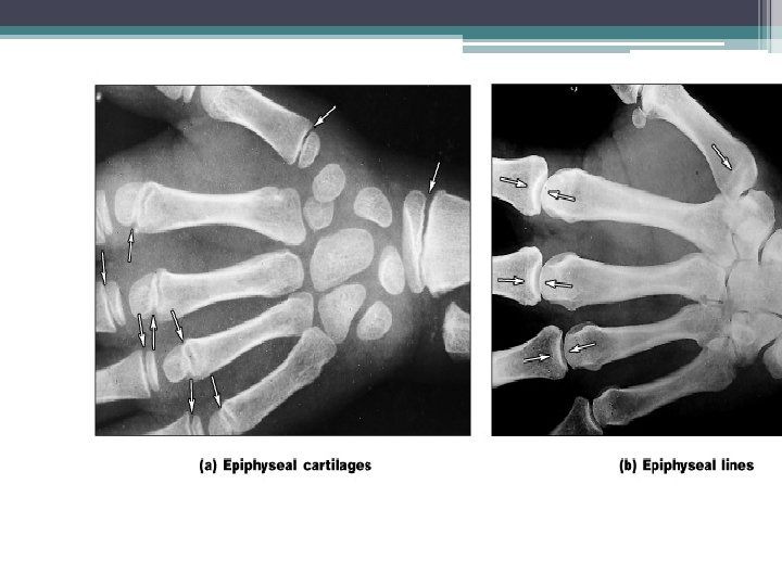
© 2013 Pearson Education, Inc. Hyaline Cartilage in Growth Plate The growth plates are where elongation of bones takes place (and the ONLY places where it can take place) because the cartilage can proliferate as fast as the bone formation behind it occurs, at least until maturity. Anatomically this is only one epiphysis of this bone; there's a second epiphysis at the other end. Human Growth Hormone? ?

© 2013 Pearson Education, Inc. Types of Skeletal Cartilages • Three types 1. Hyaline cartilage Provides support, flexibility, and resilience Collagen fibers only; most abundant type Articular skeleton, costal (ribs), respiratory, nasal cartilage 2. Elastic cartilage Similar to hyaline cartilage, but contains elastic fibers External ear and epiglottis 3. Fibrocartilage Thick collagen fibers—has great tensile strength Menisci of knee; vertebral discs

© 2013 Pearson Education, Inc. Important Point! • cartilage is not "turned into" bone, it is replaced by bone in a complicated process **hyaline cartilage forms the model for the bones in a developing embryo. Growth and reshaping occurs in two ways: interstitial growth and appositional growth.

© 2013 Pearson Education, Inc. Growth of Cartilage • Appositional growth: takes place "at the edge" of the cartilage mass • Interstitial growth takes “in the middle" of the mass of cartilage. • Calcification of cartilage- Depositing calcium ▫ Occurs during normal bone growth Youth and old age ▫ Hardens, but cacified cartilage is not bone!

© 2013 Pearson Education, Inc. Review Questions: pg 196 M. C 3, 4, 10, Short Answer: 15, 18,

Classification of Bones © 2013 Pearson Education, Inc. • 206 named bones in Adult skeleton • Divided into two groups: See fig. 7. 1 ▫ Axial skeleton and Appendicular skeleton

© 2013 Pearson Education, Inc. Classification of Bones by Shape • Long bones ▫ Longer than they are wide ▫ Limb, wrist, ankle bones • Short bones ▫ Cube-shaped bones (in wrist and ankle) ▫ Sesamoid bones (within tendons, e. g. , Patella) ▫ Vary in size and number in different individuals • Flat bones ▫ Thin, flat, slightly curved ▫ Sternum, scapulae, ribs, most skull bones • Irregular bones ▫ Complicated shapes ▫ Vertebrae, coxal bones

Figure 6. 2 Classification of bones on the basis of shape. © 2013 Pearson Education, Inc. Flat bone (sternum) Long bone (humerus) Irregular bone (vertebra), right lateral view Short bone (talus)

Functions of Bones 1. Support: Standing upright, sitting 2. Protection: soft organs 3. Movement: muscles attached 4. Storage: fat, calcium, phosphorus 5. Blood Cell Formation: hematopoiesis 6. Hormone production ▫ Osteocalcin Regulates bone formation

© 2013 Pearson Education, Inc. Bones • Are organs ▫ Contain different types of tissues Bone (osseous) tissue, nervous tissue, cartilage, fibrous connective tissue, muscle and epithelial cells in its blood vessels • Three levels of structure ▫ Gross anatomy ▫ Microscopic ▫ Chemical

© 2013 Pearson Education, Inc. Gross Anatomy: Bone Texture Compact and Spongy bone • Compact ▫ Dense outer layer; smooth and solid • Spongy: also called trabecular ▫ Less dense- open spaces

© 2013 Pearson Education, Inc. Structure of Short, Irregular, and Flat Bones • Thin plates of spongy bone covered by compact bone • Plates sandwiched between connective tissue membranes ▫ Periosteum (outer layer) and endosteum (inner layer) • No shaft or epiphyses • Bone marrow throughout spongy bone; no marrow cavity

© 2013 Pearson Education, Inc. Long Bones • Epiphyses ▫ Bone ends ▫ External compact bone; internal spongy bone ▫ Articular cartilage covers articular surfaces ▫ Between is epiphyseal line • Remnant of childhood bone growth at epiphyseal plate Diaphysis ▫ Tubular shaft forms long axis ▫ Compact bone surrounding medullary cavity

© 2013 Pearson Education, Inc. Bone Membranes: • Endosteum • Periosteum

© 2013 Pearson Education, Inc. Review Questions: # 5, 21, 22

© 2013 Pearson Education, Inc. Hematopoietic Tissue in Bones • Red marrow ▫ Found within trabecular cavities of spongy bone and in flat bones (e. g. , sternum) ▫ In medullary cavities and spongy bone of newborns ▫ Adult long bones have little red marrow Heads of femur and humerus only ▫ Yellow marrow can convert to red, if necessary
 © 2013 Pearson Education, Inc.")
Table 6. 1 Bone Markings (1 of 2) © 2013 Pearson Education, Inc.
 © 2013 Pearson Education, Inc.")
Table 6. 1 Bone Markings (2 of 2) © 2013 Pearson Education, Inc.

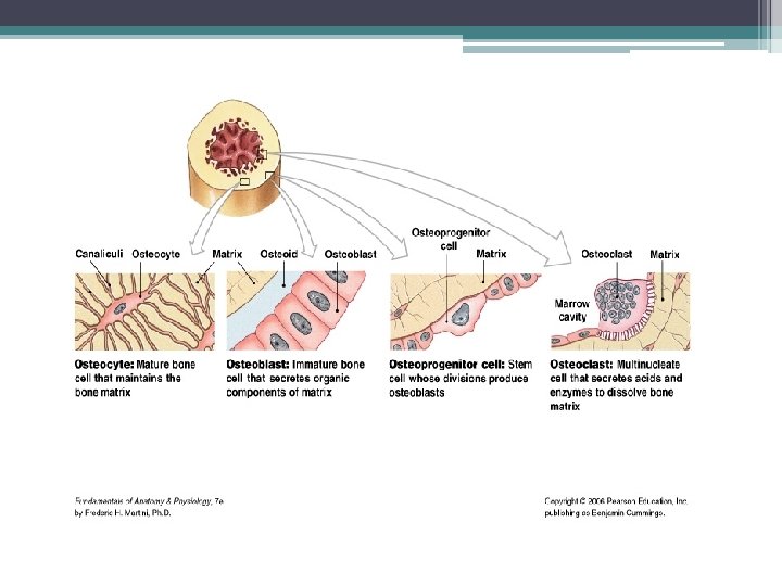
© 2013 Pearson Education, Inc. Cells of Bone Tissue

© 2013 Pearson Education, Inc. Anatomy of Compact Bone • Osteon or Haversian system ▫ Structural unit of compact bone ▫ Hollow tubes of bone matrix called lamellae Collagen fibers in adjacent rings run in different directions Withstands stress – resist twisting

Bone Tissue

Figure 6. 7 Microscopic anatomy of compact bone. Compact bone Spongy bone © 2013 Pearson Education, Inc. Central (Haversian) canal Perforating (Volkmann’s) canal Endosteum lining bony canals and covering trabeculae Osteon (Haversian system) Circumferential lamellae Lamellae Nerve Vein Artery Canaliculi Osteocyte in a lacuna Perforating (Sharpey’s) fibers Periosteal blood vessel Periosteum Lamellae Central canal Lacunae Interstitial Lacuna lamella (with osteocyte)

© 2013 Pearson Education, Inc. Chemical Composition of Bone: Inorganic Components • Hydroxyapatites (mineral salts) ▫ 65% of bone by mass ▫ Mainly of tiny calcium phosphate crystals in and around collagen fibers ▫ Responsible for hardness and resistance to compression

© 2013 Pearson Education, Inc. Bone Fun Facts • Half as strong as steel in resisting compression • As strong as steel in resisting tension • Last long after death because of mineral composition ▫ Reveal information about ancient people ▫ Can display growth arrest lines Horizontal lines on bones Proof of illness - when bones stop growing so nutrients can help fight disease

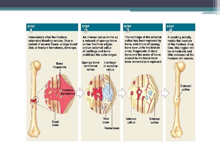
Bone Repair • • Micro-Fracture Osteoclasts digest damaged bone Osteblasts lay down new bone tissue Full repair can take up to 3 years

Osteoporosis “bone Thinning” • Women more at risk • Causes: lack of estrogen, diet, genetics, lack vitamin D

Bone Disorders • • fibrodysplasia ossificans progressiva, or FOP Osteoarthritis Rheumatoid Arthritis Scoliosis Slipped Disc Leukemia Scurvy Clef palate
- Slides: 35