
2010 ANATOMY PHYSIOLOGY C Karen Lancour National Bio
2010 ANATOMY & PHYSIOLOGY (C) Karen Lancour National Bio Rules Committee Chairman karenlancour@charter. net Patty Palmietto National Event Supervisor – A & P 2 -2 -2010
ANATOMY & PHYSIOLOGY Event Content: 2010 n n BASIC ANATOMY AND PHYSIOLOGY v Skeletal system v Muscular system v Endocrine system v Major disorders v Treatment and prevention of disorders PROCESS SKILLS - observations, inferences, predictions, calculations, data analysis, and conclusions.

Event Rules – 2010 n BE SURE TO CHECK THE 2010 EVENT RULES FOR EVENT PARAMETERS AND TOPICS FOR EACH COMPETITION LEVEL

TRAINING MATERIALS n n Training Handout – content Event Supervisor Guide – sample stations, process skills, methods Internet Resource – links to on-line courses, lab manuals, notes, sites Sample Tournament – sample stations with key
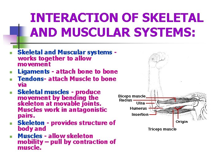
INTERACTION OF SKELETAL AND MUSCULAR SYSTEMS: n n n Skeletal and Muscular systems works together to allow movement Ligaments - attach bone to bone Tendons- attach Muscle to bone via Skeletal muscles - produce movement by bending the skeleton at movable joints. Muscles work in antagonistic pairs. Skeleton - provides structure of body and Muscles - allow skeleton mobility – pull by contraction of muscle.

Skeletal System Functions Support & shape to body n Protection of internal organs n Movement in union with muscles n Storage of minerals (calcium, phosphorus) & lipids n Blood cell production n
The Skeletal System Know the Skeletal Anatomy § Axial Skeleton § Appendicular Skeleton § Surface Anatomy of the bone § § By x-ray or diagram Structure/function of joints, muscle and ligament attachments § Including range of motion
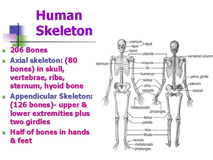
Human Skeleton n n 206 Bones Axial skeleton: (80 bones) in skull, vertebrae, ribs, sternum, hyoid bone Appendicular Skeleton: (126 bones)- upper & lower extremities plus two girdles Half of bones in hands & feet
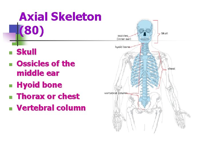
Axial Skeleton (80) n n n Skull Ossicles of the middle ear Hyoid bone Thorax or chest Vertebral column

Appendicular Skeleton (126) Upper Extremity (64) n Shoulder Girdle n Arms n Hands Lower Extremity (62) n Pelvic Girdle n Legs n Feet
Types of Bone n n n Long bones: longer than they are wide; shaft & 2 ends (e. g. : bones of arms & legs, except wrist, ankle & patella) Short bones: roughly cube-shaped (e. g. : ankle & wrist bones) Sesamoid bones: short bones within tendons (e. g. : patella) Flat bones: thin, flat & often curved (e. g. , : sternum, scapulae, ribs & most skullbones) Irregular bones: odd shapes; don't fit into other classes (e. g. : hip bones & vertebrae)
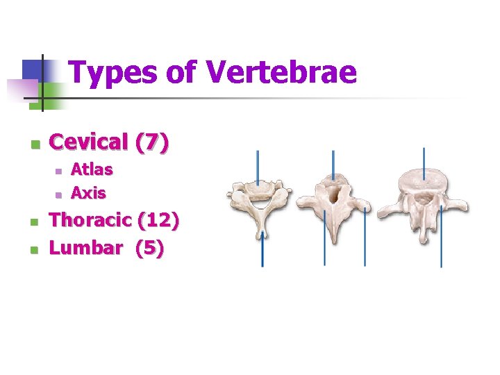
Types of Vertebrae n Cevical (7) n n Atlas Axis Thoracic (12) Lumbar (5)
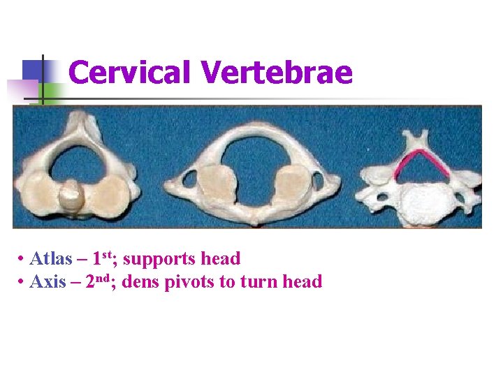
Cervical Vertebrae • Atlas – 1 st; supports head • Axis – 2 nd; dens pivots to turn head
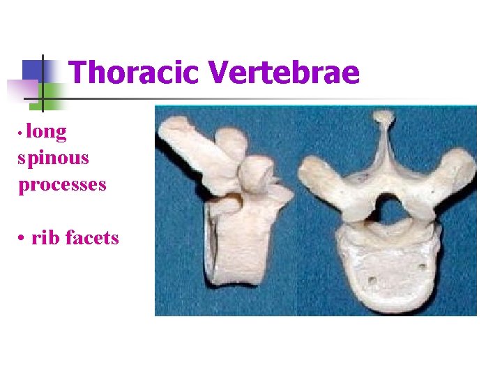
Thoracic Vertebrae • long spinous processes • rib facets
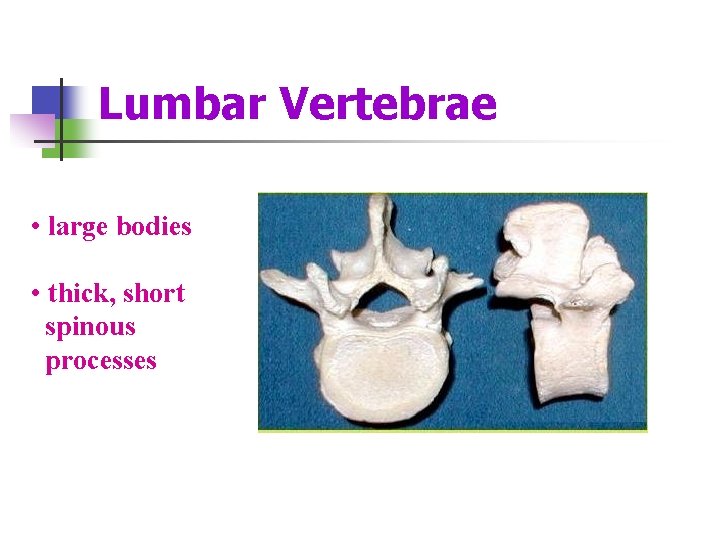
Lumbar Vertebrae • large bodies • thick, short spinous processes
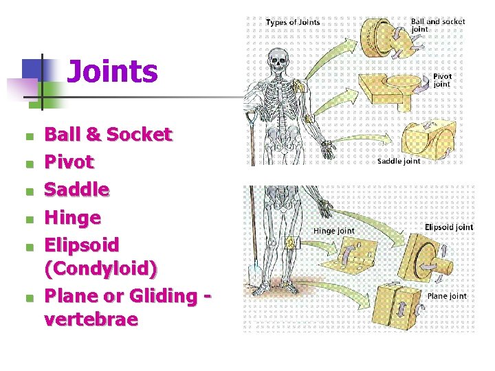
Joints n n n Ball & Socket Pivot Saddle Hinge Elipsoid (Condyloid) Plane or Gliding vertebrae
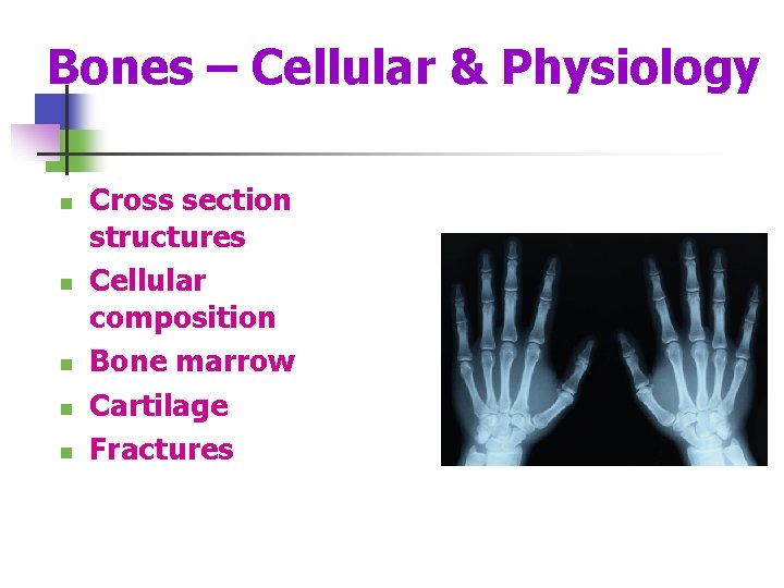
Bones – Cellular & Physiology n n n Cross section structures Cellular composition Bone marrow Cartilage Fractures

Bone Cells n n n Osteoblasts – bone forming cells synthesize and secrete unmineralized ground substance and are found in areas of high metabolism within the bone Osteocytes – mature bone cells made from osteoblasts that have made bone tissue around themselves. They maintain healthy bone tissue by secreting enzymes and controlling the bone mineral content; they also control the calcium release from the bone tissue to the blood. Osteogenic cells respond to traumas, such as fractures, by giving rise to bone-forming cells and bone-destroying cells Osteoclasts – bone absorbing cell – large cells that break down bone tissue – important to growth, healing, remodeling Bone lining cells - made from osteoblasts along the surface of most bones in an adult. Bone-lining cells are thought to regulate the movement of calcium and phosphate into and out of the bone
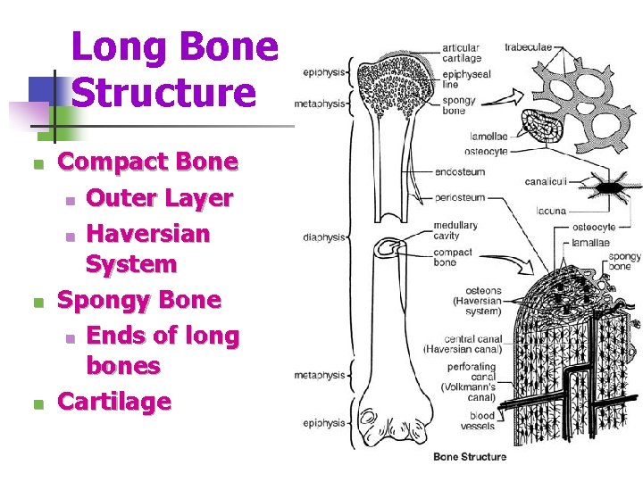
Long Bone Structure n n n Compact Bone n Outer Layer n Haversian System Spongy Bone n Ends of long bones Cartilage
Red and Yellow Bone Marrow n n n The formation of blood cells, (hematopoiesis), takes place mainly in the red marrow of the bones. In infants, red marrow is found in the bone cavities. With age, it is largely replaced by yellow marrow for fat storage. In adults, red marrow is limited to the spongy bone in the skull, ribs, sternum, clavicles, vertebrae and pelvis. Red marrow functions in the formation of red blood cells, white blood cells and blood platelets.
Cartilage – Characteristics Mostly water; no blood vessels or nerves n Tough, resilient n New cartilage forms from chondroblasts n Heal poorly n
Types of Skeletal Cartilage n n n Hyaline Cartilages: fine collagen fiber matrix- most abundant type- found in articular (movable joint) cartilages, costal cartilages (connect ribs tosternum), respiratory cartilages (in larynx & upper respiratory passageways) & nasal cartilages Elastic Cartilages: similar to hyaline cartilage, more elastic fibers (very flexible) – found in external ear & epiglottis (larynx covering) Fibrocartilage: rows of chondrocytes with thick collagen fibers; highly compressible with great tensile strength- found in menisci of knee, intervertebral discs & pubic symphysis
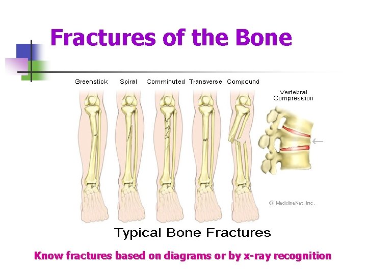
Fractures of the Bone Know fractures based on diagrams or by x-ray recognition

Bone Repair Sequence n n n Injury – broken blood vessels, hematoma Invasion of blood vessels & generalized cells (2 -3 days) Fibroblasts develop (1 week) Chondroblasts develop Callus forms (4 weeks) Remodeling with osteoclasts (8 weeks)
Disease/Injury Levels n n n Osteoarthritis Osteoporosis Fractures (via pictures and x-rays) Disc herniation Scoliosis ACL and MCL injuries
MUSCULAR SYSTEM Muscle Function: n Stabilizing joints n Maintaining posture n Producing movement n Moving substances within the body n Stabilizing body position and regulating organ volume n Producing heat– muscle contraction generates 85% of the body’s heat
Characteristics of Muscle Tissue n n Excitability- receive and respond to stimuli Contractility- ability to shorten and thicken Extensibility- ability to stretch Elasticity- ability to return to its original shape after contraction or extension
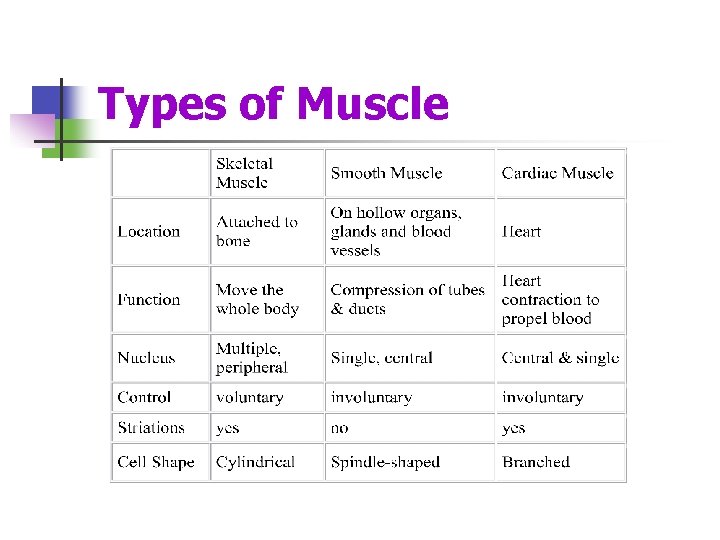
Types of Muscle
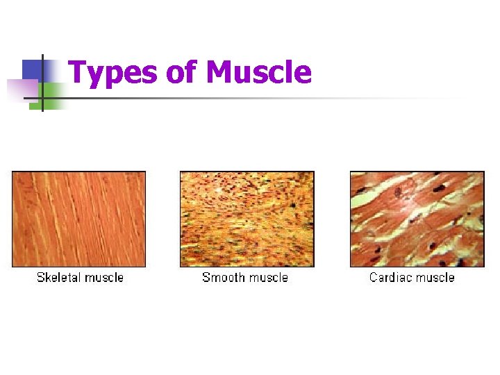
Types of Muscle
Skeletal Muscles n n n Nearly 650 muscles are attached to the skeleton. See muscle list for competitions. Skeletal muscles- work in pairs: one muscle moves the bone in one direction and the other moves it back again. Most muscles- extend from one bone across a joint to another bone with one being more stationary than another in a given movement. Muscle movement- bends the skeleton at moveable joints. Tendons - made of dense fibrous connective tissue shaped like heavy cords anchor muscles firmly to bone. Tendon injury- though very strong and secure to muscle, may be injured.
Skeletal Muscles n n n origin - Attachment to the more stationary bone by tendon closest to the body or muscle head or proximal insertion - attachment to the more moveable bone by tendon at the distal end During movement, the origin remains stationary and the insertion moves. The force producing the bending is always a pull of contraction. Reversing the direction is produced by the contraction of a different set of muscles. As one group of muscles contracts, the other group stretches and then they reverse actions.
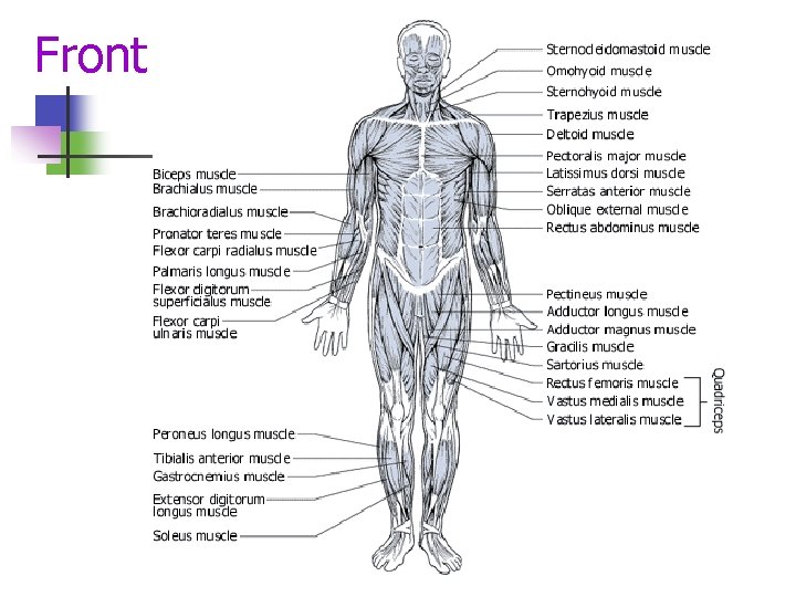
Front
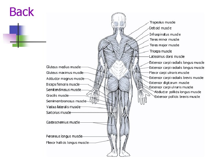
Back
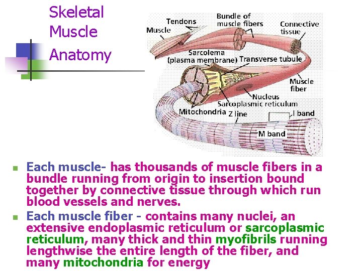
Skeletal Muscle Anatomy n n Each muscle- has thousands of muscle fibers in a bundle running from origin to insertion bound together by connective tissue through which run blood vessels and nerves. Each muscle fiber - contains many nuclei, an extensive endoplasmic reticulum or sarcoplasmic reticulum, many thick and thin myofibrils running lengthwise the entire length of the fiber, and many mitochondria for energy
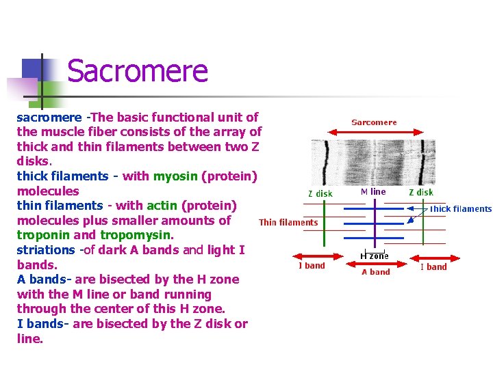
Sacromere sacromere -The basic functional unit of the muscle fiber consists of the array of thick and thin filaments between two Z disks. thick filaments - with myosin (protein) molecules thin filaments - with actin (protein) molecules plus smaller amounts of troponin and tropomysin. striations -of dark A bands and light I bands. A bands- are bisected by the H zone with the M line or band running through the center of this H zone. I bands- are bisected by the Z disk or line.
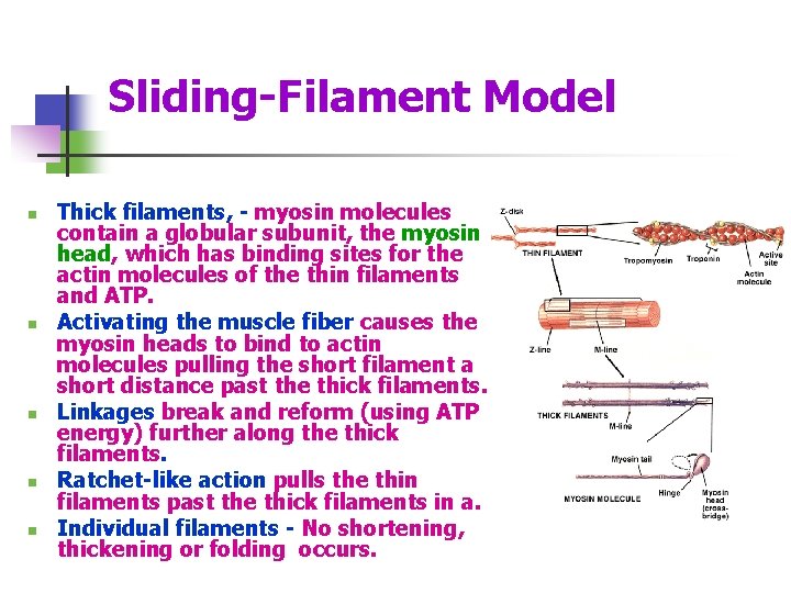
Sliding-Filament Model n n n Thick filaments, - myosin molecules contain a globular subunit, the myosin head, which has binding sites for the actin molecules of the thin filaments and ATP. Activating the muscle fiber causes the myosin heads to bind to actin molecules pulling the short filament a short distance past the thick filaments. Linkages break and reform (using ATP energy) further along the thick filaments. Ratchet-like action pulls the thin filaments past the thick filaments in a. Individual filaments - No shortening, thickening or folding occurs.
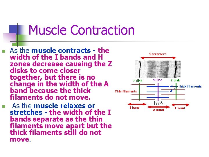
Muscle Contraction n n As the muscle contracts - the width of the I bands and H zones decrease causing the Z disks to come closer together, but there is no change in the width of the A band because thick filaments do not move. As the muscle relaxes or stretches - the width of the I bands separate as the thin filaments move apart but the thick filaments still do not move.
Muscle and Tendon Injuries n n Strains – injuries from overexertion or trauma which involve stretching or tearing of muscle fibers. They often are accompanied by pain and inflammation of the muscle and tendon. Sprain - the injury near a joint and involves a ligament Cramps – painful muscle spasms or involuntary twitches. Stress-induced muscle tension – may cause back pain and headaches.
Muscular Disorders n n n Poliomyelitis – viral infection of the nerves that control skeletal muscle movement. Muscular Dystrophies – most common caused by mutation of gene for the protein dystrophin which helps in attaching and organizing the filaments in the sacromere. Duchenne Muscular Dystrophy and Becker muscular dystrophy are the two most common types. The gene for dystrophin is on the X chromosome so the disorder is sex-linked. Myasthenia gravis – autoimmune disease affecting the neuromuscular junction. affecting the ability of the impulse to cause the muscle contraction. Administering an inhibitor of acetylcholinesterase can temporarily restore contractibility.
Exercise on Skeletal and Muscular System Skeletal System n Exercise slows decline in minerals and maintains joint mobility n Stress of exercise helps the bone tissues to become stronger n Hyaline cartilage at the ends of the bones becomes thicker and can absorb shock better n Ligaments will stretch slightly to enable greater joint flexibility Muscular System n Exercise helps muscles become more effective and efficient. n Tendons will become thicker and stronger n High intensity exercise for short duration produces strength, size and power gains in muscles n Low intensity exercise for long durations will give endurance benefits n Trained muscles have better tone or state of readiness to respond n Exercise promotes good posture enabling muscles to work effectively and helps prevent injury
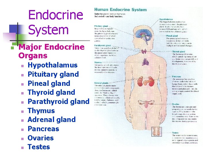
Endocrine System n Major Endocrine Organs n n n n n Hypothalamus Pituitary gland Pineal gland Thyroid gland Parathyroid gland Thymus Adrenal gland Pancreas Ovaries Testes
Hormones n n n specific chemical compound produced by a specific tissue of the body released in the body fluids carried to a distant target tissue affects a pre-existing mechanism effective is small amounts.
Classes of Hormones: peptides – short chains of amino acids (most hormones) pituitary, parathyroid, heart, stomach, liver & kidneys amines - derived from tyrosine and secreted by thyroid and adrenal cortex steroids - lipids derived from cholesterol secreted by the gonads, adrenal cortex, and placenta
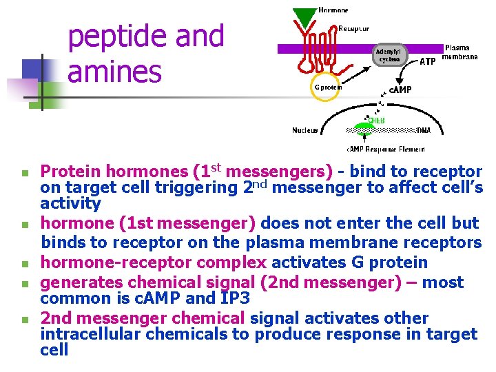
peptide and amines n n n Protein hormones (1 st messengers) - bind to receptor on target cell triggering 2 nd messenger to affect cell’s activity hormone (1 st messenger) does not enter the cell but binds to receptor on the plasma membrane receptors hormone-receptor complex activates G protein generates chemical signal (2 nd messenger) – most common is c. AMP and IP 3 2 nd messenger chemical signal activates other intracellular chemicals to produce response in target cell
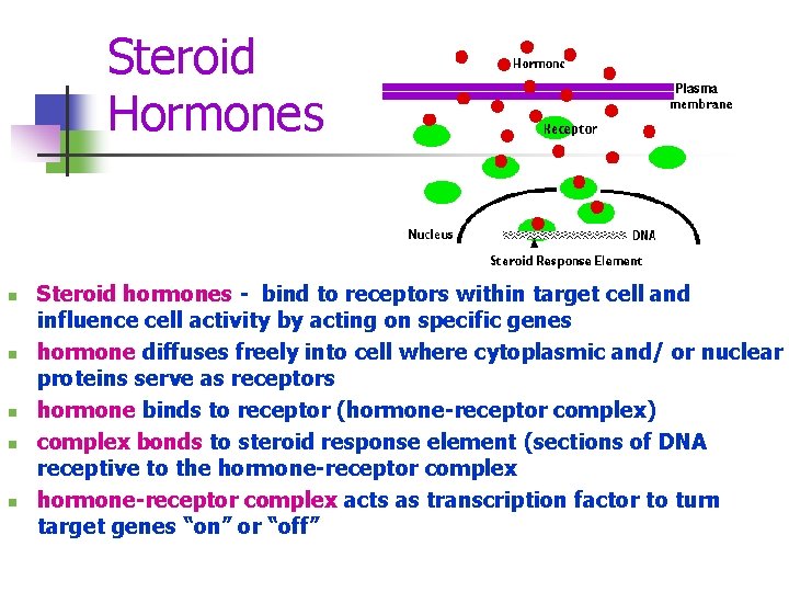
Steroid Hormones n n n Steroid hormones - bind to receptors within target cell and influence cell activity by acting on specific genes hormone diffuses freely into cell where cytoplasmic and/ or nuclear proteins serve as receptors hormone binds to receptor (hormone-receptor complex) complex bonds to steroid response element (sections of DNA receptive to the hormone-receptor complex acts as transcription factor to turn target genes “on” or “off”
Diseases of the Endocrine System Diabetes – increased levels of glucose in blood n Hypoglycemia - low blood sugar n Graves Disease – overactive thyroid n Goiter – enlarged thyroid gland n
- Slides: 46