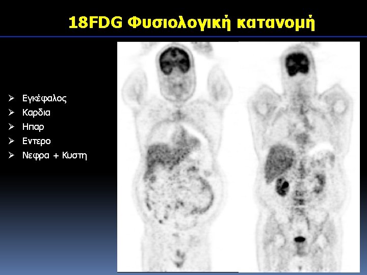
18 FFDG 18 FDG 18 FDGPET Sarcoidosis 18


")


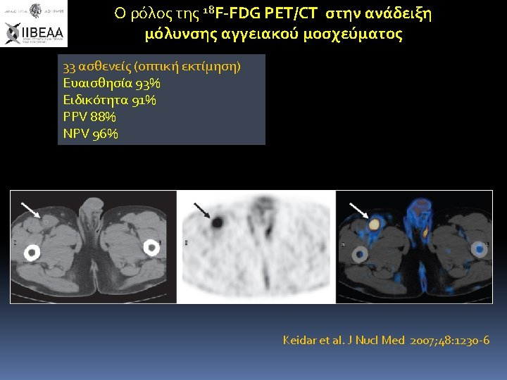


- Slides: 33
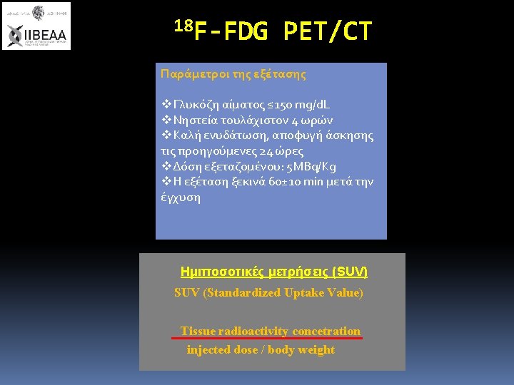
18 F-FDG
18 FDG-φλεγμονή 18 FDG-PET Sarcoidosis 18 FDG-PET Tuberculosis M. Soussan et. al. EJR, October 2012, 287276 D. Saranovic et al. JNM 53(10): 1543 -9 2012
FDG-φλεγμονή (Vaskulitis)
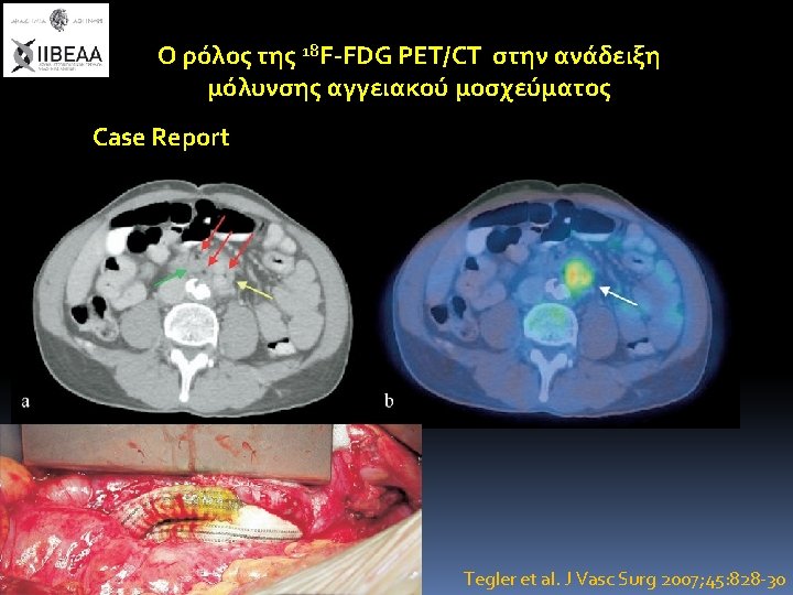
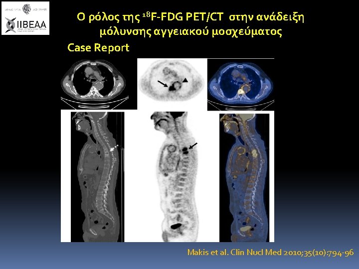
18 F-FDG PET/CT μόλυνση αγγειακού μοσχεύματος Sah BR et al. Eur J Vasc Endovasc Surg 2015; 49: 455 -64
Differential FDG-PET Uptake Patterns in Uninfected and Infected Central Prosthetic Vascular Grafts P. Berger et al. Eur J Vasc Endovasc Surg (2015) 50, 376 - 383 2007 -2013
Figure 6. Final conclusion of the PET scans Sensitivity of 77% (95% CI 62– 92%) Specificity of 89% (95% CI 77– 100%)
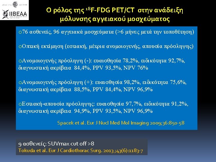
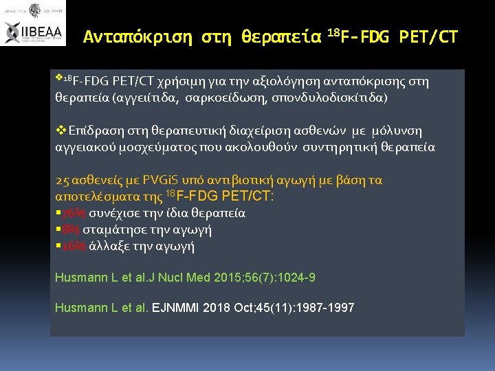
Ο ρόλος της 18 F-FDG PET/CT στην ανάδειξη μόλυνσης αγγειακού μοσχεύματος 25ασθενείς Ευαισθησία 93% Ειδικότητα 70% PPV 82% NPV 88% FDG PET analysis (four point scale) Grade Ι: uptake ≤ background (-) Grade II: uptake ≤ soft tissue (-) Grade III: moderate uptake ≥ soft tissue (+) Grade IV: strong uptake comparable with bladder (+) Brukking et al. Eur J Vasc Endovasc Surg 2010; 40, : 348 -354
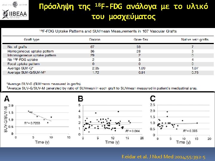
3 years after femoro-femoral Gore-Tex graft SUVmax=1, 1 16 years after aorto-bifemoral Dacron graft SUVmax=2, 5 Keidar et al. J Nucl Med 2014; 55: 392 -5
Fig. 1 This 53 -year-old farmer developed in November 2013 an infection (SUVmax 7. 5) of his polyester composite vascular graft (Gelweave Vaskutek, Aortic Valved Graft ATS) after bacteremia with Pasteurella multocida [Panel A: maximum intensity projections (top) and fused axial PET/CT images (bottom)]. PET/CT examination (Panel B) in January 2014 showed a complete metabolic response with a SUVmax of 2. 9. Panel C shows control PET/CT examination in June 2014 Husmann L et al. EJNMMI 2018 Oct; 45(11): 1987 -1997
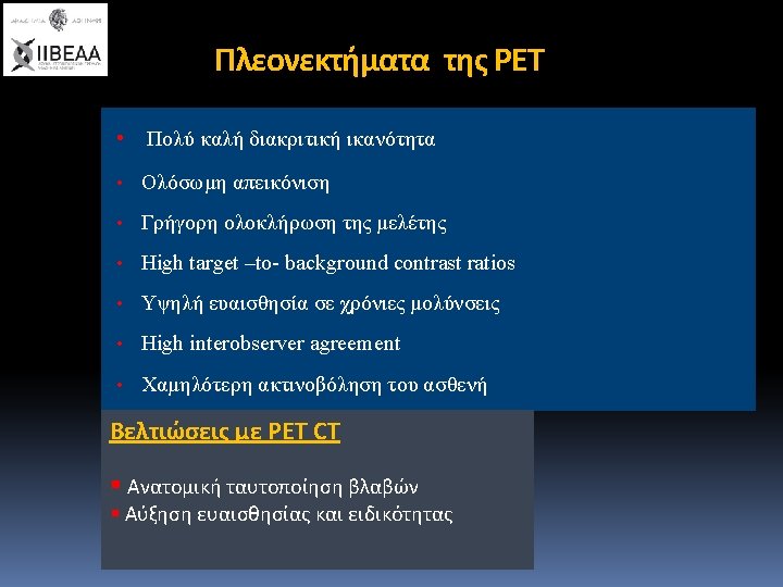
Bruggink et al. Semin Vasc Surg 2011; 24: 182 -90