14022022 The Standard Model Lesson 4 Forces and


 �This is a very short range force that holds")


 �PET scanners use antimatter annihilation to build")


- Slides: 15
14/02/2022 The Standard Model Lesson 4: Forces and Bosons
What are we learning today? �That bosons are force mediating particles. �The four fundamental forces. �Brief description of a PET scanner.
• There are four fundamental forces. • Strong • Weak • Electromagnetism • Gravity • Each force has an associated exchange particle
The Strong Force (3: 36) �This is a very short range force that holds particles of the same charge together. �It holds quarks together to form hadrons. �It’s exchange particles are called gluons because they glue the quarks together. • The strong force between the quarks in individual protons is strong enough to overcome the force of repulsion between the protons in the nucleus.
Variation of strong force with distance
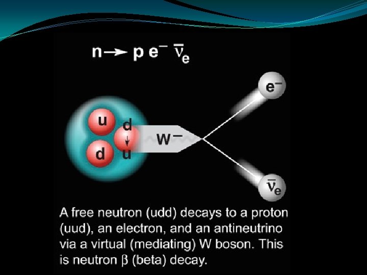
The Weak Force �This is involved in radioactive beta decay. �It’s exchange particles are W and Z bosons. �It is an extremely short range force. �Experienced in quark and lepton interactions
The Electromagnetic Force �This stops the electron from flying out of the atom. �It’s exchange particle is the photon. This is a classical analogy, and is. Time useless for attractive forces.
The Gravitational Force �This is the weakest of the four fundamental forces. �It attracts particles that have mass and is responsible for holding matter together. �It’s exchange particle is the graviton. �Gravity cannot be easily explained by the Standard Model.
Uses of Antimatter Positron Emission Tomography (PET) �PET scanners use antimatter annihilation to build up 3 D images of the functions of our body.
Radioactive glucose �A radioactive tracer that emits positrons is attached to a compound such as glucose, oxygen or water and injected into the body.
�When this tracer emits a positron it will annihilate nearly instantaneously with an electron. �This produces a pair of gamma-ray photons of specific frequency moving in approximately opposite directions to each other. Gamma ray electron positron Gamma ray
�The gamma rays are detected by a ring of scintillators, each producing a burst of light that can be detected by a photodiode. �Complex computer analysis traces tens of thousands of possible events each second and the positions of the original emissions are calculated.
�A 3 D image can then be constructed. �CT and MRI are often used to obtain a more accurate picture. �The detecting equipment in PET scanners has much in common with particle detectors. �The latest developments in particle accelerators can be used to improve this field of medical physics.
Now complete: • Page 4 Qs 8 -10 • Page 8 Revision Qs R 1 -R 3