140 mic General Microbiology Madeha AlOnazi LAB 6

140 mic General Microbiology Madeha Al-Onazi

LAB 6 Staining SIMPLE STAIN + GRAM STAIN

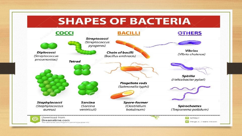
PURPOSE To recognize three basic shapes of bacterial cells.

THE THREE COMMON SHAPES OF BACTERIA

1 - Coccus Having one of the following arrangements: �Diplococcus: a pair of cocci �Streptococcus: a chain of cocci �Tetrad: a square of 4 cocci �Sarcina: a cube of 8 cocci �Staphylococcus: cocci in irregular, often grape-like clusters

2 -Bacillus. �Bacillus: a single bacillus �Streptobacillus: bacilli in chains �Coccobacillus: oval and similar to a coccus.

3 -Spiral �Vibrio: an incomplete spiral or comma-shaped �Spirillum: a thick, rigid spiral �Spirochete: a thin, flexible spiral

SIMPLE STAIN

SIMPLE STAIN : �The simple stain is a very simple staining procedure involving only one stain. �You may choose from methylene blue, safranin, and crystal violet.

The Method 1. Prepare the smear. - place a small drop of water on a clean slide. Drag the sterile inoculating needle tip through the edge of colony. - Gently spread the mixture into a circle to spread out.

SIMPLE STAIN 2. Let the smear air dry completely.

SIMPLE STAIN : 3. Heat-Fix the smear. �Smears are heat-fixed by quickly passing the slide through a flame two or three times. �This causes the microbes to stick to the slide and not get washed off during the staining process.

SIMPLE STAIN : 4. Stain the smear. �Place the slide on a rack over the sink. Flood the smear with stain and let it for 60 -90 seconds. Rinse gently and blot dry.

SIMPLE STAIN 5. Then, place a drop of oil directly on the stained smear. Turn the oil lens into position and fine focus to observe the cells.

RESULT
")
Coccus (cocci pl. )
")
Bacillus (Bacilli pl. )
")
Spirillum (Spirilli pl. )

Gram Stain - Hans Christian Gram, circa 1884, was studying the etiology of respiratory disease. - Gram’s staining procedures are done millions of times daily worldwide.

Gram’s procedure divides the bacterial organisms: Gram positive bacteria Gram negative bacteria

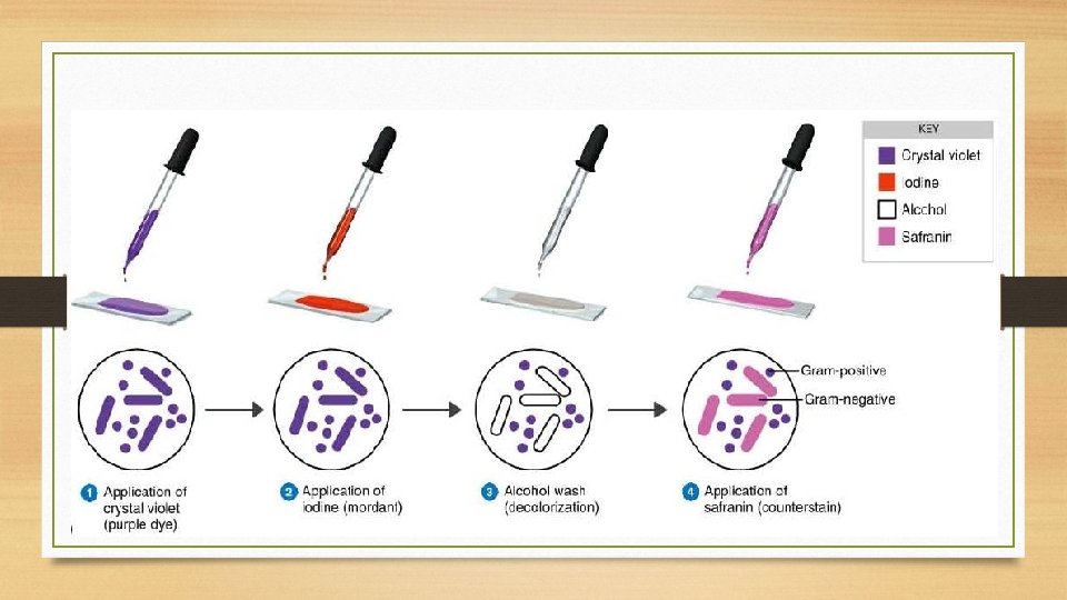
Gram Stain The gram stain is called a differential stain because it stain cell differently based on their cell wall structure.

Gram Stain - A differential technique is a process that distinguishes between a variety of microbial organisms based on. - The Gram staining technique depends upon: 1 - The ability of their cell wall to hold certain dyes to 2 - And to resist decolorization.

The Cell Wall Structure

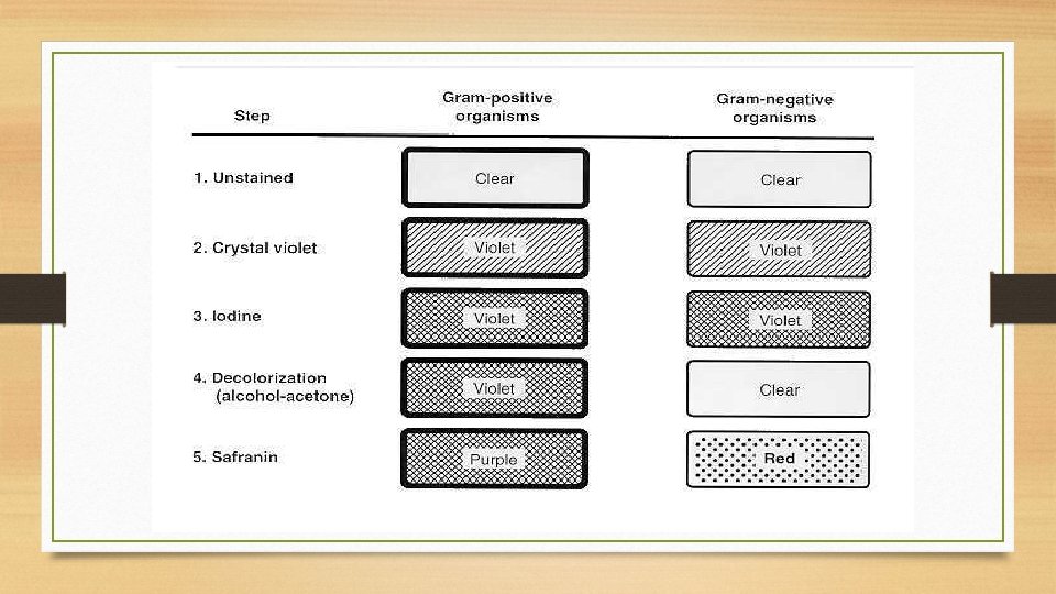
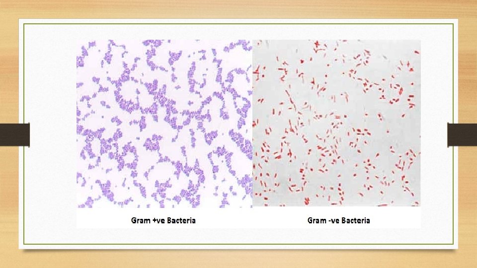
Bacterial Cell Wall Gram-positive bacteria Have a thick peptidoglycan layer surrounds the cell. The stain gets trapped into this layer and the bacteria turned purple. Gram-negative bacteria Have a thin peptidoglycan layer that does not retain crystal violet stain. Instead, it has a thick lipid layer which dissolved easily upon decolonization with Alcohol. Therefore, cells will be counterstained with safranin and turned red.
Cultures of: Staphylococcus aureus, Bacillus subtilis, Escherichia coli")
The Material 1 - Fresh (24 hrs)Cultures of: Staphylococcus aureus, Bacillus subtilis, Escherichia coli 2 - Microscopic 3 -Slides 4 - Water 5 -marker
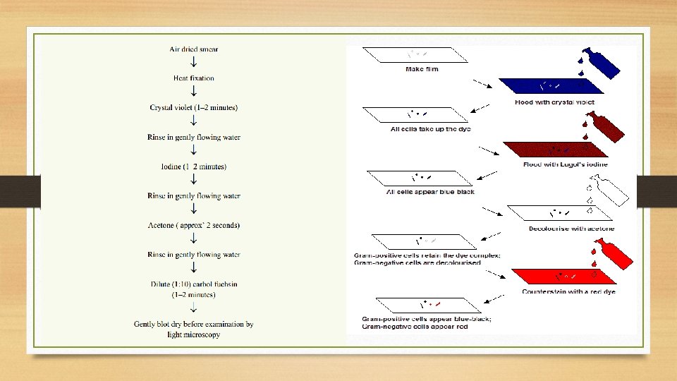
 - Gram ’s Iodine……. ( makes 1_ stain")
- Gram ’s crystal violet…………(primary stain) - Gram ’s Iodine……. ( makes 1_ stain fix to cell wall] substance that increases the reaction between the stain and the cells - Decolorizer 95% ethyl alcohol ……. (washes stain out of cell walls with high lipid content) - Gram’s safranin ………(counterstain)

The Method


Result

Shape: Cocci Arrangment: irregular clusters Colour: Violet Gram’s reaction: Gram’s +ve Name of microorganism: Staphylococci

Shape: Bacilli Arrangment: Chains Colour: Violet Gram’s reaction: Gram’s +ve Name of microorganism: Bacillus

What is the Gram stain reaction, cell morphology, and cell arrangement seen here? Answer: Gram-positive Streptococcus

What is the Gram stain reaction, cell morphology, and cell arrangement seen here? Answer: Gram-negative bacilli

https: //www. bing. com/videos/search? q=gram+stain+&&view=detail& mid=0 C 9 ACCFE 02 C 326849888&FORM= VRDGAR
- Slides: 39