130116 CELLULAR EVENTS in INFLAMMATION 1 Dr Ksheera


 is the earliest")


 Selectins")
 Selectins �Expressed on the surface of activated endothelial cell � Recognise specific carbohydrate")
 is involved in rolling �E-selectin")

 Integrins �Present on the endothelial cell surface �Activated during the process of loose")
 Immunoglobulin gene superfamily adhesion molecule �Intercellular adhesion molecule-1 (ICAM-1) and vascular cell adhesion")


 Leukotriene B 4 (LT-B 4), a")


- Slides: 32
13/01/16 CELLULAR EVENTS in INFLAMMATION - 1 Dr. Ksheera Cariappa Assistant professor
�Inflammation is defined as the local response of living mammalian tissues to injury due to any agent. �It is the body’s defence reaction in order to eliminate or limit the spread of injurious agents, followed by removal of the necrosed cells and tissues.
�Agents causing inflammation may be: 1. Infective agents like bacteria, viruses and their toxins, fungi, parasites. 2. Immunological agents like cell-mediated antigenantibody reactions. 3. Physical agents like heat, cold, radiation, mechanical trauma. 4. Chemical agents like organic and inorganic poisons. 5. Inert materials such as foreign bodies.
ACUTE INFLAMMATION �Acute inflammatory response by the host to any agent is a continuous process. �Divided into following two events: I. Vascular events. II. Cellular events.
I. VASCULAR EVENTS �Alteration in the microvasculature (arterioles, capillaries and venules) is the earliest response to tissue injury. �These alterations include: Ø haemodynamic changes Ø changes in vascular permeability
II. CELLULAR EVENTS �Cellular phase of inflammation consists of 2 processes: 1. Exudation of leucocytes 2. Phagocytosis
ACUTE INFLAMMATION- CELLULAR EVENTS “AIM IS TO DELIVER LEUCOCYTES TO THE SITE OF INJURY” LUMINAL MARGINATION ROLLING ADHESION TRANSMIGRATION ACROSS ENDOTHELIUM MIGRATION IN INTERSTITIAL TISSUES
Exudation of Leucocytes �Escape of leucocytes from the lumen of microvasculature to the interstitial tissue most important feature of inflammatory response. �In acute inflammation, polymorphonuclear neutrophils (PMNs) comprise the first line of body defense, followed later by monocytes and macrophages.
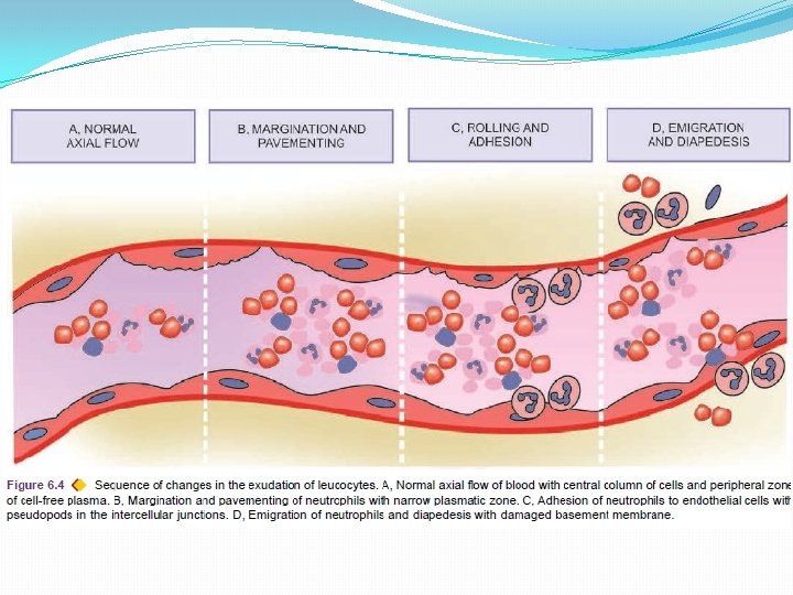
�Sequence of changes leading to migration of leucocytes are as follows: 1. Changes in the formed elements of blood 2. Rolling and adhesion 3. Emigration 4. Chemotaxis
1. CHANGES IN THE FORMED ELEMENTS OF BLOOD �Early stage of inflammation rate of flow of blood is increased due to vasodilatation. �But subsequently there is slowing or stasis of bloodstream. �With stasis changes in the normal axial flow of blood in the microcirculation take place.
�Normal axial flow consists of central stream of cells comprised by leucocytes and RBCs and peripheral cellfree layer of plasma close to vessel wall. �Due to slowing and stasis central stream of cells widens and peripheral plasma zone becomes narrower because of loss of plasma by exudation. This phenomenon is known as margination. �As a result of this redistribution, the neutrophils of the central column come close to the vessel wall; this is known as pavementing.
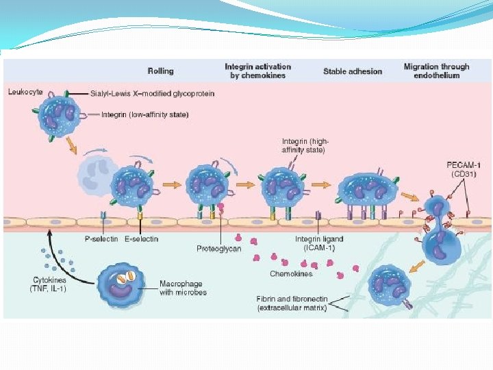
2. ROLLING AND ADHESION �Peripherally marginated and pavemented neutrophils slowly roll over the endothelial cells lining the vessel wall (rolling phase). �Followed by the transient bond between the leucocytes and endothelial cells becoming firmer (adhesion phase).
�Rolling and adhesion phases is brought about by the following 3 molecules: i) Selectins ii) Integrins iii) Immunoglobulin gene superfamily adhesion molecule
i) Selectins �Expressed on the surface of activated endothelial cell � Recognise specific carbohydrate groups found on the surface of neutrophils (s-Lewis X molecule). � 3 types of selectins are: Ø P-selectin Ø E-selectin Ø L-selectin
�P-selectin (preformed and stored in endothelial cells and platelets) is involved in rolling �E-selectin (synthesised by cytokine activated endothelial cells) is associated with both rolling and adhesion �L-selectin (expressed on the surface of lymphocytes and neutrophils) is responsible for homing of circulating lymphocytes to the endothelial cells in lymph nodes.
ii) Integrins �Present on the endothelial cell surface �Activated during the process of loose and transient adhesions between endothelial cells and leucocytes. �At the same time the receptors for integrins on the neutrophils are also stimulated. �This process brings about firm adhesion between leucocyte and endothelium.
iii) Immunoglobulin gene superfamily adhesion molecule �Intercellular adhesion molecule-1 (ICAM-1) and vascular cell adhesion molecule-1 (VCAM-1) �Allow a tighter adhesion and stabilise the interaction between leucocytes and endothelial cells. �Platelet-endothelial cell adhesion molecule- 1 (PECAM-1) or CD 31 is also involved in leucocyte migration from the endothelial surface.
3. EMIGRATION �After sticking of neutrophils to endothelium �Neutrophils move along the endothelial surface till a suitable site between the endothelial cells is found where the neutrophils throw out cytoplasmic pseudopods. �Neutrophils lodge between the endothelial cells and basement membrane �Cross the basement membrane by damaging it locally with secreted collagenases and escape out into the extravascular space; this is known as emigration. �Damaged basement membrane is repaired almost immediately.
�Simultaneous to emigration of leucocytes, escape of red cells through gaps between the endothelial cells, diapedesis, takes place. �Passive phenomenon—RBCs being forced out either by raised hydrostatic pressure or may escape through the endothelial defects left after emigration of leucocytes. �Diapedesis gives haemorrhagic appearance to the inflammatory exudate.
4. CHEMOTAXIS �Chemotactic factor-mediated transmigration of leucocytes after crossing several barriers (endothelium, basement membrane, perivascular myofibroblasts and matrix) to reach the interstitial tissues is called chemotaxis.
Chemotaxis: DEFINITION- Process Of Directed Cell Migration Along A Chemical Gradient Responsible for emigration of leucocytes towards the site of injury
CHEMOTACTIC-AGENTS EXOGENOUS BACTERIAL PRODUCTS-COMMONEST PEPTIDES OR LIPIDS ENDOGENOUS COMPLEMENT COMPONENTS-C 5 a LEUKOTRIENE B 4 CYTOKINES-IL-8
CHEMOTAXIS-MECHANISM Bind to specific receptors on leucocytes Effector molecules produced-phospholipase, tyrosinase etc Second messengers-finally leading to polymerization of actin Leucocyte moves by extending filopodia
Neutrophil Crawling on a Glass Slide It is going that way:
Boyden’s chamber experiment
�Potent chemotactic substances or chemokines for neutophils: i) Leukotriene B 4 (LT-B 4), a product of lipooxygenase pathway of arachidonic acid metabolites ii) Components of complement system (C 5 a and C 3 a in particular) iii) Cytokines (Interleukins, in particular IL-8) iv) Soluble bacterial products (such as formylated peptides).
�In addition to neutrophils, other inflammatory cells too respond and partake in inflammation. �Chemokines for them are: Ø monocyte chemoattractant protein (MCP-1) for monocytes Ø eotaxin for eosinophils Ø virally infected cells for NK cells, etc.
SUMMARY!!!!!