
13 PART A The Respiratory System Power Point
- Slides: 44
13 PART A The Respiratory System Power. Point® Lecture Slide Presentation by Jerry L. Cook, Sam Houston University ESSENTIALS OF HUMAN ANATOMY & PHYSIOLOGY EIGHTH EDITION ELAINE N. MARIEB Copyright © 2006 Pearson Education, Inc. , publishing as Benjamin Cummings
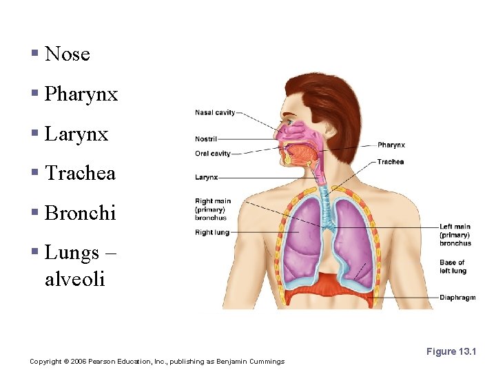
Organs of the Respiratory system § Nose § Pharynx § Larynx § Trachea § Bronchi § Lungs – alveoli Figure 13. 1 Copyright © 2006 Pearson Education, Inc. , publishing as Benjamin Cummings
Function of the Respiratory System § Oversees gas exchanges between the blood and external environment § Exchange of gasses takes place within the lungs in the alveoli § Passageways to the lungs purify, warm, and humidify the incoming air Copyright © 2006 Pearson Education, Inc. , publishing as Benjamin Cummings
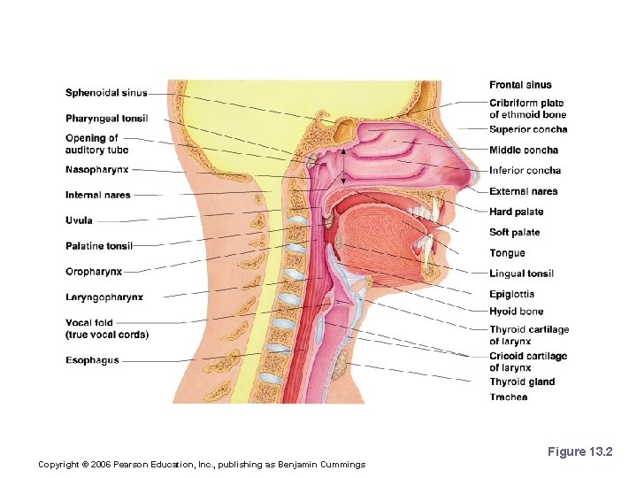
The Nose § The only externally visible part of the respiratory system § Air enters the nose through the external nares (nostrils) § The interior of the nose consists of a nasal cavity divided by a nasal septum Copyright © 2006 Pearson Education, Inc. , publishing as Benjamin Cummings
Upper Respiratory Tract Figure 13. 2 Copyright © 2006 Pearson Education, Inc. , publishing as Benjamin Cummings
Anatomy of the Nasal Cavity § Olfactory receptors are located in the mucosa on the superior surface § The rest of the cavity is lined with respiratory mucosa § Moistens air § Traps incoming foreign particles Copyright © 2006 Pearson Education, Inc. , publishing as Benjamin Cummings
Anatomy of the Nasal Cavity § Lateral walls have projections called conchae § Increases surface area § Increases air turbulence within the nasal cavity § The nasal cavity is separated from the oral cavity by the palate § Anterior hard palate (bone) § Posterior soft palate (muscle) Copyright © 2006 Pearson Education, Inc. , publishing as Benjamin Cummings
Paranasal Sinuses § Cavities within bones surrounding the nasal cavity § Frontal bone § Sphenoid bone § Ethmoid bone § Maxillary bone Copyright © 2006 Pearson Education, Inc. , publishing as Benjamin Cummings
Paranasal Sinuses § Function of the sinuses § Lighten the skull § Act as resonance chambers for speech § Produce mucus that drains into the nasal cavity Copyright © 2006 Pearson Education, Inc. , publishing as Benjamin Cummings
Pharynx (Throat) § Muscular passage from nasal cavity to larynx § Three regions of the pharynx § Nasopharynx – superior region behind nasal cavity § Oropharynx – middle region behind mouth § Laryngopharynx – inferior region attached to larynx § The oropharynx and laryngopharynx are common passageways for air and food Copyright © 2006 Pearson Education, Inc. , publishing as Benjamin Cummings
Structures of the Pharynx § Auditory tubes enter the nasopharynx § Tonsils of the pharynx § Pharyngeal tonsil (adenoids) in the nasopharynx § Palatine tonsils in the oropharynx § Lingual tonsils at the base of the tongue Copyright © 2006 Pearson Education, Inc. , publishing as Benjamin Cummings
Larynx (Voice Box) § Routes air and food into proper channels § Plays a role in speech § Made of eight rigid hyaline cartilages and a spoon-shaped flap of elastic cartilage (epiglottis) Copyright © 2006 Pearson Education, Inc. , publishing as Benjamin Cummings
Structures of the Larynx § Thyroid cartilage § Largest hyaline cartilage § Protrudes anteriorly (Adam’s apple) § Epiglottis § Superior opening of the larynx § Routes food to the larynx and air toward the trachea Copyright © 2006 Pearson Education, Inc. , publishing as Benjamin Cummings
Structures of the Larynx § Vocal cords (vocal folds) § Vibrate with expelled air to create sound (speech) § Glottis – opening between vocal cords Copyright © 2006 Pearson Education, Inc. , publishing as Benjamin Cummings
Trachea (Windpipe) § Connects larynx with bronchi § Lined with ciliated mucosa § Beat continuously in the opposite direction of incoming air § Expel mucus loaded with dust and other debris away from lungs § Walls are reinforced with C-shaped hyaline cartilage Copyright © 2006 Pearson Education, Inc. , publishing as Benjamin Cummings
Primary Bronchi § Formed by division of the trachea § Enters the lung at the hilus (medial depression) § Right bronchus is wider, shorter, and straighter than left § Bronchi subdivide into smaller and smaller branches Copyright © 2006 Pearson Education, Inc. , publishing as Benjamin Cummings
Lungs § Occupy most of the thoracic cavity § Apex is near the clavicle (superior portion) § Base rests on the diaphragm (inferior portion) § Each lung is divided into lobes by fissures § Left lung – two lobes § Right lung – three lobes Copyright © 2006 Pearson Education, Inc. , publishing as Benjamin Cummings
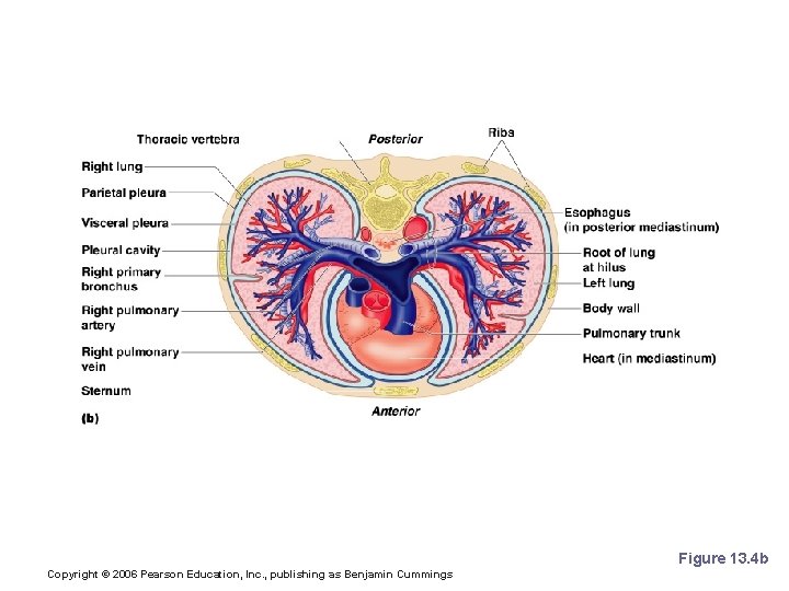
Lungs Figure 13. 4 b Copyright © 2006 Pearson Education, Inc. , publishing as Benjamin Cummings
Coverings of the Lungs § Pulmonary (visceral) pleura covers the lung surface § Parietal pleura lines the walls of the thoracic cavity § Pleural fluid fills the area between layers of pleura to allow gliding Copyright © 2006 Pearson Education, Inc. , publishing as Benjamin Cummings
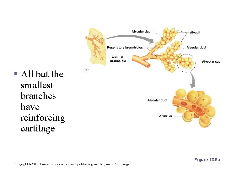
Respiratory Tree Divisions § Primary bronchi § Secondary bronchi § Tertiary bronchi § Bronchioli § Terminal bronchioli Copyright © 2006 Pearson Education, Inc. , publishing as Benjamin Cummings
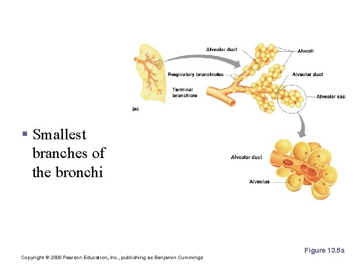
Bronchioles § Smallest branches of the bronchi Figure 13. 5 a Copyright © 2006 Pearson Education, Inc. , publishing as Benjamin Cummings
Bronchioles § All but the smallest branches have reinforcing cartilage Figure 13. 5 a Copyright © 2006 Pearson Education, Inc. , publishing as Benjamin Cummings
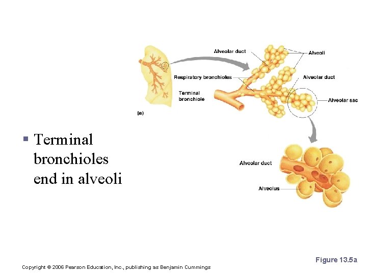
Bronchioles § Terminal bronchioles end in alveoli Figure 13. 5 a Copyright © 2006 Pearson Education, Inc. , publishing as Benjamin Cummings
Respiratory Zone § Structures § Respiratory bronchioli § Alveolar duct § Alveoli § Site of gas exchange Copyright © 2006 Pearson Education, Inc. , publishing as Benjamin Cummings
Alveoli § Structure of alveoli § Alveolar duct § Alveolar sac § Alveolus § Gas exchange takes place within the alveoli in the respiratory membrane Copyright © 2006 Pearson Education, Inc. , publishing as Benjamin Cummings
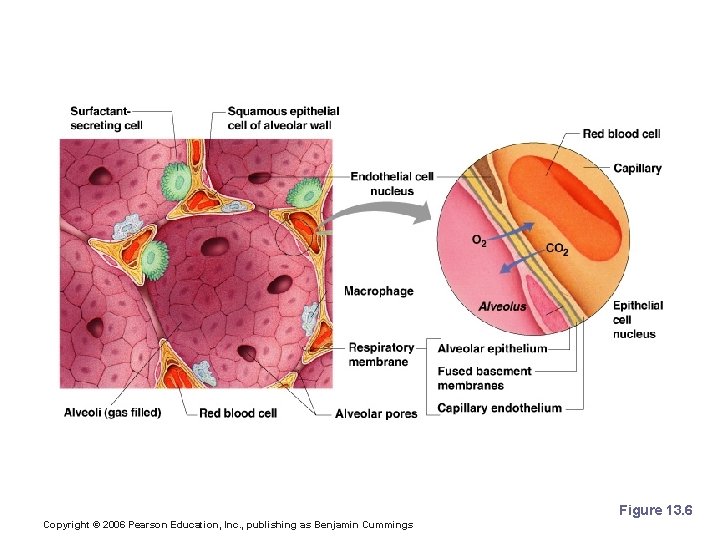
Respiratory Membrane (Air-Blood Barrier) § Thin squamous epithelial layer lining alveolar walls § Pulmonary capillaries cover external surfaces of alveoli Copyright © 2006 Pearson Education, Inc. , publishing as Benjamin Cummings
Respiratory Membrane (Air-Blood Barrier) Figure 13. 6 Copyright © 2006 Pearson Education, Inc. , publishing as Benjamin Cummings
Gas Exchange § Gas crosses the respiratory membrane by diffusion § Oxygen enters the blood § Carbon dioxide enters the alveoli § Macrophages add protection § Surfactant coats gas-exposed alveolar surfaces Copyright © 2006 Pearson Education, Inc. , publishing as Benjamin Cummings
Events of Respiration § Pulmonary ventilation – moving air in and out of the lungs § External respiration – gas exchange between pulmonary blood and alveoli Copyright © 2006 Pearson Education, Inc. , publishing as Benjamin Cummings
Events of Respiration § Respiratory gas transport – transport of oxygen and carbon dioxide via the bloodstream § Internal respiration – gas exchange between blood and tissue cells in systemic capillaries Copyright © 2006 Pearson Education, Inc. , publishing as Benjamin Cummings
Mechanics of Breathing (Pulmonary Ventilation) § Completely mechanical process § Depends on volume changes in the thoracic cavity § Volume changes lead to pressure changes, which lead to the flow of gases to equalize pressure Copyright © 2006 Pearson Education, Inc. , publishing as Benjamin Cummings
Mechanics of Breathing (Pulmonary Ventilation) § Two phases § Inspiration – flow of air into lung § Expiration – air leaving lung Copyright © 2006 Pearson Education, Inc. , publishing as Benjamin Cummings
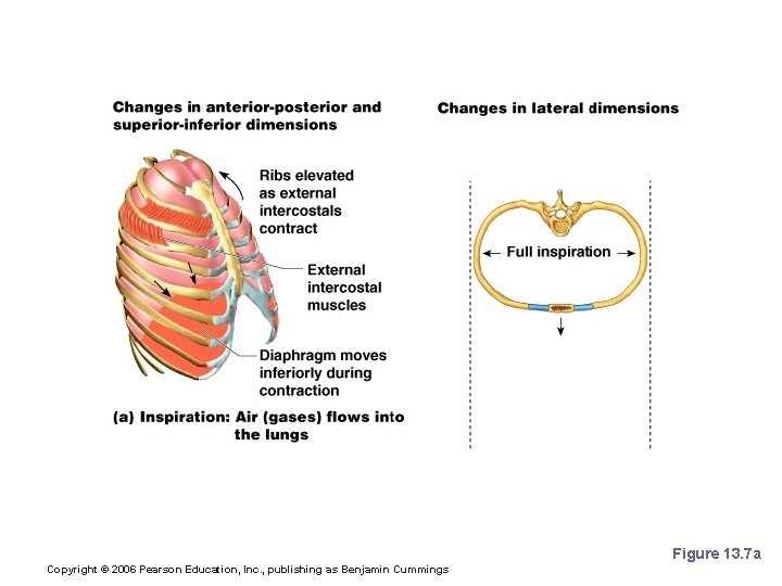
Inspiration § Diaphragm and intercostal muscles contract § The size of the thoracic cavity increases § External air is pulled into the lungs due to an increase in intrapulmonary volume Copyright © 2006 Pearson Education, Inc. , publishing as Benjamin Cummings
Inspiration Figure 13. 7 a Copyright © 2006 Pearson Education, Inc. , publishing as Benjamin Cummings
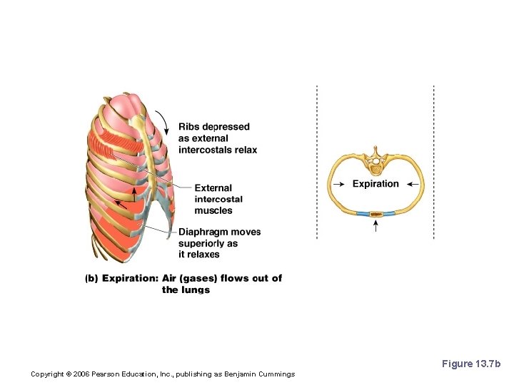
Expiration § Largely a passive process which depends on natural lung elasticity § As muscles relax, air is pushed out of the lungs § Forced expiration can occur mostly by contracting internal intercostal muscles to depress the rib cage Copyright © 2006 Pearson Education, Inc. , publishing as Benjamin Cummings
Expiration Figure 13. 7 b Copyright © 2006 Pearson Education, Inc. , publishing as Benjamin Cummings
Pressure Differences in the Thoracic Cavity § Normal pressure within the pleural space is always negative (intrapleural pressure) § Differences in lung and pleural space pressures keep lungs from collapsing Copyright © 2006 Pearson Education, Inc. , publishing as Benjamin Cummings
Nonrespiratory Air Movements § Can be caused by reflexes or voluntary actions § Examples § Cough and sneeze – clears lungs of debris § Laughing § Crying § Yawn § Hiccup Copyright © 2006 Pearson Education, Inc. , publishing as Benjamin Cummings
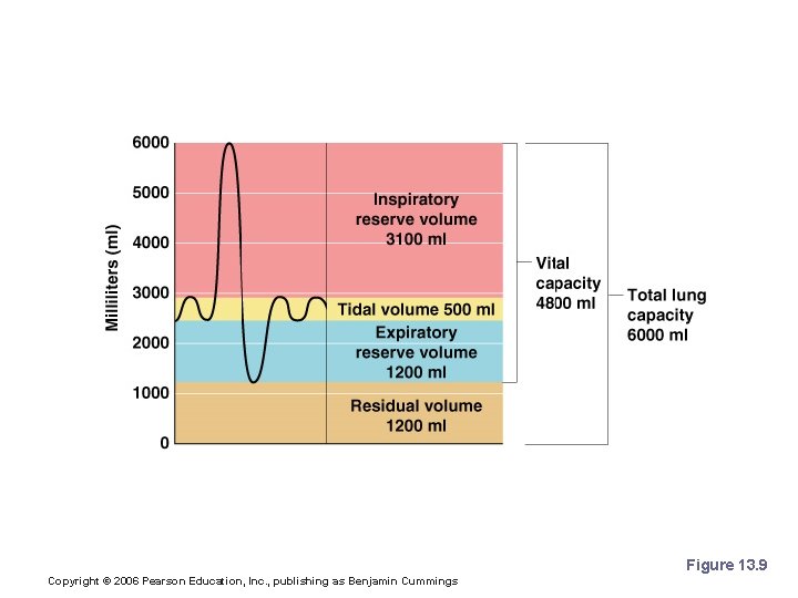
Respiratory Volumes and Capacities § Normal breathing moves about 500 ml of air with each breath (tidal volume [TV]) § Many factors that affect respiratory capacity § A person’s size § Sex § Age § Physical condition § Residual volume of air – after exhalation, about 1200 ml of air remains in the lungs Copyright © 2006 Pearson Education, Inc. , publishing as Benjamin Cummings
Respiratory Volumes and Capacities § Inspiratory reserve volume (IRV) § Amount of air that can be taken in forcibly over the tidal volume § Usually between 2100 and 3200 ml § Expiratory reserve volume (ERV) § Amount of air that can be forcibly exhaled § Approximately 1200 ml Copyright © 2006 Pearson Education, Inc. , publishing as Benjamin Cummings
Respiratory Volumes and Capacities § Residual volume § Air remaining in lung after expiration § About 1200 ml Copyright © 2006 Pearson Education, Inc. , publishing as Benjamin Cummings
Respiratory Volumes and Capacities § Vital capacity § The total amount of exchangeable air § Vital capacity = TV + IRV + ERV § Dead space volume § Air that remains in conducting zone and never reaches alveoli § About 150 ml Copyright © 2006 Pearson Education, Inc. , publishing as Benjamin Cummings
Respiratory Volumes and Capacities § Functional volume § Air that actually reaches the respiratory zone § Usually about 350 ml § Respiratory capacities are measured with a spirometer Copyright © 2006 Pearson Education, Inc. , publishing as Benjamin Cummings
Respiratory Capacities Figure 13. 9 Copyright © 2006 Pearson Education, Inc. , publishing as Benjamin Cummings