13 Characterizing and Classifying Viruses Viroids and Prions


 surrounding")


 Culture � Cells isolated")


- Slides: 47
13 Characterizing and Classifying Viruses, Viroids, and Prions
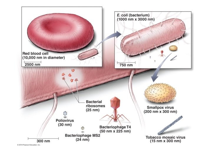
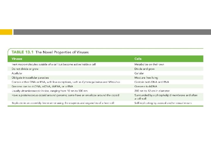
Characteristics of Viruses � Minuscule, acellular, infectious agent having either DNA or RNA � Cause infections of humans, animals, plants, and bacteria � Cause most of the diseases that plague the industrialized world � Cannot carry out any metabolic pathway � Neither grow nor respond to the environment � Cannot reproduce independently � Recruit the cell's metabolic pathways to increase their numbers � No cytoplasmic membrane, cytosol, organelles � Have extracellular and intracellular state
Characteristics of Viruses Extracellular state � � � Called virion Protein coat (capsid) surrounding nucleic acid Nucleic acid and capsid also called nucleocapsid Some have phospholipid envelope Outermost layer provides protection and recognition sites for host cells Intracellular state � � Capsid removed Virus exists as nucleic acid
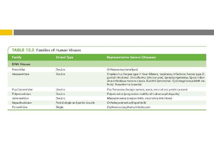
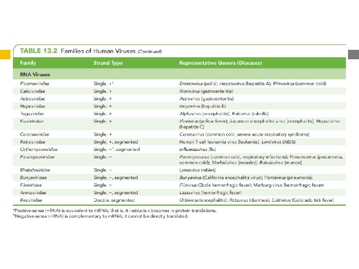
Characteristics of Viruses Genetic Material of Viruses Show more variety in nature of their genomes than do cells � Primary way scientists categorize and classify viruses � May be DNA or RNA, but never both � Can be ds. DNA, ss. DNA, ds. RNA, ss. RNA May be linear and segmented or single and circular � Much smaller than �
Characteristics of Viruses Hosts of Viruses � Most cells viruses infect only particular host's Due to affinity of viral surface proteins for complementary proteins on host cell surface � May be so specific they only infect particular kind of cell in a particular host � Generalists – infect many kinds of cells in many different hosts
Characteristics of Viruses - Capsid Morphology Capsids Provide protection for viral nucleic acid Means of attachment to host's cells Composed of proteinaceous subunits called capsomeres � Capsomere may be made of single or multiple types of proteins
Figure 13. 6 The complex shape of bacteriophage T 4. Head Tail fibers Tail Base plate
Characteristics of Viruses The Viral Envelope � Acquired release from host cell during viral replication or Envelope is portion of membrane system of host � Composed of phospholipid bilayer and proteins Some proteins are virally coded glycoproteins (spikes) � Envelope proteins and glycoproteins often play role in host recognition
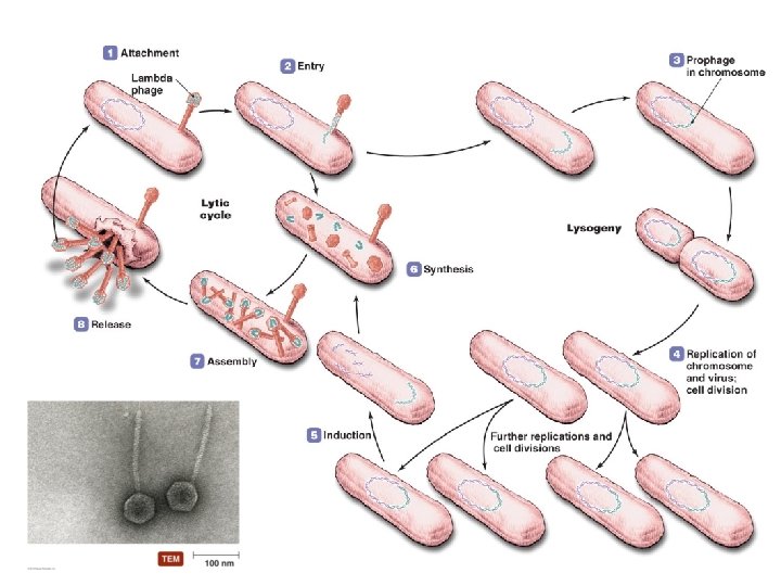
Viral Replication Dependent on hosts' organelles and enzymes to produce new virions Lytic replication � Viral replication usually results in death and lysis of host cell � Five stages of lytic replication cycle Attachment Entry Synthesis Assembly Release
Viral Replication: Overview PLAY Viral Replication: Overview
Figure 13. 8 The lytic replication cycle in bacteriophages.
Viral Replication: Virulent Bacteriophages PLAY Viral Replication: Virulent Bacteriophages
Viral Replication Lysogeny � Modified replication cycle � Infected host cells grow and reproduce normally for generations before they lyse � Temperate phages Prophages � Lysogenic Results – inactive phages conversion when phages carry genes that alter phenotype of a bacterium
Viral Replication: Temperate Bacteriophages PLAY Viral Replication: Temperate Bacteriophages
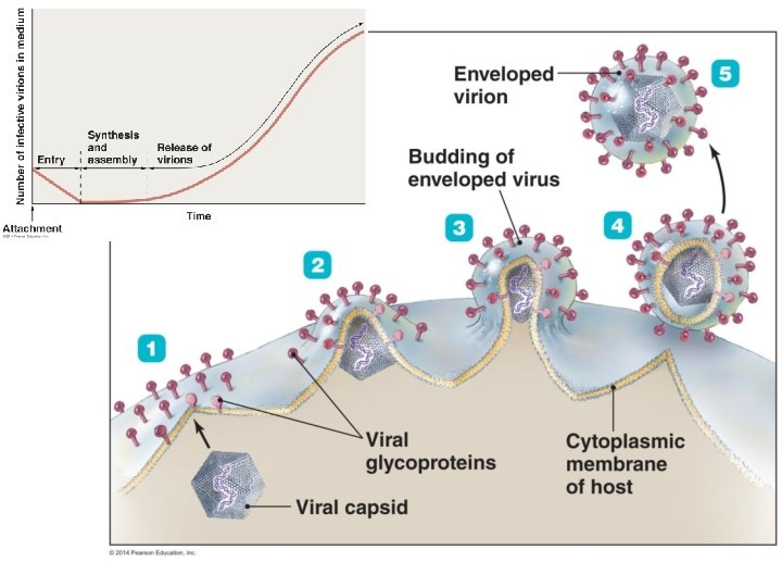
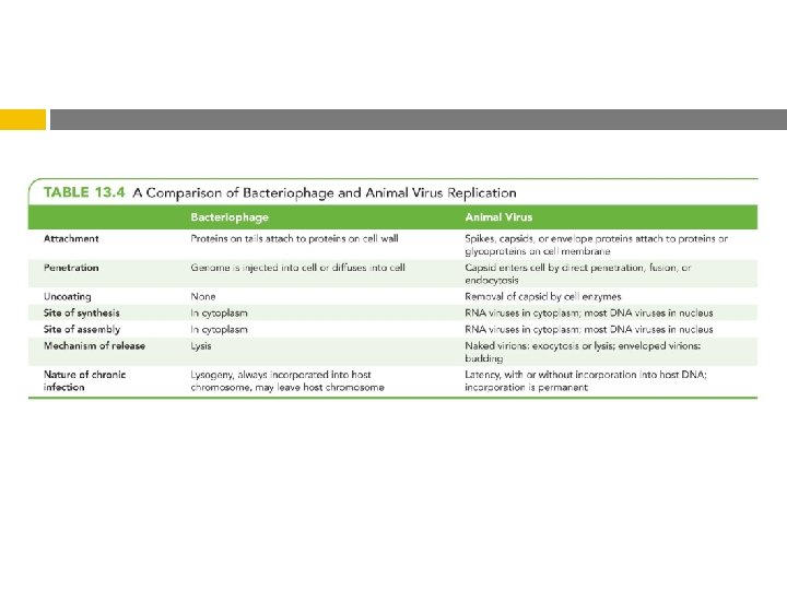
Viral Replication of Animal Viruses � Same basic replication pathway as bacteriophages � Differences result from Presence of envelope around some viruses Eukaryotic nature of animal cells Lack of cell wall in animal cells
Viral Replication of Animal Viruses � Attachment of animal viruses Chemical attraction between viral protein and cell receptor Animal viruses do not have tails or tail fibers Have glycoprotein spikes or other attachment molecules that mediate attachment
Viral Replication of Animal Viruses � Synthesis of DNA viruses of animals Each type of animal virus requires different strategy depending on its nucleic acid DNA viruses often enter the nucleus RNA viruses often replicate in the cytoplasm Must consider How m. RNA is synthesized What serves as template for nucleic acid replication
Viral Replication of Animal Viruses � Synthesis of DNA viruses of animals ds. DNA viruses Similar to replication of cellular DNA Viral genome replicated in the nucleus Viral proteins are made in the cytoplasm Some exceptions Poxvirus replication occurs in the cytoplasm Hepatitis B viruses replicate DNA from an RNA intermediary
Viral Replication of Animal Viruses � Synthesis of DNA viruses of animals ss. DNA viruses Cells do not use ss. DNA Parvoviruses have ss. DNA genomes Host enzymes produce DNA strand complementary to viral genome to form ds. DNA molecule ds. DNA used for viral replication and transcription
Figure 13. 13 Synthesis of proteins and genomes in animal RNA viruses. –ss. RNA virus Receptors on cytoplasmic membrane of host ds. RNA virus +ss. RNA –ss. RNA Transcription by viral RNA polymerase ds. RNA Transcription by RNA-dependent RNA transcriptase Complementary –ss. RNA to act as template Further transcription Translation of viral proteins, genome acts as m. RNA Further transcription Copies of –ss. RNA Complementary +ss. RNA to act as template and as m. RNA Translation of viral proteins Unwinding –ss. RNA Transcription by viral RNA polymerase to make complementary RNA strands Translation of viral proteins Copies of +ss. RNA Assembly Positive-sense ss. RNA virus +ss. RNA acts as m. RNA Assembly Negative-sense ss. RNA virus Double-stranded RNA virus
Viral Replication of Animal Viruses � Synthesis of RNA viruses of animals Retroviruses Do not use their genomes as m. RNA Use DNA intermediary transcribed by viral reverse transcriptase as template to produce viral genomes
Viral Replication of Animal Viruses � Assembly Most and release of animal viruses DNA viruses assemble in nucleus Most RNA viruses develop solely in cytoplasm Number of viruses produced depends on type of virus and size and initial health of host cell Enveloped viruses cause persistent infections Naked viruses are released by exocytosis or lysis
Viral Replication: Animal Viruses PLAY Viral Replication: Animal Viruses
Viral Replication of Animal Viruses � Latency When of animal viruses remain dormant in host cells Viruses are called latent viruses or proviruses May be prolonged for years with no viral activity Some latent viruses do not become incorporated into host chromosome Incorporation of provirus into host DNA is permanent
The Role of Viruses in Cancer Cell division is under strict genetic control � � � Neoplasia � � Genes dictate that some cells can no longer divide at all Cells that can divide are prevented from unlimited division Genes for cell division "turned off" or genes inhibiting division "turned on" Uncontrolled cell division in multicellular animal Mass of neoplastic cells is tumor Benign vs. malignant tumors � � Malignant tumors also called cancers Metastasis occurs when tumors spread
The Role of Viruses in Cancer Environmental factors that contribute to the activation of oncogenes � Ultraviolet light � Radiation � Carcinogens � Viruses
The Role of Viruses in Cancer Viruses cause 20– 25% of human cancers � � Some carry copies of oncogenes as part of their genomes Some promote oncogenes already present in host Some interfere with tumor repression Specific viruses are known to cause ~15% of human cancers Burkitt's lymphoma Hodgkin's disease Kaposi's sarcoma Cervical cancer
Culturing Viruses in the Laboratory Viruses cannot grow in standard microbiological media Cultured inside host cells Three types of media for culturing viruses � Media consisting of mature organisms � Embryonated eggs � Cell cultures
Culturing Viruses in the Laboratory Culturing Viruses in Mature Organisms � Culturing bacteria viruses in Phages grown in bacteria in liquid cultures or on agar plates Lysis of bacteria produces plaques Allows estimation of phage numbers by plaque assay
Culturing Viruses in the Laboratory Culturing Viruses in Mature Organisms � Culturing viruses in plants and animals Numerous plants and animals have been used to culture viruses Laboratory animals can be difficult and expensive to maintain Ethical concerns
Culturing Viruses in the Laboratory Culturing Viruses in Embryonated Chicken Eggs � � � Inexpensive Among the largest of cells Free of contaminating microbes Contain a nourishing yolk Fertilized chicken eggs are often used Embryonic tissues provide ideal site for growing viruses Some vaccines prepared in chicken cultures �
Culturing Viruses in the Laboratory Culturing Viruses in Cell (Tissue) Culture � Cells isolated from an organism and grown on a medium or in a broth � Cell cultures sometimes inaccurately called tissue cultures � Two types of cell cultures Diploid cell cultures Continuous cell cultures
Are Viruses Alive? Some consider them complex pathogenic chemicals Others consider them to be the least complex living entities � Use sophisticated methods to invade cells � Have the ability to take control of their host cell � Are able to replicate themselves
Other Parasitic Particles: Viroids and Prions Characteristics of Viroids � Extremely small, circular pieces of RNA that are infectious and pathogenic in plants � Similar to RNA viruses, but lack capsid � May appear linear due to hydrogen bonding Figure 13. 21 One effect of viroids on plants.
Other Parasitic Particles: Viroids and Prions Characteristics of Prions � Proteinaceous � Cellular infectious agents Pr. P Made by all mammals Normal, functional structure has -helices � Prion Pr. P Disease-causing � Prion Pr. P form has -sheets Pr. P causes cellular Pr. P to refold into prion
Prions: Overview PLAY Prions: Overview
-helices -pleated sheet Figure 13. 22 The two stable, three-dimensional forms of prion protein (Pr. P). Cellular Pr. P Prion Pr. P
Prions: Characteristics PLAY Prions: Characteristics
Other Parasitic Particles: Viroids and Prions Characteristics of Prions � Prion diseases Spongiform encephalopathies � Large vacuoles form in brain Characteristic spongy appearance BSE, v. CJD, kuru Transmitted by ingestion, transplantation, or contact of mucous membranes with infected tissues Prions destroyed by incineration or autoclaving in concentrated sodium hydroxide Figure 13. 23 A brain showing the large vacuoles and spongy appearance typical in prion-induced diseases.
Prions: Diseases PLAY Prions: Diseases