1182020 Tumours of Muscle SKELETAL MUSCLE RHABDOMYOMA RHABDOMYOSARCOMA

11/8/2020 Tumours of Muscle

SKELETAL MUSCLE • RHABDOMYOMA • RHABDOMYOSARCOMA

SKELETAL MUSCLE TUMORS Tumors of skeletal muscle differentiation are almost all malignant. Rhabdomyoma, a benign type of skeletal muscle tumor, is rare and is most often found in the heart. Rhabdomyosarcoma is the most common soft tissue sarcoma of childhood and adolescence, usually appearing before age 20. Of interest, it occurs most commonly in the head and neck or genitourinary tract, usually at sites where there is little, if any, normal skeletal muscle. This tumor occurs in three different histologic types,

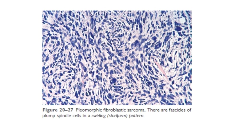
MORPHOLOGY Rhabdomyosarcoma is histologically subclassified into the embryonal, alveolar, and pleomorphic variants. The gross appearance of these tumors is variable. Some, particularly the embryonal variant when arising near the mucosal surfaces of the bladder or vagina, can manifest as soft, gelatinous, grapelike masses, designated sarcoma botryoides. In other cases they are poorly defined, infiltrating tanwhite masses. The rhabdomyoblast is the diagnostic cell in all types; it has granular eosinophilic cytoplasm rich in thick and thin filaments. The rhabdomyoblasts may be round or elongated; the latter are known as tadpole or strap cells (Fig. 20– 28) and may contain cross-striations visible by light microscopy.

The diagnosis of rhabdomyosarcoma is based on the demonstration of skeletal muscle differentiation, either in the form of sarcomeres under the electron microscope or by immunohistochemical demonstration of skeletal muscle–specific transcription factors such as myogenin and MYOD-1, and the muscle-associated intermediate filament desmin.

Rhabdomyosarcomas are aggressive neoplasms treated with a combination of surgery, chemotherapy, and radiation. Location, histologic appearance, and tumor genetics all impact the likelihood of cure, with progressively worsening rates for embryonal, pleomorphic, and alveolar variants, in that order. The malignancy is curable in almost two thirds of children; the prognosis is much less favorable in adults with the pleomorphic type.

SMOOTH MUSCLE • LEIOMYOMA • LEIOMYOSARCOMA

*. COUNTING mitoses is MUCH more important than getting a visual feel for pleormorphism and hyperchromasia in the evaluation of soft tissue tumors, as to whether they are benign or sarcomas, i. e. , you can have almost normal looking smooth muscle, but an increased mitotic rate of more than a few per 10 HPFs (High Power Fields), would be enough criteria for a sarcoma. *Conversely, you may see extreme pleomorphism, but if the mitotic rate is not increased, it is usually benign! It takes a lot of COURAGE and SAVVY to call a pleomorphic leiomyoma benign.

The MAIN difference between leiomyomas and leiomyosarcomas is the number of mitoses per high power field! How many do you see here? COUNTING mitoses is MUCH more important than getting a visual feel for pleormorphism and hyperchromasia in the evaluation of soft tissue tumors, as to whether they are benign or sarcomas. Please remember this.

VASCULAR • HEMANGIOMA • LYMPHANGIOMA • HEMANGIOENDOTHELIOMA • HEMANGIOPERICYTOMA • ANGIOSARCOMA
")
PERIPHERAL NERVE • NEUROFIBROMA • SCHWANNOMA • GRANULAR CELL TUMOR • MALIGNANT (SCHWANNOMA)

UNCERTAIN • SYNOVIAL SARCOMA • ALVEOLAR “SOFT PART” SARCOMA • EPITHELIOD SARCOMA

ALLAH HAFIZ
- Slides: 15