
11 Biological Membranes and Transport 2017 W H
11| Biological Membranes and Transport © 2017 W. H. Freeman and Company
Lipids Aggregate into Structures in Water • Three major structures are observed: – micelles – bilayers – liposomes • Structures formed depend on: – type of lipid – concentration

Micelle • Forms in the solution of amphipathic molecules that have larger, more polar head than tail – fatty acids – sodium dodecyl sulfate • Micelles are composed of a few dozen to a few thousand lipid molecules. • Aggregation of individual lipids into micelles is concentration dependent.
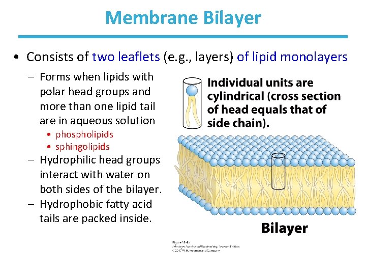
Membrane Bilayer • Consists of two leaflets (e. g. , layers) of lipid monolayers – Forms when lipids with polar head groups and more than one lipid tail are in aqueous solution • phospholipids • sphingolipids – Hydrophilic head groups interact with water on both sides of the bilayer. – Hydrophobic fatty acid tails are packed inside.

Vesicle (Liposome) • Small bilayers will spontaneously seal into spherical vesicles in a concentration-dependent manner. • Synthetic vesicle membranes can be made in vitro and can contain artificially inserted proteins. • The central aqueous cavity can enclose dissolved molecules. • They are useful artificial carriers of molecules (e. g. , drugs). • Vesicles fuse readily with cell membranes or with each other.
What Are Membranes? • Complex lipid-based structures that form pliable sheets • Composed of a variety of lipids and proteins • All cells have a cell membrane, which separates the cell from its surrounding. • Eukaryotic cells have various internal membranes that divide the internal space into compartments (i. e. , organelles).
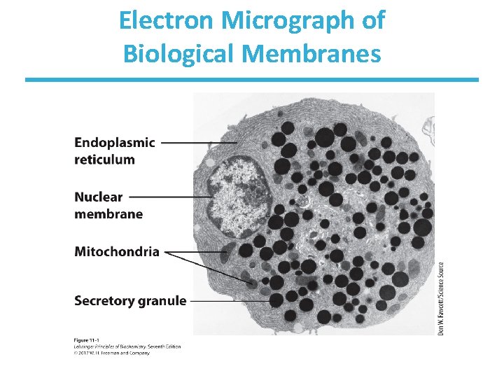
Electron Micrograph of Biological Membranes
Functions of Membranes • Define the boundaries of the cell • Allow import and export – Selective import of nutrients (e. g. lactose) – Selective export of waste and toxins (e. g. antibiotics) • Retain metabolites and ions within the cell • Sense external signals and transmit information into the cell • Provide compartmentalization within the cell – separate energy-producing reactions from energy-consuming ones – keep proteolytic enzymes away from important cellular proteins • Produce and transmit nerve signals • Store energy as a proton gradient • Support synthesis of ATP
Common Features of Membranes • Sheet-like flexible structure, 30– 100 Å (3– 10 nm) thick • Main structure is composed of two leaflets of lipids (bilayer) – with the exception of archaebacteria: monolayer of bifunctional lipids • Form spontaneously in aqueous solution and are stabilized by noncovalent forces, especially hydrophobic effect • Protein molecules span the lipid bilayer • Asymmetric – – Some lipids are found more commonly “inside. ” Some lipids are found more commonly “outside. ” Carbohydrate moieties are attached on the outer leaflet. They can be electrically polarized. • Fluid structures: two-dimensional solution of oriented lipids
Fluid Mosaic Model of Membranes • Proposed in 1972 by Singer and Nicholson (UCSD) • Lipids form a viscous, two-dimensional solvent into which proteins are inserted and integrated more or less deeply. • Proteins can either be embedded in or associated with the membrane: – Integral proteins are firmly associated with the membrane, often spanning the bilayer. – Peripheral proteins are weakly associated and can be removed easily. • Some are noncovalently attached. • Some are linked to membrane lipids.
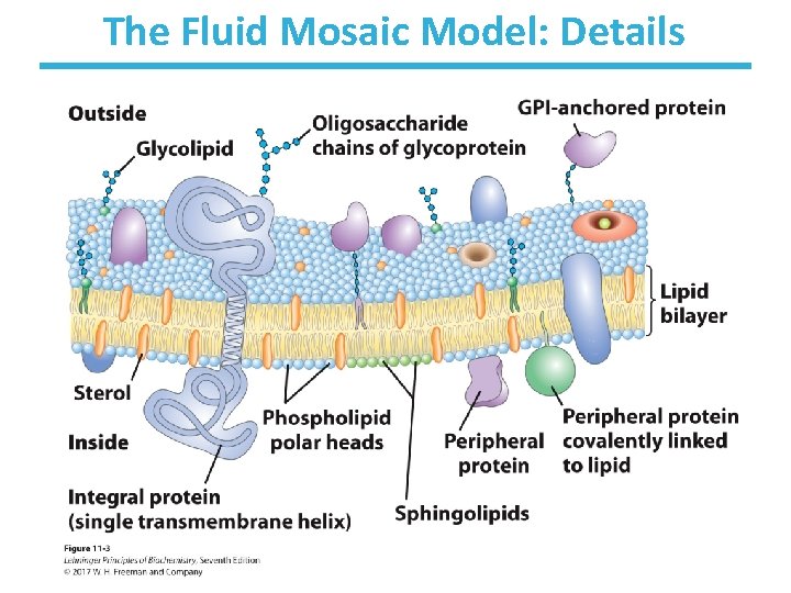
The Fluid Mosaic Model: Details
The Composition of Membranes • Lipid composition of membranes varies by: – organisms – tissues – organelles • Ratio of lipid to protein varies type of phospholipid varies abundance and type of sterols varies lack of sterols in prokaryotes cholesterol predominant in the plasma membrane, virtually absent in mitochondria - galactolipids abundant in plant chloroplasts but almost absent in animals -
Membrane Bilayers Are Asymmetric • Two leaflets have different lipid compositions. • The outer leaflet is often more positively charged. • Phosphatidylserine outside has a special meaning: – platelets: activates blood clotting – other cells: marks the cell for destruction
Functions of Proteins in Membranes • Receptors: detecting signals from outside – – light (opsin) hormones (insulin receptor) neurotransmitters (acetylcholine receptor) pheromones (taste and smell receptors) • Channels, gates, pumps – nutrients (maltoporin) – ions (K-channel) – neurotransmitters (serotonin reuptake protein) • Enzymes – lipid biosynthesis (some acyltransferases) – ATP synthesis (F 0 F 1 ATPase/ATP synthase)
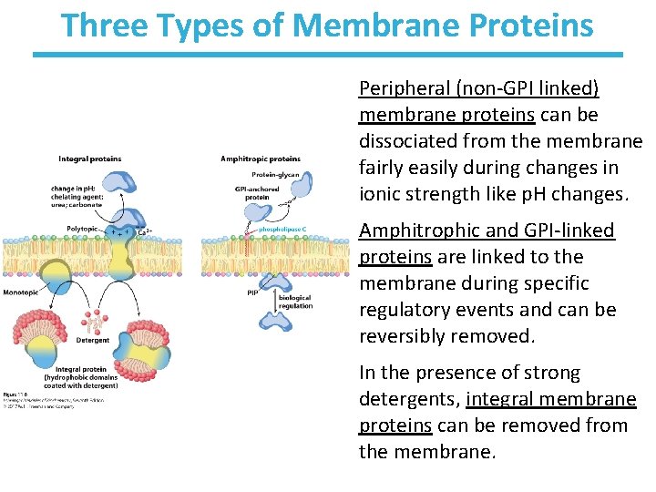
Three Types of Membrane Proteins Peripheral (non-GPI linked) membrane proteins can be dissociated from the membrane fairly easily during changes in ionic strength like p. H changes. Amphitrophic and GPI-linked proteins are linked to the membrane during specific regulatory events and can be reversibly removed. In the presence of strong detergents, integral membrane proteins can be removed from the membrane.
Peripheral Membrane Proteins • Associate with the polar head groups of membranes • Relatively loosely associated with membrane – through ionic interactions with the lipids or aqueous domains of integral membrane proteins • Removed by disrupting ionic interactions either with high salt or change in p. H • Purified peripheral membrane proteins are no longer associated with any lipids.
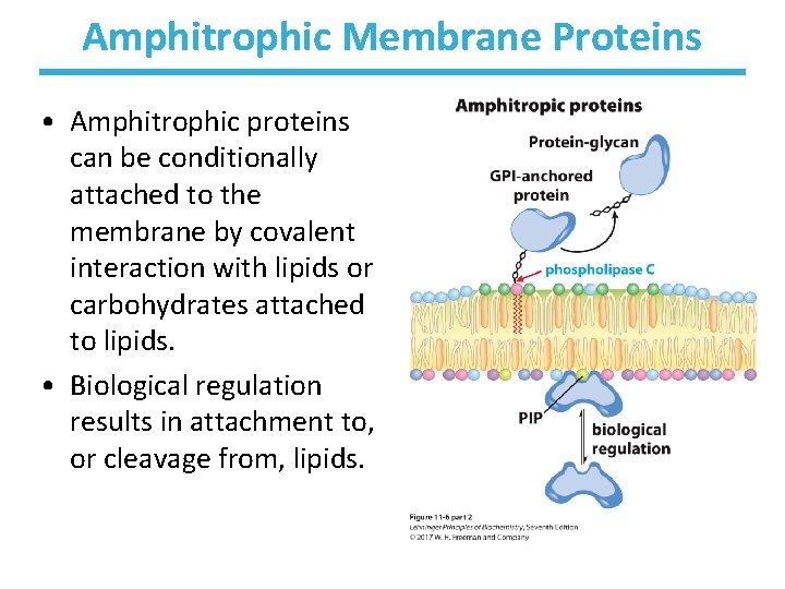
Amphitrophic Membrane Proteins • Amphitrophic proteins can be conditionally attached to the membrane by covalent interaction with lipids or carbohydrates attached to lipids. • Biological regulation results in attachment to, or cleavage from, lipids.
Lipid-linked Membrane Proteins
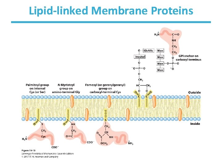
Lipid Anchors • Some membrane proteins are lipoproteins. • They contain a covalently linked lipid molecule. – – long-chain fatty acids isoprenoids sterols glycosylated phosphatidylinositol (PGI) • The lipid part can become part of the membrane. • The protein is now anchored to the membrane. – reversible process – allows targeting of proteins – Some, such as GPI anchors are found only on the outer face of plasma membrane.
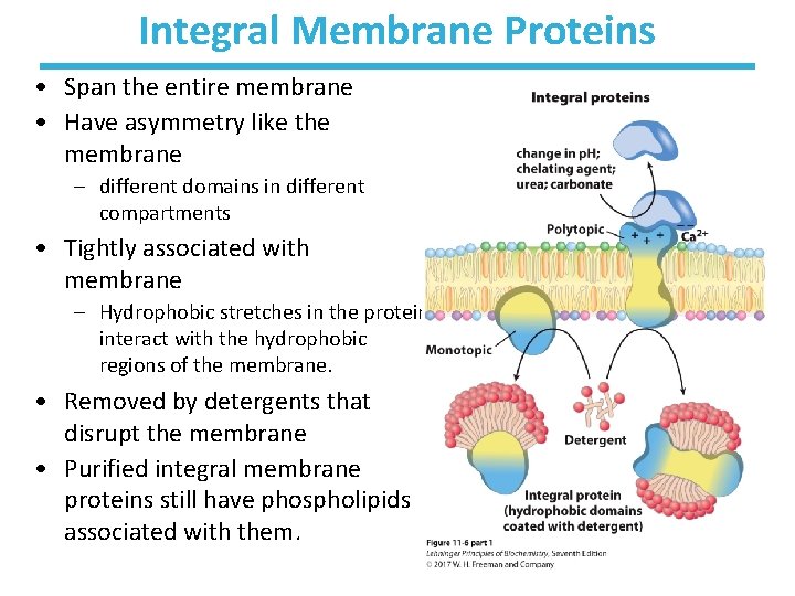
Integral Membrane Proteins • Span the entire membrane • Have asymmetry like the membrane – different domains in different compartments • Tightly associated with membrane – Hydrophobic stretches in the protein interact with the hydrophobic regions of the membrane. • Removed by detergents that disrupt the membrane • Purified integral membrane proteins still have phospholipids associated with them.
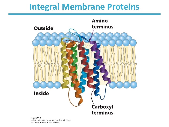
Integral Membrane Proteins
Physical Properties of Membranes • Dynamic and flexible structures • Can exist in various phases and undergo phase transitions • Not permeable to large polar solutes and ions • Permeable to small polar solutes and nonpolar compounds • Permeability can be artificially increased by chemical treatment. – when we want to get DNA into the cell
Membrane Phases • Depending on their composition and the temperature, the lipid bilayer can be in gel or fluid phase – liquid-ordered state (i. e. , “gel phase”): individual molecules do not move around – liquid-disordered state (i. e. , “fluid phase”): individual molecules can move around • Heating causes phase transition from the gel to fluid. • Under physiological conditions, membranes are more fluid-like than gel-like. – must be fluid for proper function
Organisms Can Adjust the Membrane Composition • Membrane fluidity is determined mainly by the fatty acid composition and melting point. • More fluid membranes require shorter and more unsaturated fatty acids. – Melting temperature decreases as double bonds are added. – Melting temperature increases with length of saturated fatty acids. • At higher temperatures, cells need more long, saturated fatty acids. • At lower temperatures, cells need more unsaturated fatty acids.
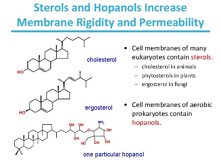
Sterols and Hopanols Increase Membrane Rigidity and Permeability • Cell membranes of many eukaryotes contain sterols. cholesterol – cholesterol in animals – phytosterols in plants – ergosterol in fungi ergosterol • Cell membranes of aerobic prokaryotes contain hopanols. one particular hopanol
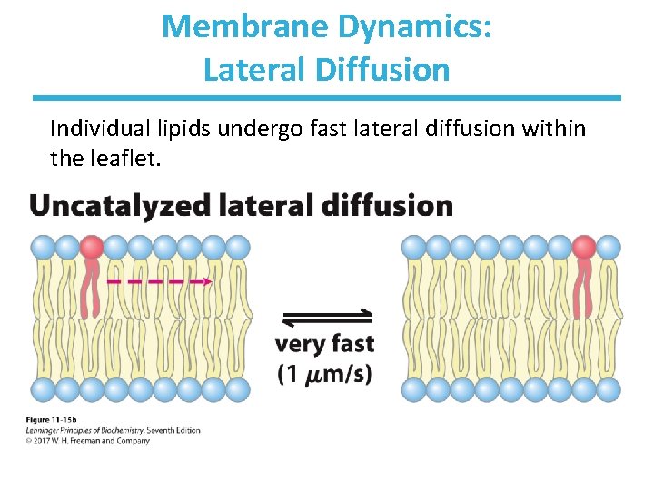
Membrane Dynamics: Lateral Diffusion Individual lipids undergo fast lateral diffusion within the leaflet.
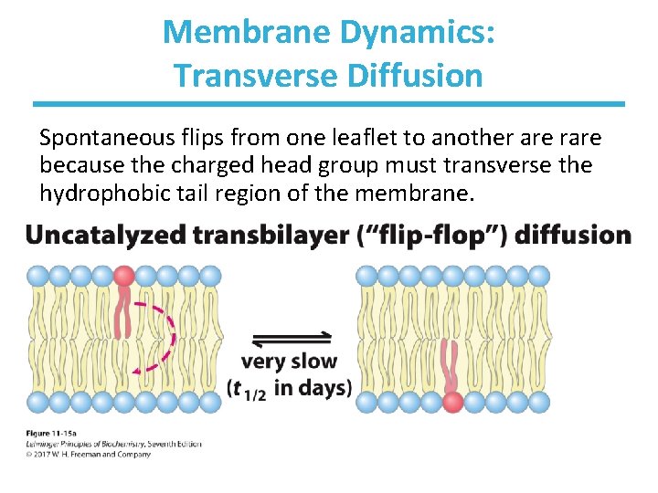
Membrane Dynamics: Transverse Diffusion Spontaneous flips from one leaflet to another are rare because the charged head group must transverse the hydrophobic tail region of the membrane.
Transport Across Membranes • Cell membranes are permeable to small nonpolar molecules that passively diffuse through the membrane. • Passive diffusion of polar molecules involves desolvation and thus has a high activation barrier. • Transport across the membrane can be facilitated by proteins that provide an alternative diffusion path. • Such proteins are called transporters or permeases.
Transport Across Membranes • Transport across a membrane must be energetically favorable. – Concentration dependence: The solute moves toward equilibrium across the membrane. – Electrochemical dependence: The solute moves toward charge equilibrium across the membrane.
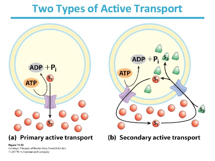
Two Types of Active Transport
Proton Transport and Chemical Energy of ATP • Energy of ATP hydrolysis can be used to drive protons through the membrane. – p. H control in the cell by F-type ATPase • Energy of the proton gradient can be used to synthesize ATP. – in chloroplast and mitochondrial membranes by ATP synthase
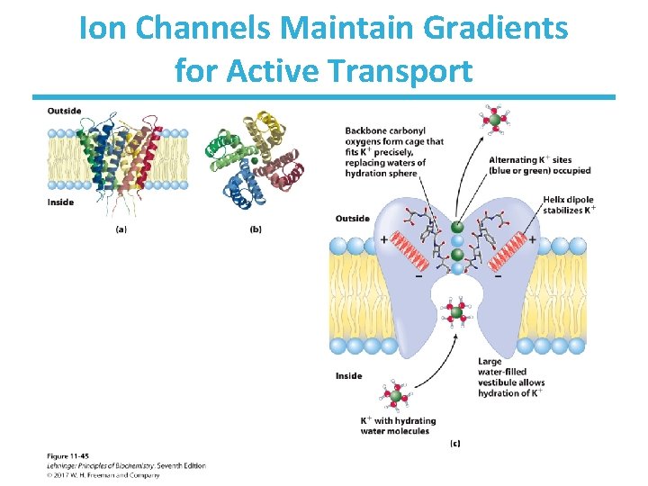
Ion Channels Maintain Gradients for Active Transport
Chapter 11: Summary In this chapter, we learned that: • lipids can form micelles, bilayers, and liposomes • membranes are composed of various lipids and proteins • properties of the bilayer depend on the lipid composition, which varies strongly from: – organism to organism – tissue to tissue – organelle to organelle • membrane proteins are found in three major classes and play a variety of structural and functional roles, especially in the transport of solutes across the membrane • active transport of solutes across membranes requires ATP but can be accomplished in many different ways
- Slides: 33