11 2 MOVEMENT Exoskeletons Exoskeletons are found in


- Slides: 19
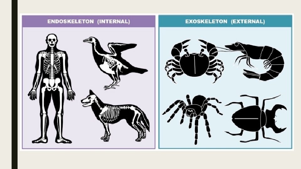
11. 2 MOVEMENT
Exoskeletons ■ Exoskeletons are found in many invertebrates including arthropods, corals and molluscs. ■ In animals with exoskeletons, the muscles for movement are attached to the inner surface of the exoskeleton.
Endoskeleton ■ A bony endoskeleton is the internal support structure of some animals including vertebrates. ■ The endoskeleton functions as an attachment site for muscles and provides a means to transmit muscular force. ■ Muscles pull on the skeleton to create movement about joints. ■ Muscles work in opposing (antagonistic) pairs to create opposing movement.
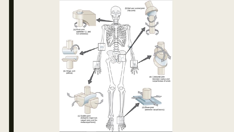
Joints ■ Bones are too rigid to bend. ■ To allow movements, the human skeletal system consists of bones held together at joints by flexible connective tissues called ligaments. ■ Joints are points of contact between bones or between cartilage and bones. ■ Joints may be classified structurally as fibrous, cartilaginous or synovial. ■ Each has there own degree of movement.
Synovial Joints ■ Synovial joints allow free movement of body parts in varying directions (one, two or three planes). ■ The elbow joint is a hinge joint and typical of a synovial joint. ■ Synovial joints are reinforced by ligaments. ■ The joint capsule encloses synovial fluid, which reduced friction and absorbs shock.
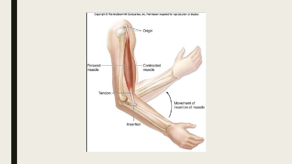
Clarification of vocabulary ■ ■ Joint: Ligament: Muscle: Tendon:
Antagonistic Muscles ■ Antagonistic muscles are muscle pairs that have opposite actions to each other. ■ Together, their opposing actions bring about movement of body parts. ■ Muscles can only pull and not push. ■ Muscles have two attachments: an origin and an insertion. ■ During contraction the insertion moves towards the origin.
Antagonistic Muscle Pairs in an Insect Leg ■ Work together to move the leg. ■ Extensor causes the leg to extend ■ Flexor causes the leg to flex(bend) ■ The contraction of the extensor muscle is what propels the insect through the air.
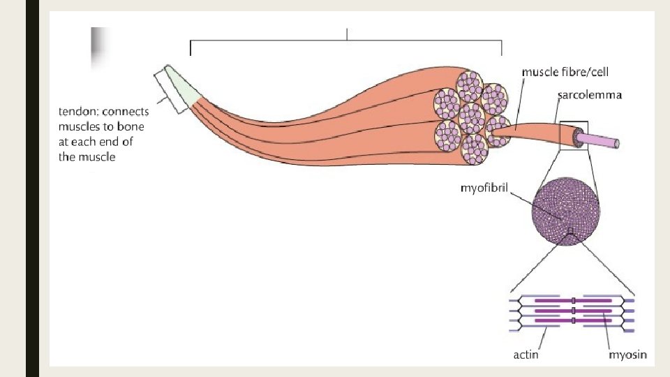
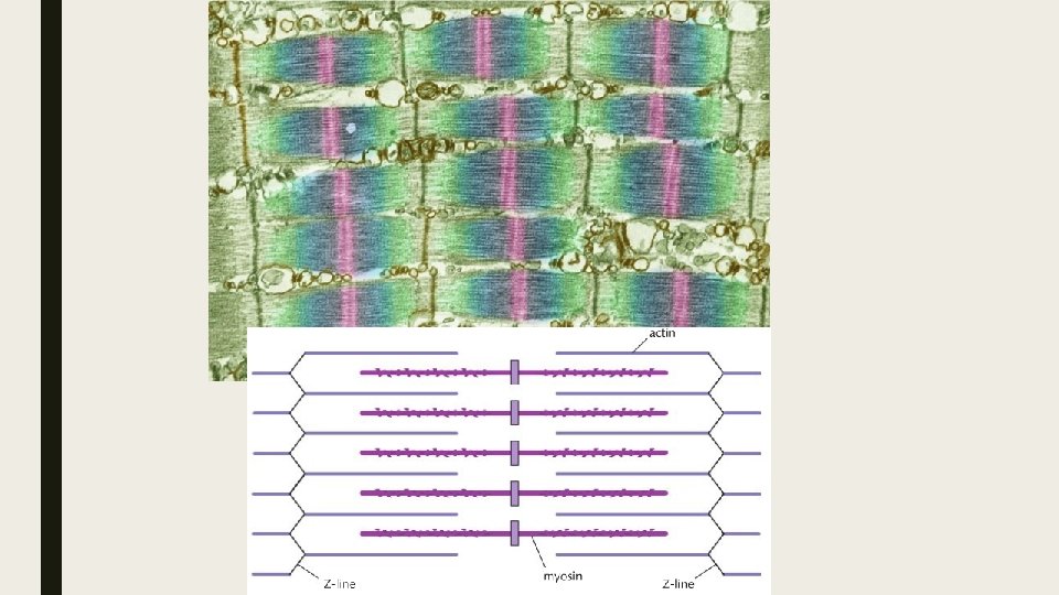
Skeletal Muscle Structure and Function ■ Skeletal muscle is organized into bundles of muscle cells (fibers). ■ The muscle fibers are made up of repeating contractile units called sarcomeres. ■ Skeletal muscles is organized into bundles of muscle cells (fibers). ■ Each fiber is a single cell with many nuclei and each fiber is itself a bundle of smaller myofibrils arranged lengthwise. ■ Each myofibril is in turn composed of two kinds of myofilaments (thick and thin), which overlap to form light and dark bands.
■ The response of a single muscle fiber to stimulation is to contract maximally or not at all; its response is referred to as the all or none. ■ If the stimulus is not strong enough to produce an action potential, the muscle fiber will not respond.
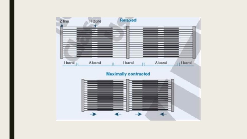
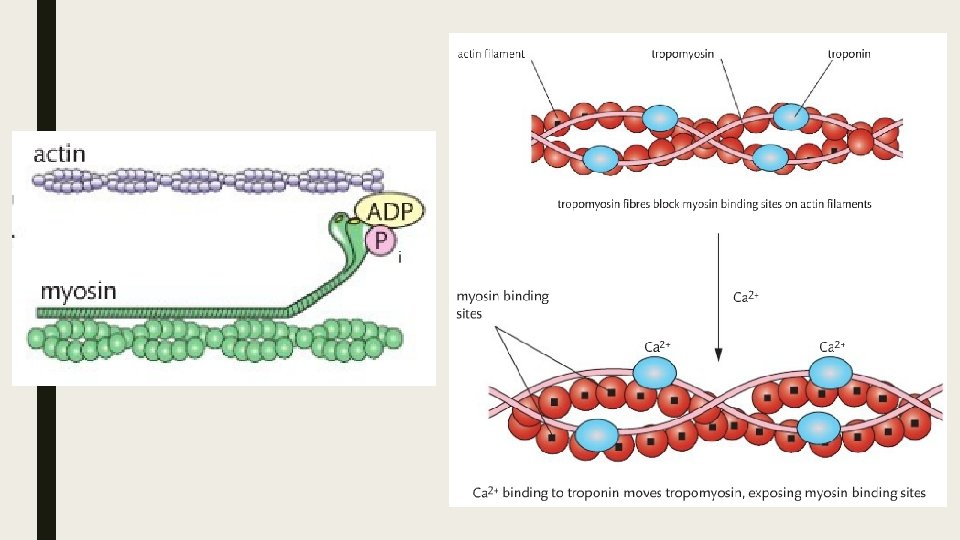
The Sliding Filament Theory Watch Me!!! ■ The sliding filament theory describes how muscle contraction occurs when the thick and thin myofibrils of a muscle fiber slide past one another. ■ Calcium ions and ATP are required. ■ The structure and arrangement of the thick and thin filaments in a muscle fiber make it possible for them to slide past each other and cause shortening (contraction) of the muscle. ■ The ends of the thick myosin filaments have cross bridges that can link to adjacent thin actin filaments. ■ When the cross bridges of the thick filament connect to the thin filaments, a shape change moves one filament past the other. ■ Two things are necessary for cross bridge formation: calcium ions, which are released from the sarcoplasmic reticulum when the muscle receives an action potential, and ATP, which is present in the muscle fiber and is hydrolyzes by ATPase enzyme on the myosin. ■ When the cross bridges attach and detach in sarcomeres throughout the muscle cell,