
10222014 1 BYAli Rezvani Ph D Molecular Medicine

10/22/2014 1

پﺮیﺘﻮﺋﻦ ﻭ پﺮیکﺎﺭﺩ ، آﻨﺎﻟیﺰ ﻣﺎیﻌﺎﺕ پﻠﻮﺭ BYAli Rezvani Ph. D. , Molecular Medicine Assistant Professor of Molecular Medicine at Clinical Biochemistry Department, Kermanshah University of Medical Sciences : 10/22/2014 2
BY: ALI REZVANI Msc Clinical Biochemistry “ Phd of Molecular Medcine 10/22/2014 3
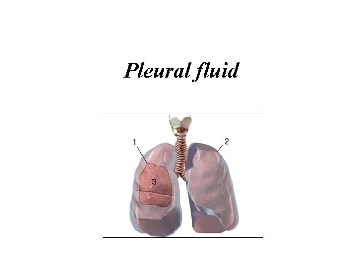
Pleural fluid
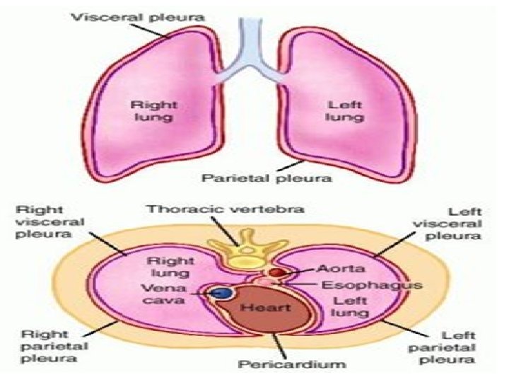
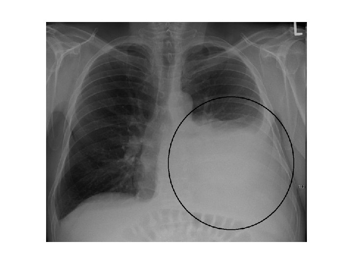
• An accumulation of fluid, called an effusion, results from an imbalance of fluid production and reabsorption. • Excessive amounts of such fluid can impair breathing by limiting the expansion of the lungs during ventilation.
Types of fluids • Four types of fluids can accumulate in the pleural space: • Serous fluid (hydrothorax) • Blood (haemothorax) • Chyle (chylothorax) • Pus (pyothorax or empyema)
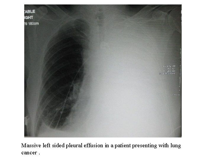
Massive left sided pleural effusion in a patient presenting with lung cancer.
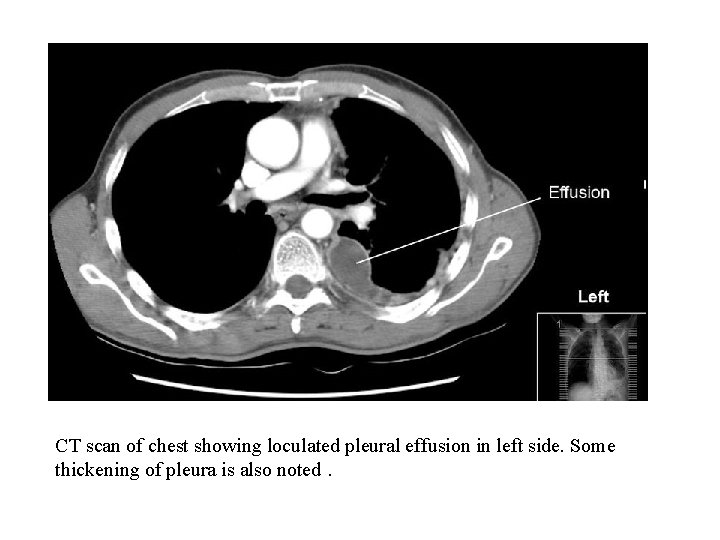
CT scan of chest showing loculated pleural effusion in left side. Some thickening of pleura is also noted.
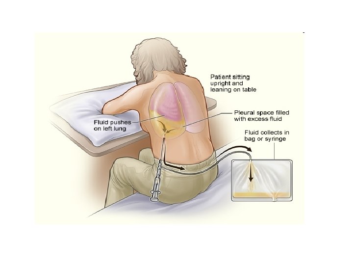
SPECIMEN COLLECTION • Thoracentesis • A needle is inserted through the back of the chest wall in the sixth, seventh, or eighth intercostal space
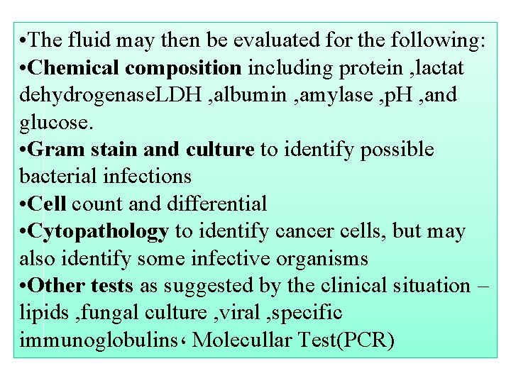
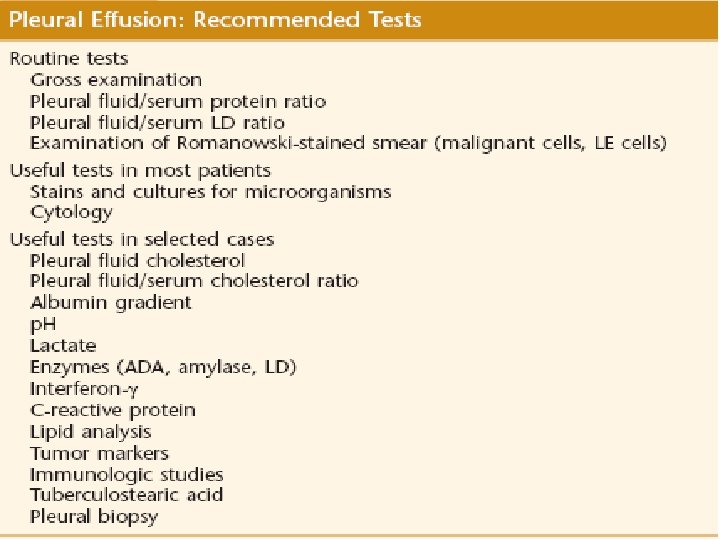
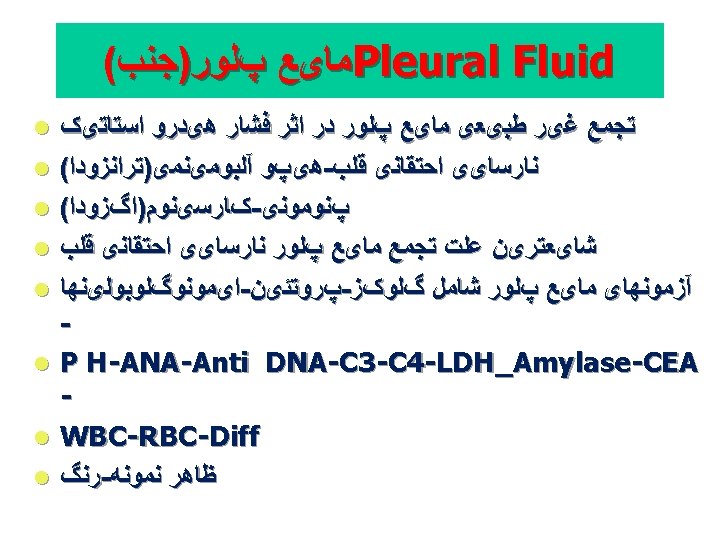
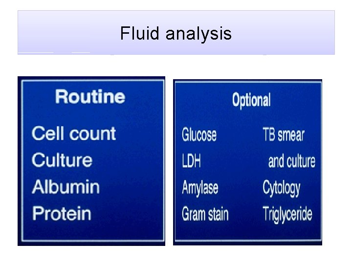
• The fluid may then be evaluated for the following: • Chemical composition including protein , lactat dehydrogenase. LDH , albumin , amylase , p. H , and glucose. • Gram stain and culture to identify possible bacterial infections • Cell count and differential • Cytopathology to identify cancer cells, but may also identify some infective organisms • Other tests as suggested by the clinical situation – lipids , fungal culture , viral , specific immunoglobulins، Molecullar Test(PCR)
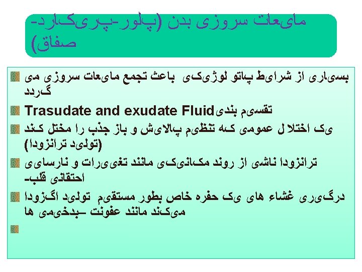
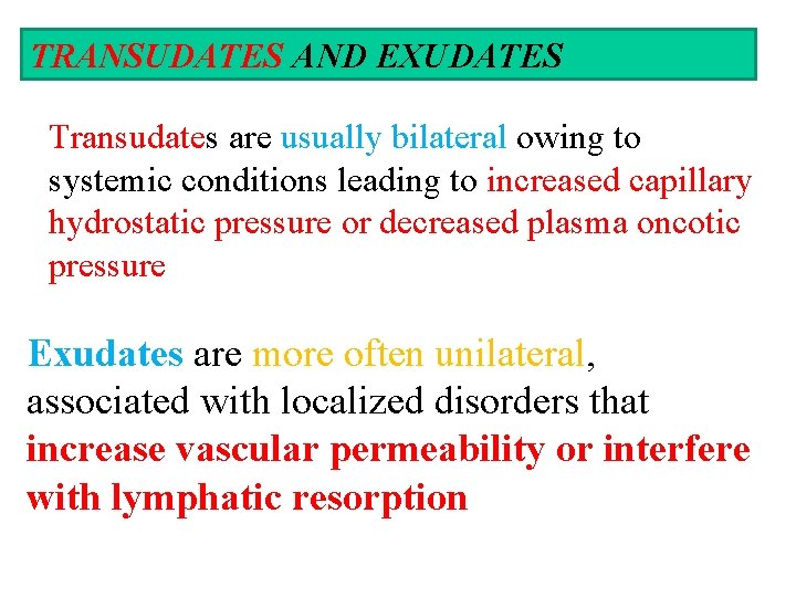
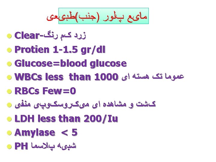
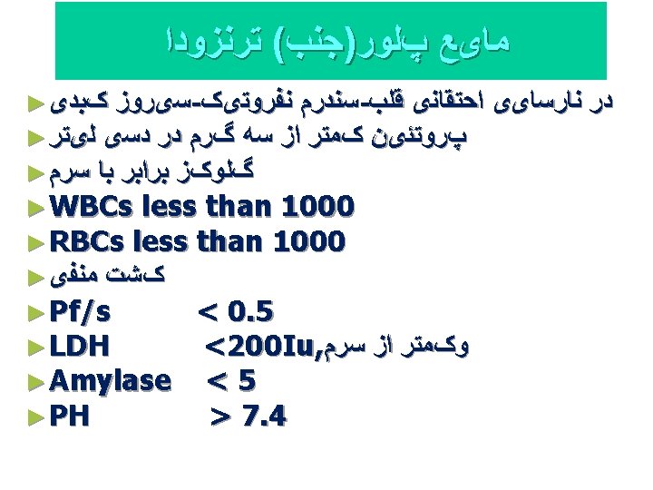
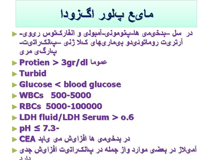
TRANSUDATES AND EXUDATES Transudates are usually bilateral owing to systemic conditions leading to increased capillary hydrostatic pressure or decreased plasma oncotic pressure Exudates are more often unilateral, associated with localized disorders that increase vascular permeability or interfere with lymphatic resorption
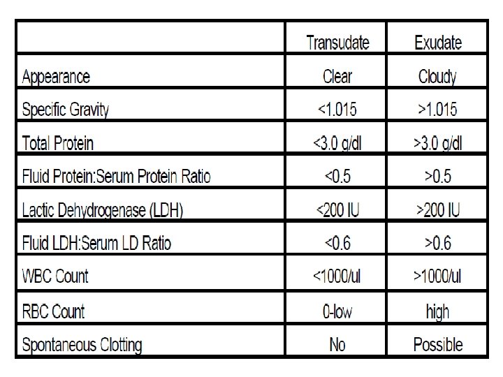
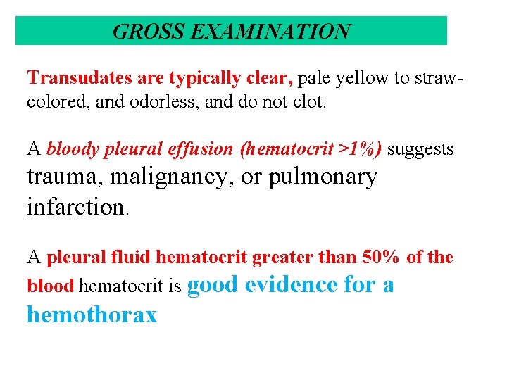
GROSS EXAMINATION Transudates are typically clear, pale yellow to strawcolored, and odorless, and do not clot. A bloody pleural effusion (hematocrit >1%) suggests trauma, malignancy, or pulmonary infarction. A pleural fluid hematocrit greater than 50% of the blood hematocrit is good evidence for a hemothorax
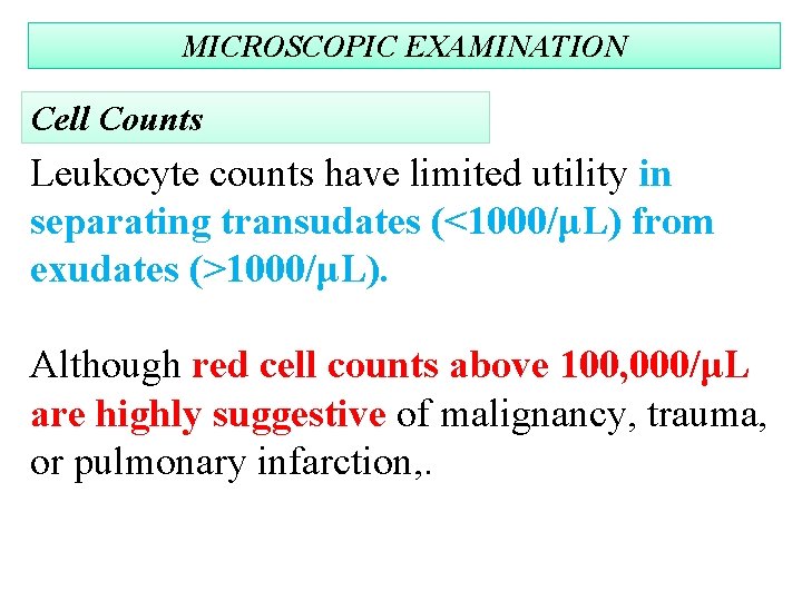
MICROSCOPIC EXAMINATION Cell Counts Leukocyte counts have limited utility in separating transudates (<1000/μL) from exudates (>1000/μL). Although red cell counts above 100, 000/μL are highly suggestive of malignancy, trauma, or pulmonary infarction, .
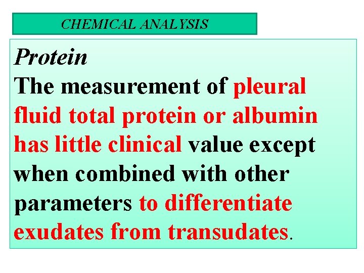
CHEMICAL ANALYSIS Protein The measurement of pleural fluid total protein or albumin has little clinical value except when combined with other parameters to differentiate exudates from transudates.
Glucose The glucose level of normal pleural fluid, transudates is similar to serum levels. Decreased pleural fluid glucose, accepted as a level below 60 mg/d. L or a pleural fluid/serum glucose ratio less than 0. 5, is most consistent and dramatic in rheumatoid pleuritis and grossly purulent parapneumonic exudates
Lactate Pleural fluid lactate levels can be a useful adjunct in the rapid diagnosis of infectious pleuritis. Levels are significantly higher in bacterial and tuberculous pleural infections than in other pleural effusions. Values greater than 90 mg/d. L (10 mmol/L) have a positive predictive value for infectious pleuritis of 94% and a negative predictive value of 100%.
Amylase: elevations above the serum level (usually 1. 5– 2. 0 or more times greater) indicate the presence of pancreatitis, esophageal rupture, or malignant effusion.
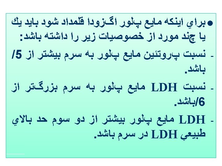
Lactate dehydrogenase Pleural fluid LD levels rise in proportion to the degree inflammation. In addition to their use in separating exudates from transudates, declining LD levels during the course of an effusion indicate that the inflammatory process is resolving. Conversely, increasing levels indicate a worsening condition requiring aggressive workup or treatment. of
Interferon-γ: Pleural fluid interferon (IFN)-γ levels are significantly increased in the pleural fluid of patients with tuberculous pleuritis. The sensitivity of levels of 3. 7 IU/L or greater is 99%, and the pecificity is 98%
p. H Pleural fluid p. H measurement has the highest diagnostic accuracy in assessing the prognosis of parapneumonic (pneumonia-related) effusions A parapneumonic exudate with a p. H greater than 7. 30 generally resolves with medical therapy alone. A p. H less than 7. 20 indicates a complicated parapneumonic, requiring surgical drainage. A p. H below 6. 0 is characteristic of esophageal rupture,
Lipids Lipid measurements are also helpful in identifying chylous effusions Thus, pleural fluid triglyceride levels above 110 mg/d. L indicate a chylous effusion; Nonchylous effusions generally have triglyceride levels below 50 mg/d. L (0. 56 mmol/L) and no chylomicrons on electrophoresis
IMMUNOLOGIC STUDIES Approximately 5% of patients with RA and 50% with SLE develop pleural effusions sometime during the course of their disease. RF is commonly present in pleural effusions associated with seropositive RA. ANA titers may be useful in the diagnosis effusion due to lupus pleuritis of

MICROBIOLOGICAL EXAMINATION Bacteria most commonly associated with parapneumonic effusions are Staphylococcus aureus, Streptococcus pneumoniae, β-hemolytic group A streptococci, enterococci, and some gram-negative bacilli. Anaerobic bacteria are isolated in a significant proportion of cases, so both anaerobic and aerobic cultures should be performed. The sensitivity of the Gram stain is approximately 50% For patients with suspected M. tuberculosis, direct staining of tuberculous effusions for acid-fast bacteria has a sensitivity of 20%– 30%, and positive cultures are found in 50%– 70% of cases
Peritonial fluid
l. Up to 50 ml Fluid normally present in peritoneal cavity l. Peritoneal effusion is called Ascites l. Laboratory criteria for dividing ascitic fluid into transudate and exudate is not well defined as it is for pleural fluid
Paracentesis : Indications Most Common causes(90% of cases): • chronic liver diseases ( cirrhosis) • Intra-abdominal malignancy • Congestive Heart Failure • Mycobacterium tuberculosis
Other causes of ascites Hypolalbuminemia • • • Nephrotic syndrome Protein-losing enteropathy malnutrition
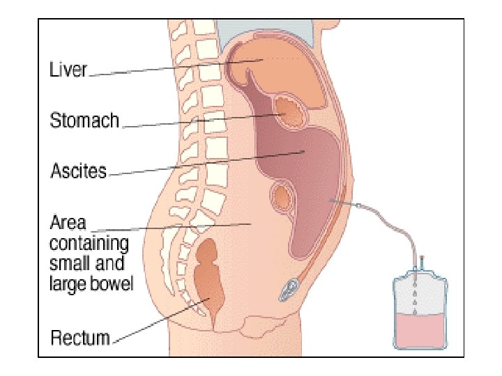
Fluid analysis
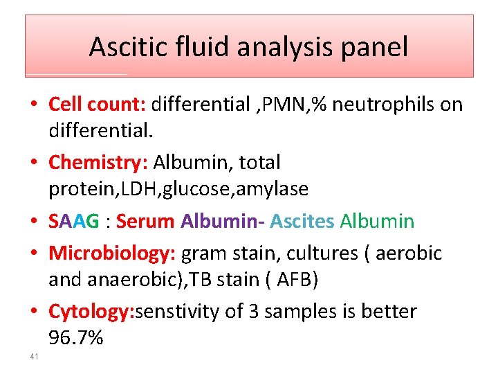
Ascitic fluid analysis panel • Cell count: differential , PMN, % neutrophils on differential. • Chemistry: Albumin, total protein, LDH, glucose, amylase • SAAG : Serum Albumin- Ascites Albumin • Microbiology: gram stain, cultures ( aerobic and anaerobic), TB stain ( AFB) • Cytology: senstivity of 3 samples is better 96. 7% 41
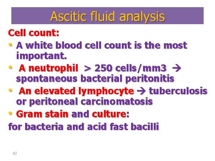
Ascitic fluid analysis Cell count: • A white blood cell count is the most important. • A neutrophil > 250 cells/mm 3 spontaneous bacterial peritonitis • An elevated lymphocyte tuberculosis or peritoneal carcinomatosis • Gram stain and culture: for bacteria and acid fast bacilli 42
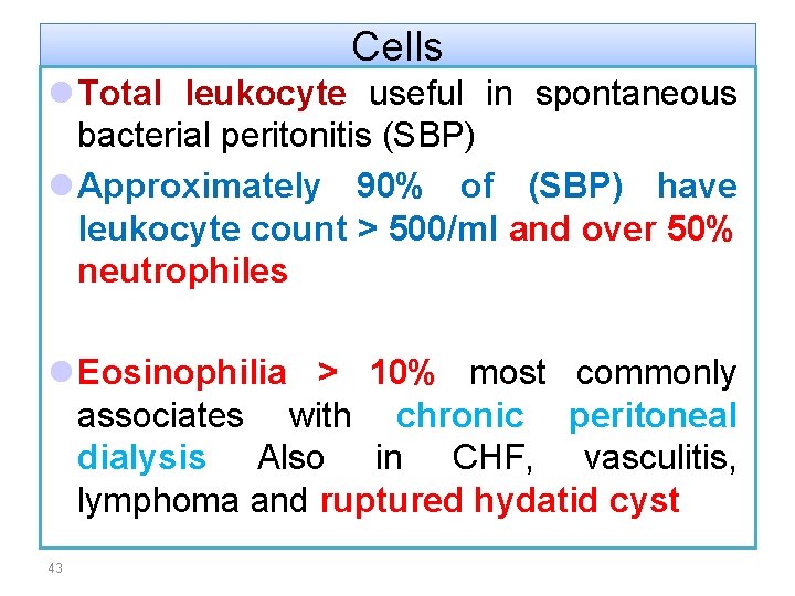
Cells l Total leukocyte useful in spontaneous bacterial peritonitis (SBP) l Approximately 90% of (SBP) have leukocyte count > 500/ml and over 50% neutrophiles l Eosinophilia > 10% most commonly associates with chronic peritoneal dialysis Also in CHF, vasculitis, lymphoma and ruptured hydatid cyst 43
• Total protein: Helpful in diagnosing spontaneous bacterial peritonitis Pts with a value<1 g/dl protein and glucose of <50 mg/dl have high risk of SBP • Cytology: for malignant cells • Amylase: to exclude pancreatic ascites
l Amylase activity in normal peritoneal fluid is similar to blood levels l l A fluid amylase level greater than three times of serum value is good evidence of pancreas- related ascitis and also in GI perforation l l Increase CEA in peritoneal washing suggest a poor prognosis of gastric Ca
l Other studies of ascitic fluid to be considered l Lactate: An ascites lactate level of >25 mg/d. L was found to be 100% sensitive and specific in predicting active SBP p. H: In the same study, the combination of an ascites fluid p. H of <7. 35 and PMN count of >500 cells/m. L was 100% sensitive and 96% specific. spontaneous bacterial peritonitis l.
Pericardial Fluid
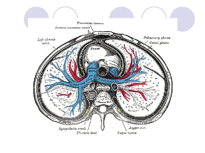
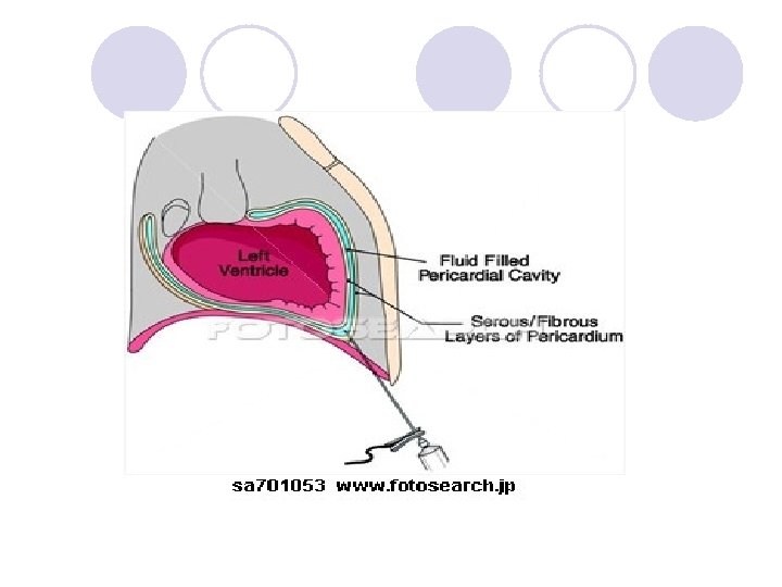
Pericardial Fluid l. Surrounding the heart is a sac known as the pericardium, which consists of two membranes.
l. In a healthy individual there is usually 15 -50 ml of clear, strawcoloured fluid. l l. However there is little data on the normal composition of pericardial fluid to serve as a reference
Pericardial effusion l A pericardial effusion is the presence of excessive pericardial fluid, this can be confirmed using an echocardiogram l Small effusions are not necessarily dangerous l Large and rapidly accumulating effusions may cause cardiac tamponade , a life-threatening complication, that puts pressure on the heart preventing the ventricles from filling correctly.
The most common causes are: a. neoplastic disease b. idiopathic pericarditis c. uremia d. following cardiac operation e. trauma
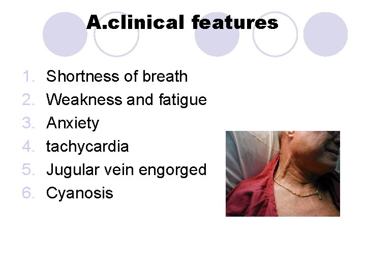
A. clinical features 1. 2. 3. 4. 5. 6. Shortness of breath Weakness and fatigue Anxiety tachycardia Jugular vein engorged Cyanosis
Pericardiocentesis l. Pericardiocentesis is a procedure used to remove the pericardial fluid from the pericardial cavity. l It is performed using a needle and under the guidance of an ultrasound
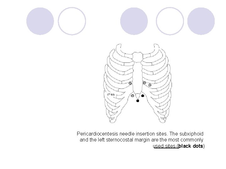
Pericardiocentesis needle insertion sites. The subxiphoid and the left sternocostal margin are the most commonly used sites (black dots)
Types l transudative( congestive heart failure , myxoedema , nephrotic syndrome) l Exudative( tuberculosis, ) l haemorrhagic( trauma, ) l Malignant (due to fluid accumulation caused by metastasis)

- Slides: 60