1 PULMONARY ARTERY CATHETER MONITORING RASOUL AZARFARIN MD


 Measurement of right heart pressure(RAP,")


![COMPONENTS OF SWAN-GANZ Normally has four[4] ports Proximal port – [Blue] used to measure](https://slidetodoc.com/presentation_image_h/343dcaff39665aa1f1c0ac341e24aa3a/image-8.jpg "COMPONENTS OF SWAN-GANZ Normally has four[4] ports Proximal port – [Blue] used to measure")

 RIJ: � � Right Atrium: 20 -25 cm Right Ventricle: 30")


 Normal mean value 8 -12 mm Hg Hypovolemia Low")

 Also known as: Pulmonary capillary wedge pressure PCWP PAW")


- Slides: 36
1
PULMONARY ARTERY CATHETER MONITORING RASOUL AZARFARIN, MD, FACC Professor of Anesthesiology Rajaie Cardiovascular Medical & Research Center
INTRODUCTION Swan-Ganz catheter has been in use for almost 30 years Initially developed for the management of acute myocardial infarction Now, widespread use in the management of a variety of critical illnesses and surgical procedures 3
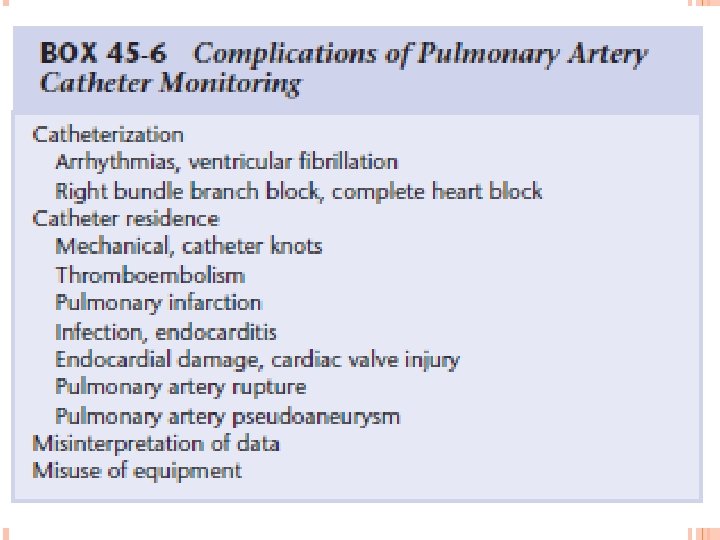
PAC Advantages � Provide lot of important haemodynamic parameters � Sampling site for Sv. O 2 Disadvantages � Costly � Invasive � Multiple complications (eg arrhythmia, catheter looping, balloon rupture, PA injury, pulmonary infarction etc) � Mortality not reduced and can be even higher Crit Care Med 2003; 31: 2734 -2741 JAMA 1996; 276 889 -897 4
INDICATION Haemodynamic measurement(cardiac output, stroke volume, systemic vascular resistance) Measurement of right heart pressure(RAP, PAP) Acute pulmonary hypertension Pulmonary embolism Cardiac tamponade Estimation or preload/left heart filling(PAOP) Derivation of oxygen variables(VO 2, DO 2) 5
INDICATIONS n Diagnostic indications: n n n n Shock states Differentiation of high vs low pressure pulmonary edema Primary pulmonary hypertension Intracardiac shunts Cardiac tamponade Pulmonary embolus Monitoring and management of complicated acute myocardial infarction n n n Assessing hemodynamic response to therapies Management of multiorgan failure Severe burns Hemodynamic instability after cardiac surgery Assessment of response to treatment in patients with primary pulmonary hypertension Therapeutic indications: n Aspiration of air emboli
PAC remains used for the management of patients with RVF , PH , weaning failure from cardiac origin and management of complex circulatory conditions in which the knowledge of PAP, PAOP and oxygenation parameters is considered to be important
COMPONENTS OF SWAN-GANZ Normally has four[4] ports Proximal port – [Blue] used to measure central venous pressure/RAP and injectate port for measurement of cardiac output Distal port – [Yellow] used to measure pulmonary artery pressure Balloon port – [Red] used to determine pulmonary wedge pressure; 1. 5 special syringe is connected Infusion port – [White] used for fluid infusion 8
9
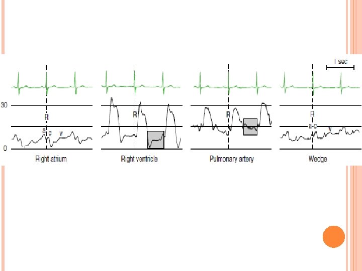
DISTANCES (ROUGH ESTIMATES) RIJ: � � Right Atrium: 20 -25 cm Right Ventricle: 30 -35 cm PA: 40 -45 cm Wedge 45 -55 If LIJ or R Ext Jugular, add 5 -10 cm If Femoral: add 15 cm If Antecubital: add 30 -35 cm Common problems: coiling in the right atrium or right ventricle – deflate balloon, withdrawn to 20 and refloat 10
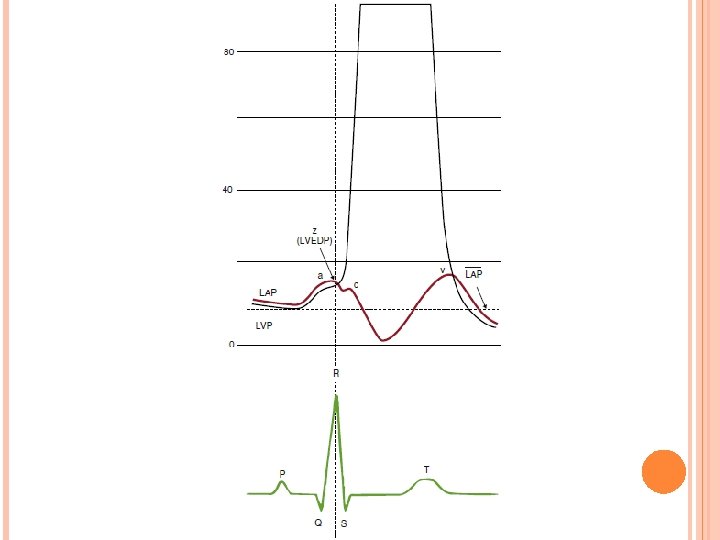
PULMONARY ARTERIAL PRESSURE MONITORING 11
CONFORMATION
PULMONARY ARTERY OCCLUSIVE PRESSURE (PAOP) Normal mean value 8 -12 mm Hg Hypovolemia Low (< 8 mm Hg) hypervolemia High (>12 mm Hg) 17
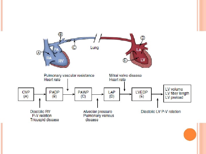
PAOP n Reflects left ventricular end diastolic volume n n Assumes a static column of blood from ventricle to catheter during diastole and consistent compliance Abnormalities are viewed in the context of SV/SI If high (>18) implies left ventricular dysfunction, especially if SV is low n If low (< 8) implies hypovolemia especially if SV is low n
PULMONARY ARTERY OCCLUSIVE PRESSURE (PAOP) Also known as: Pulmonary capillary wedge pressure PCWP PAW Wedge pressure Wedge 19
PAP WAVEFORM Similar in appearance to arterial waveform Waves Systolic pressure Dicrotic notch End diastolic pressure 21
PAP Normal values PA systolic pressure = 20 -30 mm Hg PA diastolic pressure = 8 -12 mm Hg About 25/10 Low Volume depletion Drugs High Pulmonary HTN Pulmonary Embolus Mitral stenosis LV failure Septal wall defects 22
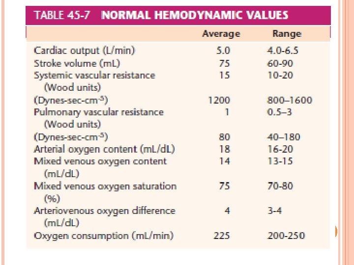
WHAT DOES A PAC TELL US? n Direct measurements n Calculated data Stroke volume (SV/SVI) n Cardiac output (CO/CI) n Vascular resistance (SVR, PVR) n Oxygen delivery CVP n PA (systolic and diasotolic) n PAOP (wedge) n Sv. O 2 (mixed) n n n Extended calculations CCO n Stroke work n End diastolic volume, EF n
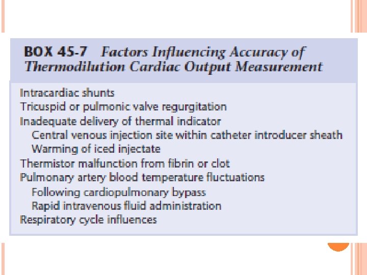
THERMODILUTION CARDIAC OUTPUT MONITORING A known volume of iced or room temperature fluid is injected as a bolus into the proximal (right atrium) lumen of the PAC, and the change in the pulmonary artery blood temperature is recorded by thermistor at the catheter tip
Thermodilution Cardiac Output Monitoring has become the de facto clinical standard for measuring CO because of its ease of implementation and the long clinical experience with its use in various settings
CO WAVEFORMS
Following the trend in cardiac output is probably more clinically useful than emphasizing any absolute value
FICK METHOD n CO = VO 2 / [Ca. O 2 – Cv. O 2] * 10 n S a. O 2 n and Sv. O 2 often substituted CO = VO 2 / [Sa. O 2 – Sv. O 2] * Hgb * 1. 34* 10 n VO 2 is not usually measured Can use 3. 5 m. L/kg or 125 m. L/m 2 n If metabolic rate is abnormal, the calculation may be incorrect n
Papillary muscle ischemia or Functional mitral regurgitation: PAC monitoring is well suited to detect this event by revealing the onset of new regurgitant v waves in the pulmonary artery or wedge pressure traces
CONTINUOUS CARDIAC OUTPUT n Newer generation catheter Uses continuous cardiac output measurements without need for bolusing n Allows for right heart “volumetric” data n n RVEDV, RVEF, and RVSV RVSW and RVSWI Also provides continuous Sv. O 2 measurements
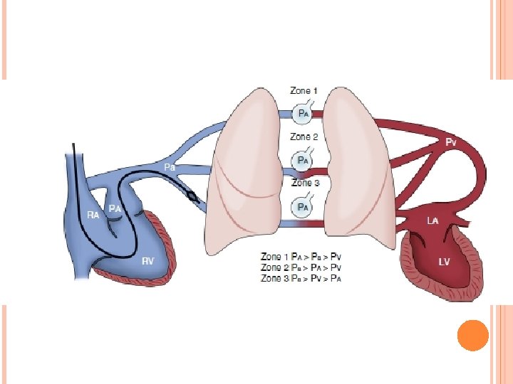
POINTS TO REMEMBER n Limitations in hemodynamic monitoring n Ventricular filling pressures do not always accurately reflect ventricular filling volume n n n The PAOP is normally slightly (1 -5 mm Hg) less than the PAD pressure n n n The pressure-volume relationship depends upon ventricular compliance If compliance changes, the pressure-volume relationship changes This relationship stills exists with pulm hypertension due to LV failure However, with an ↑ PVR or tachycardia (>125 bpm) this relationship may breakdown and the PAD becomes significantly higher than the PAOP The PAOP may not equal LVEDP when n n there is high alveolar pressures when the catheter tip is above the left atrium severe hypovolemia tachycardia (130 bpm) in mitral stenosis.
35
36
THANK YOU 37