1 NMR principles NMR parameters 2 outline 3

 : A=Z+N Nominal atomic mass The number of neutrons The number")

 : m=-1/2 △E 0 E m=+1/2 B 0")
 at 11. 74 T")
 Energy high X-RAY ULTRAVIOLET low INFRARED VISIBLE 400 nm short FREQUENCY RADIO")

 Z")
 defines the location of a nmr line along the rf")
 characterize scalar interactions (through-bond) between nuclei linked via a")

 is the fractional change in")
 describes the rate at")


 (B) (C ) (D ) MIXING t 2 t 1")
 H Hβ O 8. 25 Hα NH C Cβ Hβ 177.")
 Hγ 1 Hβ 121. 1 O 8. 44 N 63. 0")
 Hβ 3 Hβ 2")

 Val Gln Hα Ala NOESY Thr Ala’ Amide protons")

 Bax and grzesiek a H 140 C H 35")

 program Etotal =")
- Slides: 43
1 NMR principles NMR parameters 2 outline 3 4 5 NMR experiments Determination of NMR solution structure Conclusion 中研院生醫所 N 133 Tel. : 02 -26523035
Nuclear spin (I) : A=Z+N Nominal atomic mass The number of neutrons The number of protons (atomic number)
A nucleus with an even mass A and even charge Z, and therefore also an even N, will have a nuclear spin I of zero. (12 C, 16 O and 18 O). A nucleus with an even mass and odd charge (both Z and N odd) will exhibit an integer value of I. [2 H(I=1), 14 N(I=1) and 10 B(I=3)]. A nucleus with odd mass (Z odd and N even, or Z even and N odd) will have nuclear spin with an I value that we can express as n/2, where n is an odd integer. [1 H(I=1/2), 13 C(I=1/2) and 17 O(I=5/2)]. Nuclei with I= 0 NMR cannot be detected. Nuclei with I≠ 0 NMR can be detected. Different isotopes of the same element have different nuclear spins, some of which are detectable by NMR, others of which are not.
Number of spin states (2 I+1) : m=-1/2 △E 0 E m=+1/2 B 0 E=-m. B 0(γh/2π) B 0: magnetic field strength h : Planck’s constant m: spin quantum number γ: magnetogyric ratio A nucleus with spin I can have 2 I+1 spin states. Each of these states has its own spin quantum number m(m=-I, I+1, ‧‧‧, I-1, I). For nuclei with I=1/2, only two states are possible: m=+1/2 and m=-1/2.
Properties of nuclei in NMR studies Isotop Spi. Frequency (MHz) at 11. 74 T Nature abundance (%) Relative sensitivity e n 1 H 1/2 2 H 1 3 H 1/2 533. 3 12 C 0 ------- 13 C 1/2 125. 7 14 N 1 36. 1 99. 63 1. 01× 10 -3 15 N 1/2 50. 7 0. 37 1. 04× 10 -3 16 O 0 17 O 5/2 67. 8 19 F 1/2 470. 4 100 0. 83 31 P 1/2 202. 4 100 6. 63× 10 -2 500. 0 76. 7 ------ 99. 98 1. 0 1. 5× 10 -2 9. 65× 10 -3 0 1. 21 98. 89 1. 108 ~ 100 3. 7× 10 -2 -----1. 59× 10 -2 ------2. 91× 10 -2
Frequency (ν) Energy high X-RAY ULTRAVIOLET low INFRARED VISIBLE 400 nm short FREQUENCY RADIO VIBRATIONAL INTRARED 2. 5μ 200 nm MICROWAVE 15μ NUCLEAR MAGNETIC RESONANCE 1 m 5 m 800 nm Wavelength (λ) long A Portion of the Electromagnetic Spectrum Showing the Relationship of the Vibrational Infrared to Other Types of Radiation.
NMR tube and holder 600 MHz NMR magnet Triple-resonance NMR probe NMR sample and probe
The normal detection pulse sequence delay FID pulse Data acquisition ( detection ) Z (|| B 0) Z M M 1 Y X B 1 M 2 X Y
1. Chemical shift (δ) defines the location of a nmr line along the rf axis. It is measured relative to a reference compound. In frequency units the chemical shift is proportional to the applied static magnetic field, and therefore chemical shifts are customarily quoted in parts per million (ppm) units. CH 3 H H (CH 3)4 Si CH 3 Li CH 4 H 8 downfield high frequency 6 4 2 Chemical shift δ(ppm) Increased shielding Increased deshielding 0 -2 upfield low frequency
2. Spin-spin coupling constant (J) characterize scalar interactions (through-bond) between nuclei linked via a small number of covalent bonds in a chemical structure. J is field independent and is customarily quoted in hertz (Hz ). (Quartet) δCH 2 J J J (Triplet) δCH 3 J J
Dihedral Angles: ψ, Ψ, ω, χ1, χ2 Cγ χ2 Cβ χ1 N Cα ω ψ Ψ C’ N O
3. Nuclear Overhauser enchancement or Nuclear Overhauser effect (NOE) is the fractional change in intensity of one NMR line when another resonance is irradiated in a double irradiation experiment. Nuclear Overhauser effects are due to dipolar interactions (through-space) between different nuclei and are correlated with the inverse sixth power of the internuclear disran. Nuclear Overhauser enchancement 簡稱NOE, NOE 效應是由原子 間的偶極-偶極作用(dipolar-dipolar interaction) 所造成, 其強度和 兩原子的距離的六次方成反比, 一般來說, 當兩個氫原子的距離小於 5Å (10 -10 M), 他們的NOE 效應便可在NOESY光譜上所觀察到, 而 距離越小, 所觀察到的NOE 效應便越強, 因此, 以已知距離的一對氫 原子的NOE為標準( 例如苯環上的相鄰氫原子), 我們便可推得所有 NOE所代表的距離. NOE / NOEstd = rstd / r 6 6
4. Longitudinal relaxation time or spin-lattice relaxation time (T 1) describes the rate at which the magnetization returns to thermodynamic equilibrium orientation along the static magnetic field after a rf pulse. 5. Transverse relaxation time or spin-spin relaxation time (T 2) describes the decay rate of the effective magnetization observed in the x, y plane after a rf pulse. Half height
6. Labile protons: -NH ; -OH ; -SH Most of time, NMR signals of these labile protons can not be seen, due to their fast exchange with H 2 O. O H H O C N C C + D 2 O O D H O C N C C R R However, exchange rate study of amide proton can be used to identify if the amide protons form H-bond or are shielded from the solvent.
Structural information Interproton distances : NOE α R-6 Dihedral angles: J-coupling and Karplus equations Chemical Shift Index (CSI): Chemical shift of 1 Hα, 13 Cα, 13 Cβ, 13 C’ Hydrogen bonding: Amide proton exchange rates
Recommended atom identifiers for the twenty common amino acids follow the 1969 IUPAC-IUB guidelines.
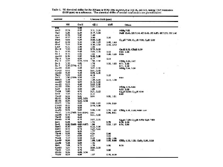
Groups of Hydrogen Atoms in the Common Amino Acid Residues with similar Random Coil 1 H Chemical Shiftsa. NH(W)* NH(bb)* 2 H(H) NH(sc)* Ring 10 Code CH 3 β(a) β(b) β(a) α, β(S. T) CH 3 5 1 δ(ppm) α, β(S. T) Ring 2 H(H) δ(ppm) 0. 9 -1. 4 1. 6 -2. 3 2. 7 -3. 3 1. 2 -3. 3 3. 9 -4. 8 6. 5 -7. 7 -8. 6 Comments βH of V, I, L, E, Q, M, P, R, K βH of C, D, N, F, Y, H, W Other aliphatic CH All αH, βH of S and T Aromatic CH of F, Y, W; 4 H of H 2 H of h in the p. H range 1 -11 NH(sc)* NH(bb)* NH(W)* 6. 6 -7. 6 8. 1 -8. 8 10. 2 Side chain NH of N, Q, K, R Backbone NH Indole NH of W
Parameters for regular polypeptide conformations β βp α-helix 310 -helix π-helix Polyproline II Polyglycine II Bond Angle (deg) Ψ ω ψ +135 -178 -139 +113 180 -119 -47 180 -57 -26 180 -49 -70 180 -57 +158 0 -83 +149 180 -78 +150 180 -80 Residues per turn 2. 0 3. 6 3. 0 4. 4 3. 33 3. 0 Translation per residue 3. 4 3. 2 1. 50 2. 00 1. 15 1. 9 3. 12 3. 1 Adapted from G. N. Ramachandran and V. Sasisekharan, Adv. Protein Chem. 23, 283 -437(1968); IUPAC-IUB Commission on biochemical Nomemclature, Biochemistry 9, 3471 -3479 (1970).
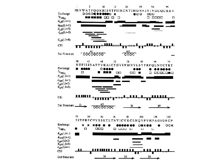
Short sequential and medium-range 1 H-1 H distances, vicinal coupling constants, and amide hydrogen exchange rates. Parameter dαN(i, i) dαN(i, i+1) dαN(i, i+2) dαN(i, i+3) dαN(i, i+4) d. NN(i, i+1) d. NN(i, i+2) dβN(i, i+1) dαβ (i, i+3) dαα (i, j) dαN (i, j) d. NN (i, j) 3 J HNα(HZ) NH exchange rate α-helix 2. 6 3. 5 4. 4 3. 4 4. 2 2. 8 4. 2 2. 5 -4. 1 (≦ 4) slow 310 -helix 2. 6 3. 4 3. 8 3. 3 (>4. 5) 2. 6 4. 1 2. 9 -4. 4 3. 1 -5. 1 (≦ 4) slow β 2. 8 2. 2 βP 2. 8 2. 2 4. 3 4. 2 3. 2 -4. 5 3. 7 -4. 7 2. 3 3. 2 3. 3 (≧ 9) slow 4. 8 3. 0 4. 0 (≧ 9) slow dαα(i, j), dαN (i, j) and d. NN (i, j) refer to interstrand distances. The first four residues in the α-helix and the first three residues in the 3 10 -helix will have fast amide proton exchange rates. Every second residue in the flanking strand will have slow amide proton exchange rates
The characteristic patterns of short-range NOEs involving amide, alpha, beta protons observed for ideal α-helices, 310 -helices and β-strand. α-helix 310 -helix β-strand 1234567 4444444 9999999 d. NN(i, i+1) dαN(i, i+3) dαβ (i, i+3) dαN(i, i+2) d. NN(i, i+2) dαN(i, i+4) 3 J HNα(HZ) The thickness of the lines is an indication of the intensity of the NOEs The values of J coupling are approximate.
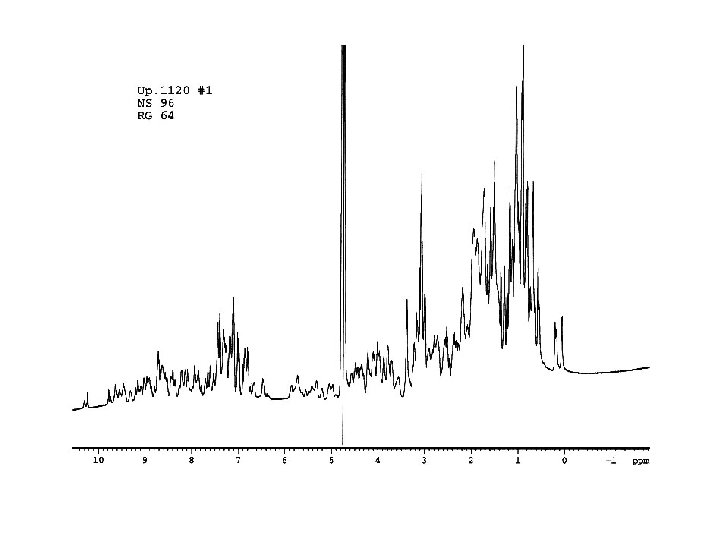
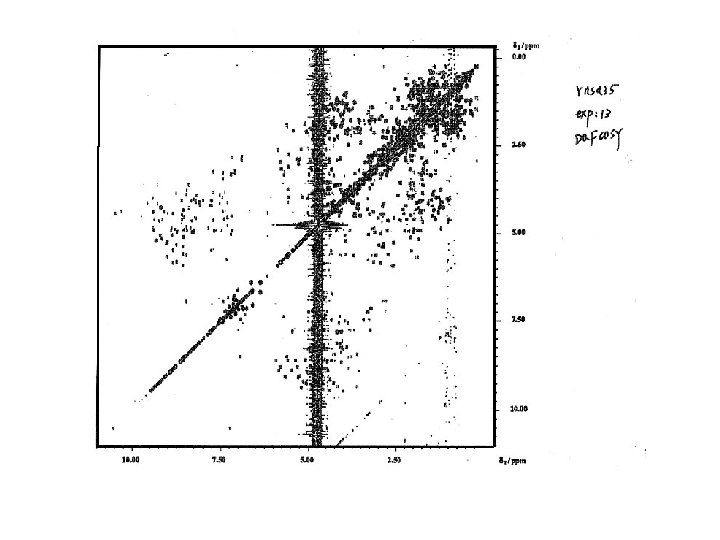
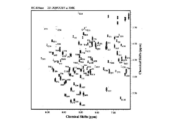
Resonance assignment strategies for small proteins Spin system identification : DQF-COSY and TOCSY experiments Sequence-specific assignment: NOESY experiment For protein < 10 k. Da, 2 D homonuclear experiments may be sufficient for resolving overlapping NMR resonances.
PREPARATION EVOLUTION (A ) (B) (C ) (D ) MIXING t 2 t 1 t 1 DETECTION τM t 2 (A) The elements of a generalized two- dimensional NMR experiment (B) DQFCOSY experiment (C) NOESY experiment (D) TOCSY experiment
Ala H (ppm) H Hβ O 8. 25 Hα NH C Cβ Hβ 177. 1 52. 5 Hβ H (ppm) Cα Hα Hβ H (ppm) 122. 5 N COSY 4. 35 19. 0 Hα Hβ 1. 39 NH TOCSY H (ppm)
Val H (ppm) Hγ 1 Hβ 121. 1 O 8. 44 N 63. 0 2. 13 Hα Cβ Hγ 2 0. 94 COSY H (ppm) 177. 1 Hγ 1 4. 18 31. 7 Hγ 1 Cγ 2 Hγ 2 NH C Cα Hβ Hα Hγ 2 Hβ Cγ 1 Hα Hγ 1 0. 97 NH TOCSY H (ppm) Hγ 2 H (ppm) H Hγ 2
Leu Hδ 2 Hδ 1 Hγ H O H (ppm) Hβ 3 Hβ 2 8. 42 Hα N C 55. 7 177. 1 NH Cα H (ppm) Hβ 2 Hα 1. 64 Hγ 4. 38 41. 9 Hδ 1 Cδ 2 0. 90 Hδ 2 1. 65 Cβ Cγ Hδ 2 Hβ 3 Hδ 1 Hγ 1. 65 Hβ 3 Hβ 2 0. 94 Hα Cδ 1 Hδ 2 Hδ 1 COSY Hδ 1 NH TOCSY H (ppm) 121. 8
Trp O 8. 09 N 120. 5 C 58. 3 Hδ 1 7. 24 3. 32 Cβ Cδ 1 Hε 1 C 2 C 3 Nε 1 N 1 10. 22 28. 3 Hβ 3 Cγ Hα 174. 8 Cα Hβ 2 Hα NH COSY 4. 70 H (ppm) 3. 19 Cδ 2 Cε 2 Hζ 2 Cζ 2 7. 50 C 4 C ε 3 Hβ 3 7. 65 Hβ 2 C 7 C 6 C 5 Cζ 3 Cη 2 H η 2 7. 24 Hα Hζ 3 7. 17 NH TOCSY H (ppm) Hβ 3
Fingerprint region Phe Hα (ppm) Val Gln Hα Ala NOESY Thr Ala’ Amide protons (ppm) + H O H H O H H O NH 3 C C N C C N C C R 1 R 2 R 3 R 4 R 5 R 6 F V Q A T α-helical NOEs dαN(i, i+3), d. NN(i, i+1) A’
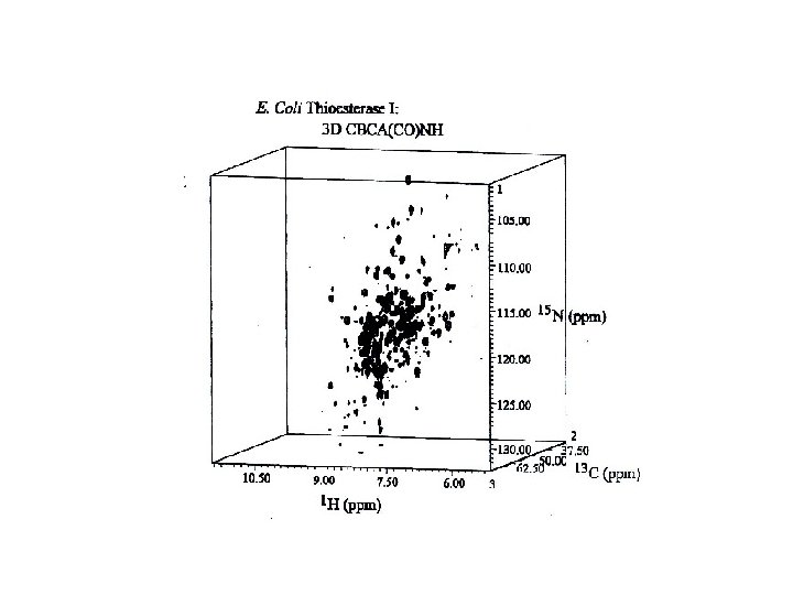
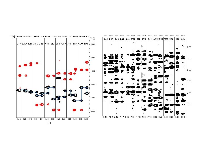
Methods for resolving overlapping NMR resonances 2 D/3 D homonuclear NMR experiments such as 2 D-DQFCOSY, 2 D-TOCSY, 2 DNOESY, 3 D-NOESY-TOCSY. 2 D/3 D heteronuclear NMR experiments such as 2 D-15 N-HSQC, 3 D-15 N-NOESYHSQC and triple-resonance experiments ( 1 H, 13 C, 15 N).
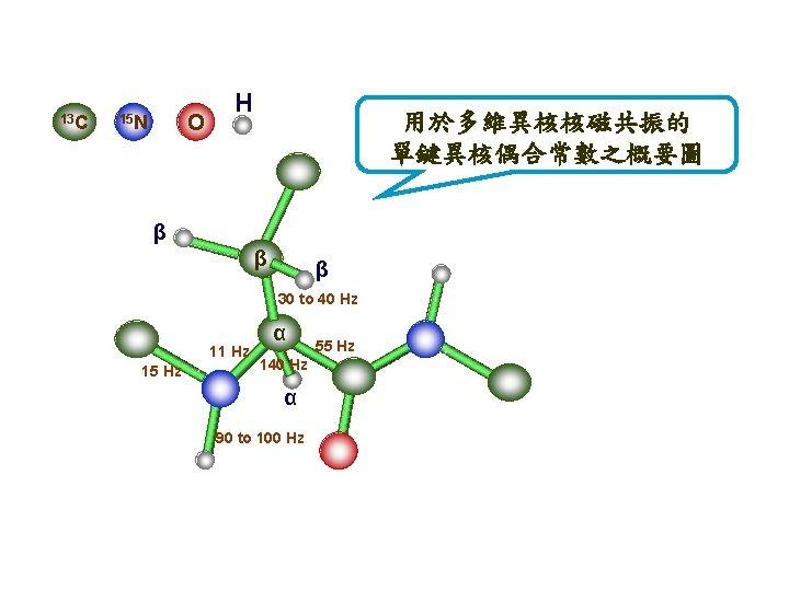
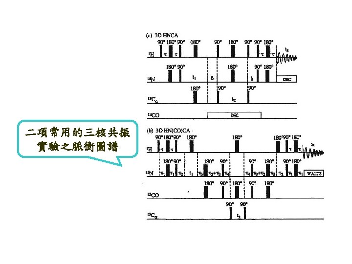
Act. Chem. 26 134 (1993) Bax and grzesiek a H 140 C H 35 N H b C 140 H H Cβ H -7 55 C O H Cβ H 15 N H 11 92 Cα C H O H Cβ H N Cα C H H O a) A dipeptide segment of a protein backbone with the approximate values for the J couplings which are essential for the assignment procedure in isotopically enriched proteins. (b-e) Schematic diagrams of the nuclei that are correlated in the b) HNCO, …….
c d e H Cβ H N C C H H O H C H H Cβ H N C C N Cα C H H O c) HNCA, d) HBHA(CBCACO) NH and e) CBCANH experiments. Nuclei for which the chemical shift is measured in the 3 D experiment are marked by solid circles. Nuclei involved in the magnetization transfer pathway, but not observed, are marked by open circles. Magnetization transfer in these experiments is marked by curved solid lines, and the direction of the transfer is marked by arrows.
Three-dimensional structure determination by simulated annealing using X-PLOR ( CNS ) program Etotal = Ebond + Eangle + Eimproper + Erep + Enoe + Etor Keep the correctness of protein geometry The energy terms of experimental data