1 MUSCLE ENERGETICS TETANUS and TETANY Tetanus Neurological


 CAUSES– 1 -Lack")


- Slides: 38
1
MUSCLE ENERGETICS
TETANUS and TETANY • Tetanus Neurological disorder that results from a decrease inhibitory input to alpha motor neurons
Mechanism of Action ---Tetanus Cause—Bacteria Clostridium tetani Toxin ---- Tetanospasmin Mechanism of action—Retrograde intraneuronal transport. (Retroaxonally)
Symptoms-Locked jaw -Opisthotonus -Breathing problems. -Sudden powerful and painful contraction of muscles -Irritability, muscle cramps. -Weakness -Difficulty in swallowing -Sardonic smile
OPISTHOTONUS
TETANUS • • Antibiotic to kill the bacteria. Injecting antibiotics that bind the toxin. Muscle relaxants. Mechanically ventilating the lungs Prophylaxsis— Immunization
TETANY • Low serum calcium levels (8. 5 -10. 5 mg/dl) CAUSES– 1 -Lack of calcium 2 -Excess of phosphate 3 -Underfunctioning of thyroid gland.
Mechanism of Action --Tetany • Decrease Calcium ion concentration --Sodium channels become activated by very little increase in membrane potential.
TETANY SYMPTOMS— 1 - Hyperflexia 2 - Carpopedal spasm 3 -Cramps 4 -Laryngospasm Treatment---1 -I/V calcium 2 -Oral calcium +Vitamin D 3 -Surgical removal of parathyroid glands
TYPES OF SKELETAL MUSCLE FIBERS • SLOW-TYPE 1 OXIDATIVE RED FAST- TYPE II GLYCOLYTIC WHITE
TYPES OF SKELETAL MUSCLE FIBER SLOW-TYPE I OXIDATIVE RED FAST-TYPE II GLYCOLYTIC WHITE Myosine with low ATPase activity Myosine with high ATPase activity Cross bridge cycling is slow Cross bridge cycling is fast Maximal shortening velocity is low Maximal shortening velocity is high Glycogen content is low High Enzymes for anaerobic glycolysis are low High Mitochondria--- High Capillary density---- High Myoglobin content---- High Oxidative metabolism capacity is high Low
TYPES OF SKELETAL MUSCLE FIBER SLOW TYPE-I OXIDATIVE RED FAST TYPE II GLYCOLYTIC WHITE Diameter moderate Diameter large Calcium pumping capacity of SR is moderate Calcium pumping capacity of SR is high Speed of contraction slow Speed of contraction fast Latent period long Latent period short Fatigue resistance Fatigable
Some Diseases related to Muscles • • • Tetanus Tetany Rigor Mortis Myasthenia Gravis Dystrophy (Duchenne’s Muscular Dystrophy)
RIGOR MORTIS • Several hours after death all the muscles of the body go into a state of contracture. • Loss of all ATP • Muscle protein deteriorates ----15 to 25 hours later. (Autolysis). • All these events occur more rapidly at higher temperatures.
Function of Skeletal Muscles • Movement • Posture (both agonist & antagonist function together, Co- activation) • Heat Production • Interaction with the Environment (Expression) • Exert Rapidly adaptable force • Voluntary control
Some terms Related to Muscles Disuse Atrophy • Atrophy { Denervation Atrophy • Hypertrophy • Fibrillation • Fasciculation
• Atrophy Decrease in the diameter of nerve fiber Decrease in the amount of contractile proteins. Disuse atrophy : Muscle is not used for a prolong period of time. Denervation atropy: 1. Neurons are destroyed 2. Neuromuscular junction becomes non functional. (ATP dependent ubiquitin – proteasome pathway)
• Hypertrophy Increase in the diameter of nerve fiber. Increase in the amount of contractile proteins Muscle is loaded during the contractile process
• Fibrillation Rapid , irregular and unsynchronized contraction of muscle fiber. • Fasciculation Small local, involuntary muscle contraction and relaxation which may be visible under the skin
Smooth Muscles
Objectives • At the end of this lecture you should be able to know types of smooth muscles and their anatomical location. • Arrangement of myofilaments and sarcoplasmic reticulum • Steps involved in Contraction • Functional differences
SMOOTH MUSCLE • Diameter – 1 -5 micrometers • Length --- 20 -500 micrometers
Smooth Muscles Generally Divided into Two Types 1. Multi-unit. Composed of : discrete, Independent fibers innervated by separate single nerve ending e. g. iris and piloerector muscles Multi unit Smooth M Fiber
Unitary Smooth Muscle 2. Unitary: 100 -1000 fibers but contract as a unit. due to presence of gap junctions e. g. in most of the viscera
Input Influencing Smooth muscle Contractile activity • Spontaneous electrical activity in the plasma membrane of the muscle cell. • Neurotransmitters released by autonomic neurons. • Hormones • Locally induced changes in the chemical composition of ECF surrounding the cell (acidity, oxygen, osmolarity, ion concentration) • Stretch.
Arrangement of Actin & Myosin Smooth M Fiber
Neuromuscular Junction of Smooth Muscle
Non striated Dense bodies--- Z-lines Protein Calmodulin--- Troponin complex. No Typical branching end feet as in Sk. Muscles Multiple Varicosities. No Schwann cells at Varicosities. Diffuse junctions. • Contact junctions • Neurotransmitter is released at Varicosities –may be Ach or Noradrenalin. • • •
Sarcoplasmic Reticulum Smooth Muscle S. R
Action Potential in smooth Muscle RMP… -50 -60 mv Action Potential in Unitary Smooth Ms e. g. Viscera Two forms: 1. Slow waves 2. Spike Potential ---10 -50 m. sec duration 3. Action potential with plateau. 4. Junctional potential
Calcium channels • Far greater voltage gated calcium channels • Few voltage gated sodium channels
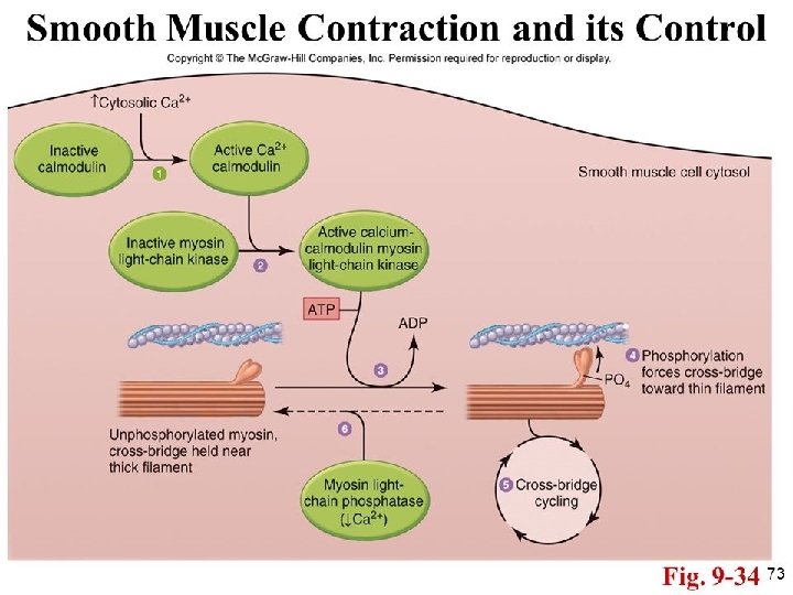
Steps involved in the Contraction of Smooth Muscle • Ca++ binds with Calmodulin • Calmodulin-Ca++ combinedly activates Myosin Kinase (phosphorylating Enzyme) • One of the Light chain of Myosin Head (Regulatory chain) is Phosphorylated in response to this myosin kinase. • The head of Myosin gets capability to repetitively attach & detach with active sites of Actin filaments. Slow cycling of Cross Bridges than skeletal muscles. • Dephosphrylation–Myosin phosphtase in sarcoplasm
Latch Phenomenon • The energy required to maintain full force of contraction , (once developed full contraction initially) in smooth muscles is very negligible (1/300 that of Sk. Muscle) • This phenomenon allows long term maintenance of tone in many smooth muscles without much expenditure of energy.
Comparison of Smooth and Skeletal muscle contraction • Slow cycling of the myosin cross bridges. • Low energy requirement to sustain smooth muscle contraction. • Slowness of onset of contraction and relaxation of the total smooth muscle tissue. • Maximum force of contraction (4 -6 Kg /cm. sq) • Latch mechanism • Stress relaxation of smooth muscle