1 FORCEPS 2 VACUUM DELIVERY 3 CAESARIAN SECTION


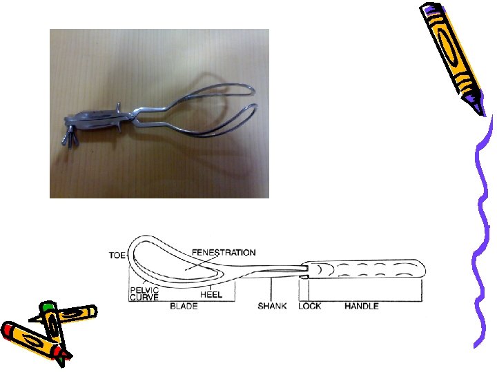
-introduced the pelvic curve • Smellie (1751)- reinforced pelvic curve &")


 • Suitable presentation & position: -.")
 • • • Documentation: – All instrumental deliveries should")
 1. Identification of blades & their")
 3. Traction: – Steady & intermittent")


 Emergency caesarian section (Unplanned) Advantages are: Patient with empty")


 •")


- Slides: 57
1. FORCEPS 2. VACUUM DELIVERY 3. CAESARIAN SECTION
Abnormal Labour Forceps and Vacuum Delivery
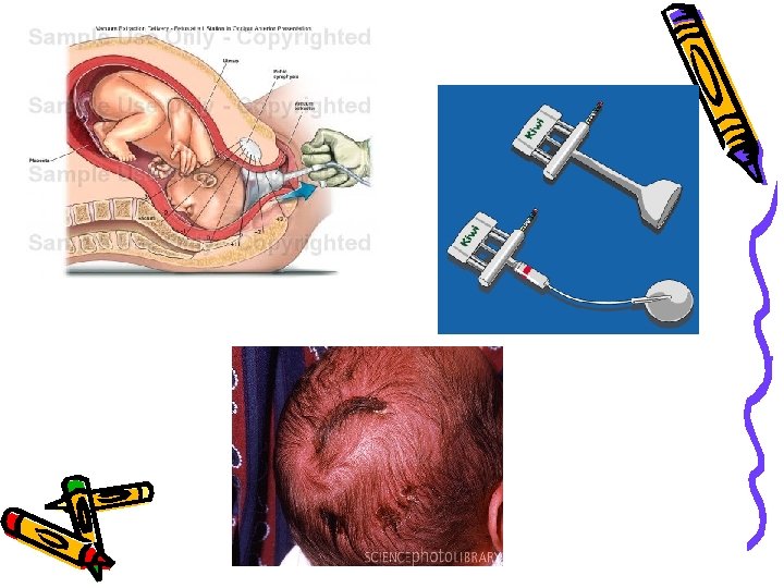
VACUUM /VENTOUSE
INDICATIONS MATERNAL • Exhaustion • Prolonged second stage • Cardiac / pulmonary disease FETAL • Failure of the fetal head to rotate • Fetal distress • Should not be used for preterm, face presentation or breech
MNEMONIC • A – Anesthesia adequate appropriate positioning & access • B – Bladder catheterization • C – Cervix fully dilated / membranes ruptured • D – Determine position, station, pelvic adequacy • E – Equipment inspect vacuum cup, pump, tubing, check pressure
MNEMONIC • F – Fontanelle position the cup over the scalp, avoid fontanelle -ve pressure ↑ 10 cm H 2 O initially & between cont sweep finger around cup to clear maternal tissue ↑ pressure to 60 cm H 2 O with the next contraction • G – Gentle traction pull with contractions only traction in the axis of the birth canal ask the mother to push during cont
MNEMONIC • H – Halt traction progress halt traction if no progress with three aided contractions vacuum pops off three times pulling for 30 min without significant • I – Incision consider episiotomy if laceration imminent • J – Jaw remove vacuum when jaw is reachable or delivery assured
COMPLICATIONS • Vacuum –assisted delivery is less traumatic to the mother & fetus than forceps • Ventouse should be the instrument of choice • Maternal Vaginal laceration due to entrapment of vaginal mucosa between suction cup & fetal head
Comparative Advantages of Vacuum Extractors and Forceps Vacuum extractors Easier to learn Quicker delivery Less maternal genital trauma Less maternal discomfort Fewer neonatal craniofacial injuries Less anesthesia required Forceps Fewer neonatal injuries, including cephalohematoma, retinal hemorrhage Higher rate of successful vaginal delivery
FETAL COMPLICATIONS • Scalp injuries abrasion & lacerations 12. 6% scalp necrosis 0. 25 -1. 8% • Cephalohematoma 25% jaundice /anemia • Intracranial hemorrhage 2. 5% • Subgaleal hematoma
FETAL COMPLICATIONS • Birth asphyxia 2. 6 -12% related to extraction force & time Some studies showed decrease birth asphyxia • Retinal hemorrhage Forceps SVD • Neonatal jaundice 50% 31% 19%
FORCEPS • HISTORY • WILLIAM CHAMBERLAIN – • Fled from France in 1569 & practiced forceps delivery as a family secret in Southampton. This was kept as a family secret for over 100 yrs and four generations. • He had two sons. • Peter I - had greater distinction & attended notable women in society. • Peter II - who had several sons, died in 1626.
HYSTORY • Levret (1747)-introduced the pelvic curve • Smellie (1751)- reinforced pelvic curve & introduced English lock and used in aftercoming head. • Tarnier (1877)-introduced axis traction. • Barton and Kjielland - introduced the two specialized forceps.
Functions • Traction: -This is the most important function. Pull required in a primigravida is 18 kgs & in a multipara it is 13 kgs. • Compression effect: -This is minimal when properly applied & should not be more than necessary to grasp the head. However it has some pressure effect on the wellossified base of the skull.
Functions • Rotation of head: -This occurs with the use of Kejilland's forceps and also in low forceps cephalic application with the occiput in the 2 or 10 'o' clock position. • Protective cage: - When applied on a premature baby it protects from the pressure of the birth canal. When applied on the aftercoming head it lessens the sudden decompression effect.
Indications forceps delivery • Delay in second stage: -. – Due to uterine inertia. – Failure of progress of labour- if no progress occurs for more than 20 to 30 minutes, with the head on the perineum. Definition of prolonged second stage of labour redefined by A. C. O. G. (1988/1991): – Nullipara • <3 hrs with regional anaesthesia • <2 hrs without regional anaesthesia – Multipara • <2 hrs with regional anaesthesia • <1 hr without regional anaesthesia 19
Indications forceps delivery • Foetal indications: - – Foetal distress in second stage when prospect of vaginal delivery is safe: • Abnormal heart rate pattern • Passage of meconium • Abnormal scalp blood ph – Cord prolapse in second stage – Aftercoming head of breech – Low birth wt. Baby – Post maturity 20
Indications forceps delivery • Maternal indication: – – – Maternal distress Pre-eclampsia Post caesarian pregnancy Heart diseases Intra partum infection Neurological disorders where voluntary efforts are contraindicated or impossible 21
Prerequisites (to be fulfilled before forceps application. ) • Suitable presentation & position: -. – Vertex, anterior face or aftercoming head are the ideal positions. • Cervix must be fully dilated. • Membranes must be ruptured. • Baby should be living. • Uterus should be contracting & relaxing. • Bladder must be empty. 12 October 2002 Forceps Delivery Prof. S. N. Panda 22
Preliminaries (before forceps application ) • • • Documentation: – All instrumental deliveries should be dictated in medical record as any surgical procedure & it should include: Consent of the patient, indication for operation, anaesthesia, personnel involved, type of instrument, difficulties & remedies, resulting maternal & foetal complications or injuries and blood loss. Anaesthesia: – Pudendal block or Labio-perineal infiltration for outlet forceps. – Regional or General anaesthesia for low & mid forceps. Catheterisation: Internal examination: – To asses the state of cervix & membranes, presentation & position, pelvic outlet Episiotomy: – Should be done either before application of forceps or during traction when the perineum bulges. 12 October 2002 Forceps Delivery Prof. S. N. Panda 23
Technique (of low & outlet forceps application ) 1. Identification of blades & their application– The instrument should be placed in front of the pelvis with the tip pointing upwards and pelvic curve forwards. First the left blade should be applied guided by the right hand & then the right blade with the left hand. 2. Locking of blades: – The blades should articulate with ease indicting correct application. 24
Technique (of low & outlet forceps application ) 3. Traction: – Steady & intermittent traction to be applied during contraction, first downwards (horizontal), backwards, forwards & lastly upwards. – In outlet forceps - Only two fingers are to be introduced. Traction is applied straight horizontal, upward & then forwards. – Removal of blades - Right blade should be removed first. 25
Complications / Dangers Complications/dangers of forceps delivery: - are mostly due to faulty technique rather than the instrument. • Maternal– Injury-. • Extension of the episiotomy involving anus & rectum or vaginal vault. • Vaginal lacerations and cervical tear if cervix was not fully dilated. – Post partum haemorrhage –. • Due to trauma, Atonic uterus or Anaesthetisia. – Shock –. • Due to blood loss, dehydration or prolonged labour. – Sepsis –. • Due to improper asepsis or devitalisation of local tissues. – Anaesthetic hazards. – Delayed or long-term sequel –. • Chronic low backache, genital prolapse & stress incontinence. 26
. Complications / Dangers • Fetal– Asphyxia. – Trauma • • • Intracranial haemorrhage. Cephalic haematoma. Facial / Brachial palsy. Injury to the soft tissues of face & forehead. Skull fracture – Remote-cerebral palsy. – Foetal death-around 2%. 27
REMEMBER • RESPECT INDICATION • DON’T HESITATE TO APPLY FORCEPS WHEN NEEDED • DON’T WASTE TIME • DO IT GENTLY
• http: //www. youtube. com/watch? v=KY td 1 mg. BO 1 Q
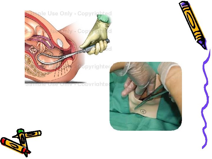
CESARIAN SECTION • Cesarean Section is removal of a fetus from the uterus by abdominal and uterine incisions, after 28 weeks of pregnancy. • It is called hysterotomy, if removal is done before 28 weeks of pregnancy.
The five Most Common Causes of Cesarean Section • CS on Request • Routine repeat cesareans. • Dystocia (non-progressive labor). • Abnormal fetal presentation eg breech , transverse , cord presentation. • Fetal distress.
Reasons suggested for the increase in caesarean section rates • • • Advancing maternal age, -Socioeconomic factors, - Reduced parity Improvements in surgical techniques -- Decreased morbidity and mortality The obstetrician’s experience and type of training Choose the time and day of delivery Procedures as high forceps and difficult mid forceps are abandoned in favour of Caesarean Section (C. S. ) The introduction of epidural anaesthesia has reduced the anaesthetic risks of the procedure. This has led to a lower threshold for doing a Caesarean section in the second stage of labour rather than performing rotational/high cavity forceps deliveries which led to maternal and neonatal morbidity. The increased use of electronic fetal monitoring has increased our awareness of fetal distress although the majority of babies are born in good condition despite an abnormal CTG and/or low p. H at fetal blood sampling. The reduction in the number of rotational forceps deliveries has led to a deskilling of obstetricians who do not feel confident to carry out these procedures. The evidence that breech presentation babies have a reduced morbidity and mortality if delivered by elective Caesarean section An increasing demand from women for elective Caesarean sections with no medical reason.
Avoiding First C-Section Should Be Priority • Avoiding primary cesarean sections unless there is a medical necessity
Timing Of CS • Elective cesarean delivery • elective caesarean section may be justified, but decisions must take into account the risk to the infant associated with delivery before 39 weeks' gestation • It is now clear that respiratory distress syndrome is indeed seen in "term" infants and is a considerable source of morbidity and mortality in this group • Emergency cesarean section • In cases of suspected or confirmed acute fetal compromise, • delivery should be accomplished as soon as possible. • The accepted standard is within 30 minutes.
Elective caesarian section (Planned operation) Emergency caesarian section (Unplanned) Advantages are: Patient with empty stomach and surgeon usually with full breakfast Best anesthetist available at that time Best assistant and nursing staff. Disadvantages are : If wrong judgment, premature child may be born. Cervix may not be dilated and hence poor drainage of lochia Lower segment is not formed and hence uterine incision in lower part of upper segment. Working under adverse circumstances: Patient may be with full stomach and surgeon may be with empty belly Odd working hours either of day or night Anesthetist, assistant and nursing staff may not be of your choice Advantage is : Mature child as patient is in labor Cervix is open, better drainage of lochia. Lower segment is well formed
Preoperative testing and preparation for CS • Pregnant women should be offered a haemoglobin assessment before CS to identify those who have anaemia. Although blood loss of more than 1000 ml is infrequent after CS (it occurs in 4 to 8% of CS) it is a potentially serious complication. • Pregnant women having CS for ante partum haemorrhage, abruption, uterine rupture and placenta praevia are at increased risk of blood loss greater than 1000 ml and should have the CS carried out at a maternity unit with on-site blood transfusion services. • Prescribe antibiotics (one dose of first-generation cephalosporin or ampicillin) • Assess risk for thromboembolic disease (offer graduated stockings, hydration, early mobilisation and low molecular weight heparin) • To reduce the risk of aspiration pneumonitis: Empty stomach, an antacid (sodium citrate 0. 3% 30 m. L or magnesium trisilicate 300 mg) + Cimetidine IV 1 hr before CS • an indwelling urinary catheter to prevent over-distension of the bladder
Anaesthesia • 1 General anaesthetic. • 2 Regional anaesthesia ( Epidural block. - Spinal block ). • 3 Infiltration of local anaesthetic agents. • Regional anaesthesia is regarded as considerably safer than general anaesthesia with respect to maternal mortality • Regional anesthesia is generally preferred because it allows the mother to remain awake, experience the birth, and have immediate contact with her infant. It is usually safer than general anesthesia. Many practitioners prefer spinal or CSE to epidural techniques because of more rapid onset and better blockage of pain
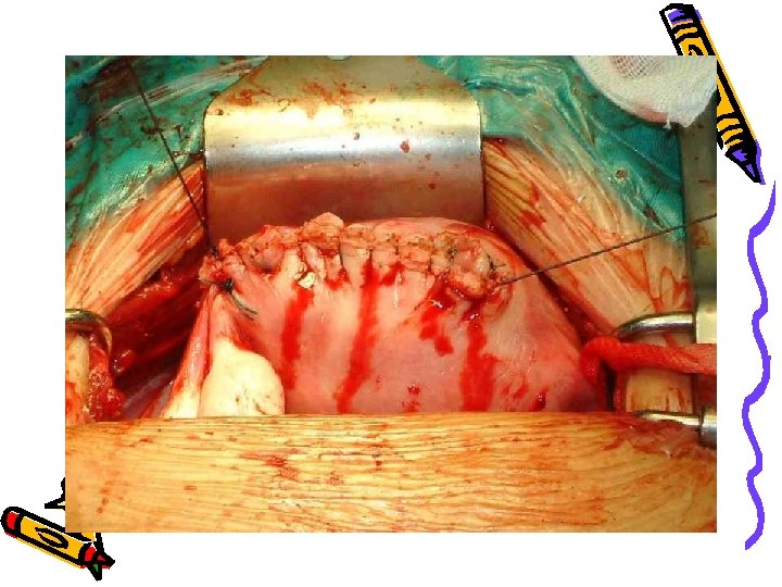
Abdominal entry
Visceral Peritoneal Incision • Place a bladder retractor over the pubic bone . • Use forceps to pick up the loose peritoneum covering the anterior surface of the lower uterine segment and incise with scissors . • Extend the incision by placing the scissors between the uterus and the loose serosa and cutting about 3 cm on each side in a transverse fashion. • Use two fingers to push the bladder downwards off of the lower uterine segment. Replace the bladder retractor over the pubic bone and bladder.
DELIVERY OF THE BABY • • To deliver the baby, place one hand inside the uterine cavity between the uterus and the baby’s head. With the fingers, grasp and flex the head. Gently lift the baby’s head through the incision taking care not to extend the incision down towards the cervix. With the other hand, gently press on the abdomen over the top of the uterus to help deliver the head. • If the baby’s head is deep down in the pelvis or vagina Ask an assistant (wearing high-level disinfected gloves) to reach into the vagina and push the baby’s head up through the vagina. Then lift and deliver the head
Give Newborn To Pediatrition
• http: //www. youtube. com/watch? v=ea np. Noc 0 q 8 U
The placenta was manually removed or spontaneously delivered • At CS, the placenta should be removed using controlled cord traction and not manual removal as this reduces the risk of endometritis. • Spontaneous delivery of the placenta may reduce blood loss and decrease the chance of postoperative endometritis • By Keeping gentle traction on the cord and massage (rub) the uterus through the abdomen. • Deliver the placenta and membranes
Uterine repair – chromic catgut vs vicryl - continuous vs interrupted sutures peritoneal closure vs non-closure (Pelvic, parietal, both ) Non-closure associated with less post-op fever but no significant effect on wound infection or endometritis. New trial fewer adhesions in closure
Prophylactic antibiotics with cesarean section (immediately after the cord is clamped versus preoperative) • Give a single dose intravenously of prophylactic antibiotics after the cord is clamped and cut: • - ampicillin 2 g IV OR cefazolin 1 g IV provides adequate prophylaxis. • No additional benefit has been demonstrated with the use of multipledose regimens. • however, no consensus on the optimal timing of administration and doses • There is also no evidence that the transplacental passage of prophylactic ampicillin increases immediate or delayed neonatal infections
The laparotomy pads put in abdominal Cesarean section cavity are all removed & counted doubly by surgeon himself and then by nurse.
Ambulation after cs • Ambulation started earlier in the simplified technique group (6 -8 hours postop vs 10 -12 hours post-op). • Ambulation enhances circulation, encourages deep breathing and stimulates return of normal gastrointestinal function. Encourage foot and leg exercises and mobilize as soon as possible, usually within 24 hours
Cesarean Hysterectomy • Hysterectomy is carried out after caesarean section in the same sitting for one of the following reasons: • Uncontrollable postpartum haemorrhage. • Unrepairable rupture uterus. • Operable cancer cervix. • Couvelaire uterus. • Placenta accreta cannot be separated. • Severe uterine infection particularly that caused by Cl. welchii. • Multiple uterine myomas in a woman not desiring future pregnancy although it is preferred to do it 3 months later.
Repeated CS is safer than VBAC • should we be promoting VBAC which may carry greater risks • to the individual for the purposes of reducing “an undesirable statistic”? • In our country where family sizes are now voluntarily limited, • is it in the woman’s interests to try for a VBAC?
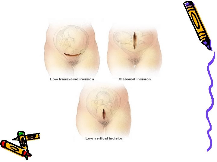
1. 2. 3. 4. 5. 6. 7. Causes of a weak scar Improper haemostasis Imperfect coaptation Inversion of decidua Extension of the angles Infection during healing Placental implantation Overdistension of the uterus The most weak scar is that of the upper segment of the uterus
Assessment of scar integrity • Hysterogram – Defect in the lateral view • Ultrasonic measurement – Scar defects – Scar thickness • Cut-off value of 3. 5 mm at 36 weeks (NPV of 99. 3% (Rozenberg et al 1996) • Manual exploration • Bleeding • Third stage troubles
Impending scar rupture • • • Pain over the scar Maternal tachycardia Fetal distress Poor progress Vaginal bleeding
Consider CS complications • Endometritis if excessive vaginal bleeding • Thromboembolism if cough or swollen calf • Urinary tract infection if urinary symptoms • Urinary tract trauma (fistula) if leaking urine