1 7 The nucleus The nucleus contains most


 Peroxisomes that protect the cytoplasm from the impact of destructive")


 consisting of protein fibers and various")


- Slides: 43
1. 7. The nucleus • The nucleus contains most of the genetic information and serves as the center of regulatory activity. The nucleus provides a site for genetic transcription that is segregated from the location of translation in the cytoplasm, allowing levels of gene regulation. Nucleus is the main region in which genetic information (DNA) is kept and preserved within the cell. Most eukaryote cells have only one nucleus, however some of them (including plant cells) may have more nuclei.
• DNA molecules in the eukaryote cells combine with proteins to form units called chromosomes. All species have specific chromosome numbers. While chromosome number found in some species may be few, it may be thousands in some other species.
• The main function of the cell nucleus is to control gene expression and mediate the replication of DNA during the cell cycle.
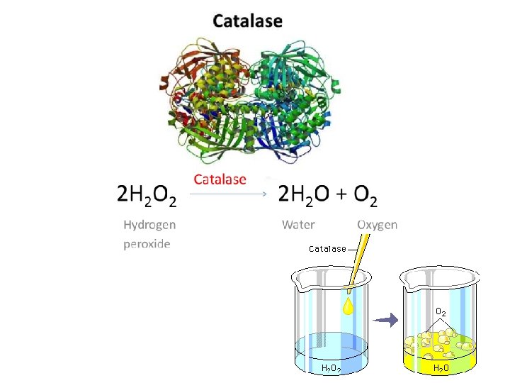
1. 8. Peroxisomes (=microbodies) Peroxisomes that protect the cytoplasm from the impact of destructive molecules and free radicals. Peroxisomes are especially important for the process of photosynthesis.
• Peroxisomes are composed of a single membrane that surrounds the finely granular peroxisome matrix and they contain a series of special enzymes. Characteristic enzyme of peroxisomes is catalase. This enzyme breaks down H 2 O 2
• H 2 O 2 that is produced during cell metabolism is highly toxic for the cell. For example, diluted H 2 O 2 solution that has been used to clean wounds since it kills harmful bacteria. ***** However if concentrated H 2 O 2 solution is used, it also damages the living cells and may delay wound healing!!! Peroxisomes that are found in plants protect against loss of photosynthesis products during photorespiration.
In germinating fat-storing seeds, peroxizomes participate in lipid mobilization; in leaves of C 3 plant (plants that produce three-carbon compound phosphoglycerate as the first stable photosynthetic intermediate), they play a key role in photorespiration; and in some legume root nodules, they are involved in the conversion of recently fixed N 2 into nitrogen-rich organic compounds.
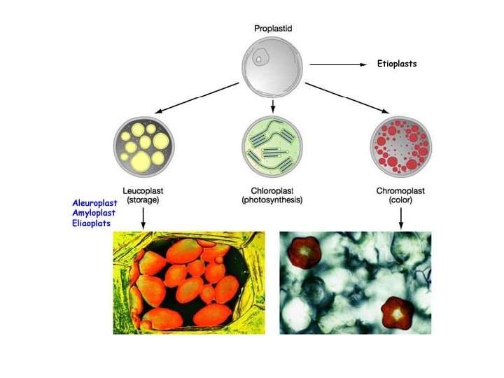
1. 9. Plastids are major organelles found only in plant cells. They are responsible for photosynthesis, for the storage of a wide variety of products and for the synthesis of key molecules required for the basic architecture and functioning of plant cells. Like mitochondria, plastids are enclosed in a pair of membranes. Plastids also resemble mitochondria in being semiautonomous and containing the genetic machinery required to synthesize a few of their own proteins.
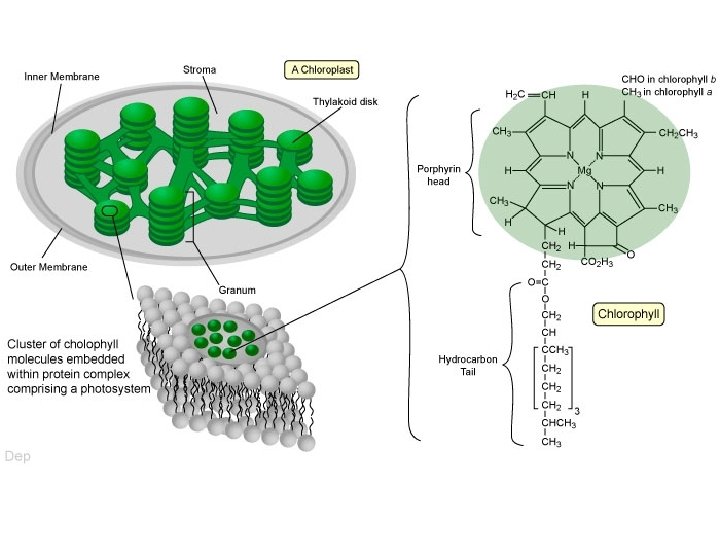
• Among the plastids, chloroplasts are very important since they have a role in photosynthesis. They are found in leaves and other green parts of the plant. Chlorophyll, the main photosynthetic pigment of plants is found within chloroplasts. • Plant and algal cells may contain one or more chloroplasts; an average 40 -50 chloroplasts are per cell as a general rule.
1. 10. Mitochondria are found in nearly all eukaryotic cells. These essential organelles house the respiratory machinery that generates ATP by way of the citric acid cycle and associated electron transfer chain. Mitochondria also supply various compounds, including organic acids and amino acids that are used as building blocks in synthetic reactions elsewhere in the cell.
• Like plastids, mitochondria are also bounded by two membranes. • The inner membrane which is much larger in area than the outer, folds into cristae that, extend deeply into the mitochondrial matrix. The inner membrane (including cristae) contains membrane-associated protein and mitochondrial lipid. • The matrix is composed of soluble enzymes, mitochondrial DNA (mt DNA) and ribosomes.
2. The Cell Wall • The main function of the cell wall is to provide strength against pressure that cell membrane is exposed to. Cell wall also provide protection against attacks microorganisms. And it also gives shape to cell. of pathogenic
• The plant cell wall is a dynamic compartment that changes throughout the life of the cell. The new primary cell wall is formed in the cell plate during cell division and rapidly increases its surface area during cell expansion in some cases by more than a hundredfold.
• Primary cell wall: Thin and flexible portion of the wall, made of cellulose and pectin. • Middle lamella: thin layer between primary walls of adjacent cells. • Secondary cell walls: found in some cells (between the plasma membrane and the primary cell wall and it is rich in lignin for strength).
During the growth process, plant cells only have a single layered cell wall called primary cell wall. This primary cell wall expands with the growing cell. During the growth and volume increase, cell wall components are loosened as controlled and central vacuole performs water uptake. The increasing water pressure within the vacuole provides the strength that is requires for the stretching of the cell and the cell wall. When plant growth or volume increase stops, secondary cell wall substances start to accumulate on the inner surfaces of the primary wall. The cell loses its ability to expand after the formation of the secondary wall.
Both primary and secondary cell walls permit the passage of water and water soluble substances. In addition, some mature plant cells (e. g. vessels) accumulate a layer of lignin in time. Lignine does not let the water to pass, it provides hardness to the cell membrane and also provides resistense to microorganism attacks. Since it does not pass water, tissues containing lignine work as water pipes.
The middle lamella forms the interface between the primary walls of neighbouring cells. Finally at differentiation, many cells elaborate within the primary wall a second cell wall, building complex structures uniquely suited to the cells function.
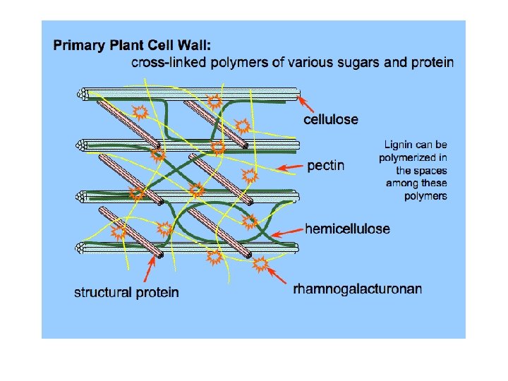
• The cell wall is a highly organized composite of many different polysaccharides, proteins and aromatic substances. • Polysaccharides, polymers of sugar, are the principal components of the cell wall and form its main structural framework. • Polysaccharides are long chains of sugar molecules covalently linked at various positions, some being decorated with side chains of various lengths. Almost all cell wall sugars are aldoses. Many sugars empirical formula is CH 2 On which derive from carbohydrate.
• Cellulose is the principal scaffolding component of all plant cell walls. Cellulose is the most abundant plant polysaccharide, accounting for 15% to 30% of the dry mass of all primary cell walls and an even larger percentage of secondary walls. Cellulose is very strong and durable since it exists in the form of microfibrils and these microfibrils attain a stable structure with numerous hydrogen bonds.
• Non-cellulosic components of the cell: Hemicellulose is a polymer that is produced with glucose and other sugars and this polymer binds cellulose fibrils together.
• Pectin is a mixture of heterogeneous, branched and highly hydrated polysaccharides rich in D-galacturonic acid. It forms a gel-like matrix by attaching itself to calcium ions and water. This matrix substance fills the spaces between the cellulose fibrils and between the cells. • Pectin extracts obtained from the plants have the property of gelling, therefore they are used in the making of jam, sugar and other foods.
***Their functions in brief: • determining wall porosity and providing charged surfaces that modulate wall p. H and ion balance; • regulating cell-cell adhesion at the middle lamella; • serving as recognition molecules that alert plant cells to the presence of symbiotic organisms, pathogens and insects.
Membrane Transport • The selective movement and redistribution of ions and small organic molecules is essential for plant growth and cellular homeostasis. Because of this, plants have evolved numerous proteins that facilitate the transport of minerals, sugars, metabolites and other compound through the limiting membranes of cells and organelles. These membrane transporters are selective for the compound being transported and activities are often tightly regulated. In plant cells, membrane transport underpins a wide range of essential processes, including the following: • Turgor generation, Nutrient acquisition, Waste product excretion Metabolite distribution, Compartmentalization of metabolites, Energy transduction
Protein Sorting and Vesicle Traffic • A typical plant cell contains 5000 to 10 000 different polypeptide sequences and billions of individual protein molecules. If such a cell is to function properly, it must direct these proteins to specific metabolic compartments, cytoplasmic structures and membrane systems. Accurate protein sorting is required at all times, both when cellular structures are formed in dividing and differentiating cells and when proteins in mature structures are degraded and replaced. Examples of the proteins that must be sorted include soluble enzymes, intrinsic membrane proteins and structural proteins in the cell wall matrix. •
• Most proteins have hydrophilic surfaces and therefore do not readily pass through the hydrophobic core of lipid bilayer. Translocation through a membrane involves a protein-like pore or channel through which a protein passes, not in its globular form but it an extended or unfolded configuration. Cytosolic chaperones interact with newly synthesized proteins, keeping them unfolded so they can pass through a protein pore to an appropriate compartment or membrane.
The first sorting event for all proteins made in the cytosol separates them into two groups. Proteins in the first group are released in the cytosol and may either remain in that compartment or be targeted to a variety of destinations, including plastids, mitochondria, peroxisomes and nuclei.
• By contrast, proteins in the second group are targeted to the ER by signal peptides located in the N terminus. Translation of a signal peptide causes the ribosome to bind to the ER during protein synthesis. ER studded with such ribosomes is referred to as rough ER. Proteins synthesized on the rough ER enter the secretory pathway, an intracellular system of vesicles and cisternae (flattened sacs) that includes the ER, Golgi complex, tonoplast and plasma membrane. These proteins can then be secreted or targeted to the various compartments of the secretory system. • The transport of the both soluble and membrane proteins is mediated by vesicles that carry proteins from one compartment to the next. Thus, vesicles are constantly being formed, transported and fused with a compartment different from the one in which they originated. Such transport implies that vesicles can recognize their destination according to the cargo they carry.
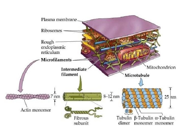
3. The Cytoskeleton • Spatial organization within the eukaryotic cell and directed movements of the cell or its contents are mediated by the cytoskeleton, a network of filamentous protein polymers that permeates the cytosol. The cytoskeleton comprises three major families of proteins: intermediate filaments, actin and tubulin. • Functions of cytoskeleton: • Anchorage • Motility • Information • Polarity
• In addition, a cytoskeleton (cell skeleton) consisting of protein fibers and various motor proteins that provide intracellular motion within the cytoplasm is also present.
• Cell movements results from the activities of the cytoskeleton and related motor proteins. When we compare plants with animals, we usually say that they do not move actively; however intracellular structures and materials move just like animal cells. This movement is carried out via similar mechanisms.
Components of the cytoskeleton: Cytoskeleton is made up of three types of long, thin fibers. Microtubules are just like empty tubes; microfilaments are thinner and the third type is intermediate fibers having a fiber thickness in between. Walls of microtubules consist of numerous tubuline proteins. They are involved in transportation within the cell.
Microfilaments consist of two actin fibers that are entwined with each other. These are thinnest class of fibers. Microfilaments have a positive and a negative end. They use negative end to bond to some cell structures and use the positive end to grow.
Intermediate filaments are made up of different proteins according to their types. These fibers are slightly thicker than microfilaments and they provide structural support to the cells.
For example, intermediate filaments form a net just below the nucleic membrane and help to preserve the shape of the nucleus.
Red: actin filaments Green: microtubules
Types of motor proteins: Three types of motor proteins are present. These are small proteins called kinesin, dynein and myosin. They convert chemical energy to kinetic energy and each of them are responsible from different types of special movements that is beneficial for the cell.
• Myosins move rapidly, kinesin moves slower but is stable (these are motor proteins that carry proteins along the microtubules), and dyneins perform sliding. • Each cytoskeleton fiber is found with a different motor protein. Kinesin and dynein attach to the microtubules and myosin interact with microfilaments.
Myosin-microfilament system is the main protein in skeletal muscles of animals. Myosin movement along the microfilaments forms the basis for muscle contraction. Plants do not have a muscle tissue just like animals, however plant cells have mini muscles formed of microfilaments and myosins. Microfilament/myosin combination is responsible from cytoplasm flow. It also plays an important role in the division of plant cells.