1 2 Assessment and Resuscitation INITIAL ASSESSMENT Initial


 of an acutely ill or")
cardiac output 2)hemoglobin concentration 3)hemoglobin-oxygen saturation. The last-mentioned depends on")


 leads to inadequate oxygen delivery and a resulting metabolic acidosis. Shock")


 usually have a primary respiratory arrest. The outcome")


- Slides: 38
1
2
Assessment and Resuscitation
INITIAL ASSESSMENT Initial assessment (the ABCs-airway, breathing, and circulation) of an acutely ill or injured child requires rapid identification of physiologic derangements in tissue perfusion and oxygenation. immediate resuscitation must be implemented before pursuing the usual information needed to develop a differential diagnosis
Oxygen delivery depends on 1)cardiac output 2)hemoglobin concentration 3)hemoglobin-oxygen saturation. The last-mentioned depends on 1)air movement 2)alveolar gas exchange 3) pulmonary blood flow 4)oxygen-hemoglobin binding characteristics.
HISTORY In the resuscitation phase, access to historical information may be limited. Characterization of onset of symptoms, details of events, and a brief identification of underlying medical problems should be sought by members of the team not actively involved in the resuscitation. Attempts at identifying historical issues that affect the ABCs are useful but should not delay intervention if tissue oxygenation and perfusion are markedly impaired.
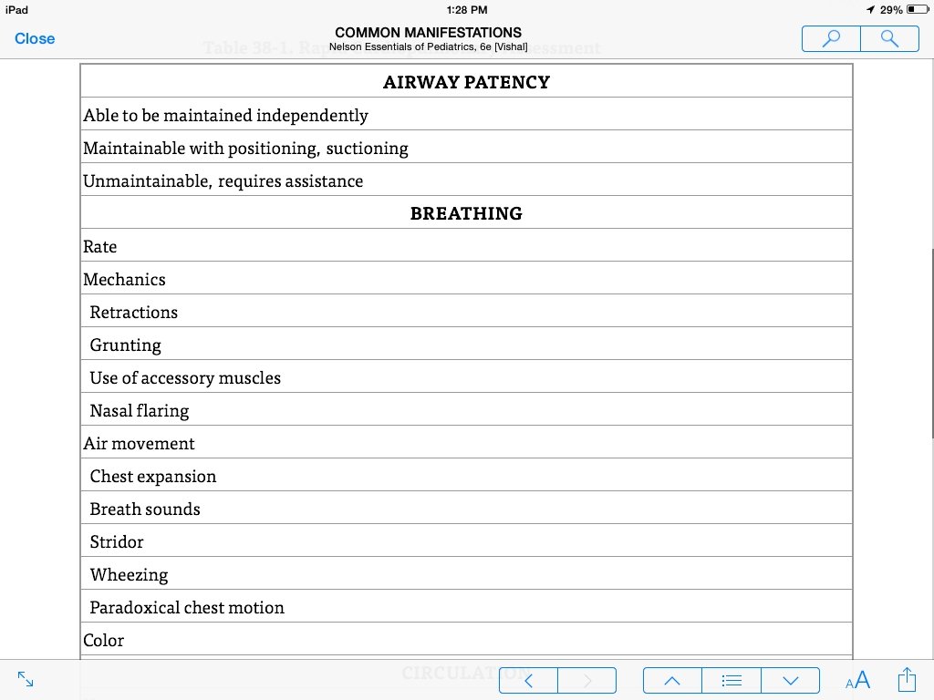
PHYSICAL EXAMINATION Initial examination must focus rapidly on the ABCs to address the issues of oxygen delivery to tissues systematically. Airway patency is the first to be addressed, including assessment of the neurologically injured child's ability to protect the airway. Protection of the cervical spine also should be initiated at this step in any child with traumatic injury or who presents with altered mental status of uncertain etiology. Assessment of breathing includes auscultation of air movement application of a pulse oximeter (when available) to identify current oxygenation status.
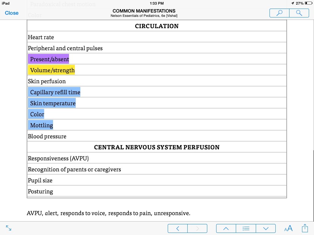
PHYSICAL EXAMINATION Circulatory status is assessed by palpation for distal and central pulses, focusing on the presence and quality of the pulses. Bounding pulses and a wide pulse pressure are often the first sign of the vasodilatory phase of shock and require immediate resuscitation measures. Weak, thready, or absent pulses are indicators for fluid resuscitation, initiation of chest compressions, or both. .
When assessment of the ABCs is complete a more complete physical examination is performed. The sequence of this examination depends on whether the situation involves an acute medical illness or trauma. In trauma patients, the examination follows the ABCDE pathway. D stands for disability and prompts assessment of the neurologic system and evaluation for major traumatic injuries. E stands for exposure; the child is disrobed and searched for evidence of any life-threatening or limb-threatening problems. For the acutely ill and the injured child, the subsequent physical examination should identify evidence of organ dysfunction starting with areas suggested in the chief complaint and progressing to a thorough and systematic investigation of the entire patient
10
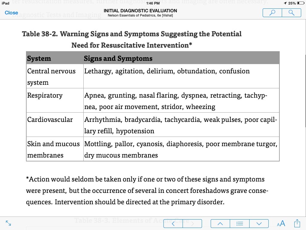
COMMON MANIFESTATIONS The physiologic responses to acute illness and injury are mechanisms that attempt to correct inadequacies of tissue oxygenation and perfusion. When initial changes, such as increasing heart and respiratory rates, fail to meet the body's needs, other manifestations of impending cardiopulmonary failure occur. Respiratory failure, the most common cause of acute deterioration in children, may result in inadequate tissue oxygenation and in respiratory acidosis. Signs and symptoms of respiratory failure (tachypnea, tachycardia, increased work of breathing, abnormal mentation) progress as tissue oxygenation becomes more inadequate.
Inadequate perfusion (shock) leads to inadequate oxygen delivery and a resulting metabolic acidosis. Shock is characterized by signs of inadequate tissue perfusion (pallor, cool skin, poor pulses, delayed capillary refill, oliguria, and abnormal mentation). The presence of any of these symptoms demands careful assessment and intervention to correct the abnormality and to prevent further deterioration.
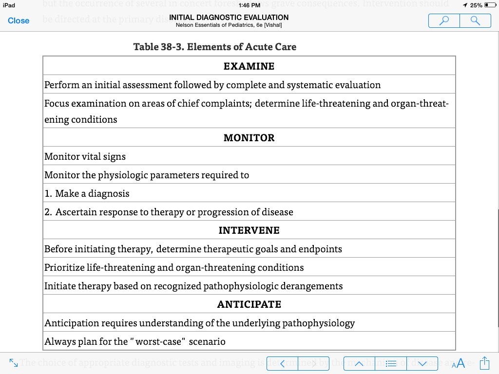
INITIAL DIAGNOSTIC EVALUATION Screening Tests During the initial phase of resuscitation, monitoring vital signs and physiologic status is the key screening activity Continuous monitoring with attention to changes may indicate response to therapy or further deterioration requiring additional intervention. During the initial rapid assessment, diagnostic evaluation often is limited to pulse oximetry and bedside measurement of glucose levels. The latter is important in any child with altered mental status or at risk for inadequate glycogen stores (infants, malnourished patients). After resuscitation measures, further diagnostic tests and imaging are often necessary.
The choice of appropriate diagnostic tests and imaging is determined by the mechanism of disease and results of evaluation after initial resuscitation The initial evaluation of major trauma patients is focused on identifying evidence of hemorrhage and organ and tissue injury. For an acutely ill child with respiratory distress, a chest x-ray is important. Appropriate cultures should be obtained when sepsis is suspected. A complete blood count may also be helpful despite poor sensitivity and specificity of the white blood cell and band counts. Children with historical or physical evidence of inadequate intravascular volume should have serum electrolyte levels obtained, including bicarbonate, blood urea nitrogen, and creatinine.
RESUSCITATION Resuscitation is focused on correcting identified abnormalities of oxygenation and perfusion and preventing further deterioration. Oxygen supplementation may improve oxygen saturation but may not completely correct tissue oxygenation. When oxygen supplementation is insufficient or air exchange is inadequate, assisted ventilation must be initiated.
Inadequate perfusion is best managed initially by providing a fluid bolus. Isotonic crystalloids (normal saline, lactated Ringer solution) are the initial fluid of choice. A bolus of 10 to 20 m. L/kg should be delivered in monitored conditions. repeated boluses until circulation has been re-established. Because most children with shock have noncardiac causes, fluid administration of this magnitude is well tolerated.
If hemorrhage is known or highly suspected, administration of packed red blood cells is appropriate Monitoring for deteriorating physiologic status during fluid resuscitation (increase in heart rate, decrease in blood pressure) identifies children who may have cardiogenic shock. Fluid resuscitation increases preload, which may worsen pulmonary edema and cardiac function If deterioration occurs, fluid administration should be interrupted, and resuscitation should be aimed at improving cardiac function.
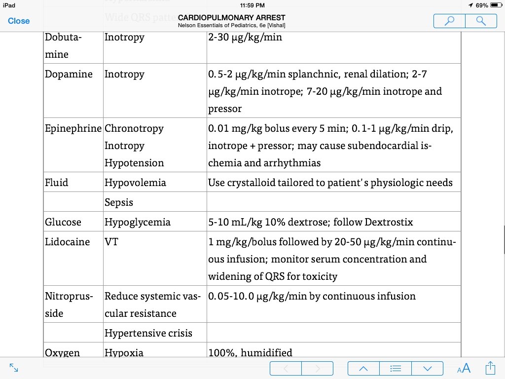
introduction of vasoactive substances is the next step. The choice of which agent to use depends on the type of shock present. Hypovolemic shock (when further volume is contraindicated) and distributive shock benefit from drugs that increase systemic vascular resistance (drugs with α-agonist activity, such as epinephrine or norepinephrine). The treatment of cardiogenic shock is more complex. To improve cardiac output by : increasing the heart rate, drugs with positive chronotropy are used (epinephrine, norepinephrine, and dopamine). Afterload reduction, using drugs such as dobutamine, nitroprusside, or milrinone, also may be needed. Measuring mixed venous oxygen saturation, central venous pressure, and regional oxygen saturations helps guide therapy.
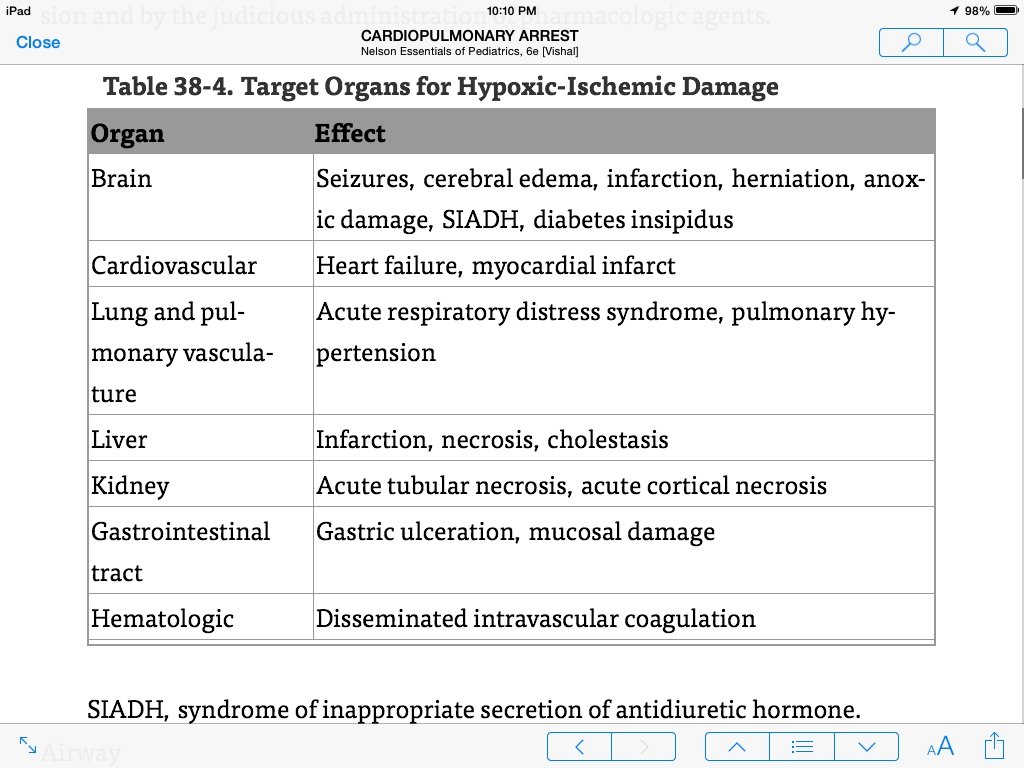
CARDIOPULMONARY ARREST Children who need (CPR) usually have a primary respiratory arrest. The outcome of cardiopulmonary arrest in children is poor; only 2% to 10% survive, and most survivors have permanent neurologic disability. The ability to anticipate or recognize pre-cardiopulmonary arrest conditions and initiate prompt and appropriate therapy not only is lifesaving, but also preserves the quality of life
Hypoxia has a central role in most of the events leading to cardiopulmonary arrest in children. The goal in resuscitating a pediatric patient to optimize 1) cardiac output 2)tissue oxygen delivery, which may be accomplished by using artificial ventilation and chest compression and by the judicious administration of pharmacologic agents.
Airway In children, airway patency often is compromised by a loss of muscle tone, allowing the mandibular block of tissue, including the tongue, bony mandible, and the soft surrounding tissues, to rest against the posterior pharyngeal wall. The head tilt-chin lift maneuver should be used to open the airway in children with no sign of head or neck trauma. In children with signs of head or neck trauma, the jaw thrust maneuver should be used.
Pediatric patients requiring resuscitation should be endotracheally intubated. Before intubation, the patient should be ventilated with 100% oxygen using a bag and mask. Cricoid pressure should be used to minimize inflation of the stomach. Many conscious patients may benefit from the use of induction medications (sedatives, analgesics, and paralytics) to assist intubation but caution is necessary to prevent further cardiovascular compromise from vasodilating effects of many sedatives. The correct size of the tube may be estimated according to the size of the child's mid-fifth phalanx or the following formula: 4 + (patient age in years/4).
the adequacy of ventilation and the position of the tube must be assessed: Use of end-tidal carbon dioxide (CO 2) monitoring devices may assist with validation of endotracheal placement, but low levels of detected CO 2 may be secondary to lack of pulmonary circulation. Adequate chest wall movement and auscultation of the chest to detect bilateral and symmetric breath sounds provide information on adequacy of air entry. If air exchange is not heard or cyanosis continues, the position or patency of the tube must be re-evaluated through direct visualization of tube location via laryngoscopy or, in patients with sufficient circulation, indirectly through the use of end-tidal CO 2 measurement.
Breathing The major role of endotracheal intubation is to protect or maintain the airway and ensure the delivery of adequate oxygen to the patient. Because hypoxemia is the final common pathway in pediatric cardiopulmonary arrests, providing oxygen is more important than correcting the respiratory acidosis. The clinician should deliver 100% oxygen at an initial rate of 12 to 20 breaths/min through the endotracheal tube, using a tidal volume necessary to produce adequate chest rise and relieve cyanosis. Care should be taken not to hyperventilate the patient.
Chest compressions should be initiated if a pulse cannot be palpated or the heart rate is less than 60 beats/min with signs of poor systemic perfusion. the child should be supine on a hard, flat surface. In infants and children, the area for compression is the lower half of the sternum. If two health care providers are available, encircling the chest with both hands to provide compressions is preferable for infants. Effective CPR requires a compression depth of one third to one half of the anteriorposterior diameter of the chest with complete recoil after each compression. The compression rate should be at least 100/min with breaths delivered 8 to 10 times per minute. If an advanced airway is in place compressions should not pause for ventilation; both should continue simultaneously.
The availability of automated external defibrillators outside the hospital setting may be useful in the relatively small number of children who are in ventricular fibrillation or pulseless ventricular tachycardia. Current recommendations include use in children older than 1 year of age.
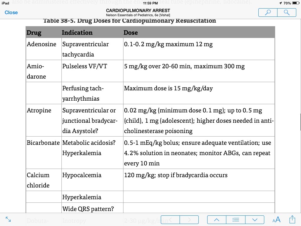
Drugs Administration of drugs through a central venous line is preferred, although this is difficult to achieve in cases of unanticipated cardiopulmonary arrest. The effectiveness of peripheral intravenous administration is limited by poor circulation. If intravascular access is not present or rapidly established, administration through an intraosseous route is recommended. Some drugs can also be administered effectively through the endotracheal tube (epinephrine, lidocaine).
the most important agent used during CPR is oxygen. Short periods of exposure to high oxygen tensions cause little pulmonary toxicity high oxygen tensions may be required to reverse some of the reactive pulmonary vascular responses to poor cardiac output. Epinephrine, a catecholamine with mixed α-agonist and β-agonist properties constitutes the mainstay of drug therapy for CPR. The α-adrenergic effects are most important during acute phases of resuscitation, causing an increase in systemic vascular resistance that improves coronary blood flow. Vasopressin, an endogenous hormone, causes constriction of capillaries and small arterioles and may be useful. but vasopressin may be considered in children failing standard medication administration.
The use of sodium bicarbonate is currently not recommended as a first-line approach. In pediatric patients, acidosis is more often respiratory, rather than metabolic, and administration of sodium bicarbonate can exacerbate the acidosis by inducing production of CO 2. Sodium bicarbonate may be judiciously used to correct identified excessive intake of acids or losses of bases, however oxygen delivery and elimination of CO 2 must be established first. Side effects include hypernatremia, hyperosmolality, hypokalemia, metabolic alkalosis (shifting the oxyhemoglobin curve to the left and impairing tissue oxygen delivery), reduced ionized calcium level, and impaired cardiac function.
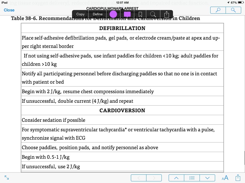
Prompt electrical defibrillation is indicated when ventricular fibrillation or pulseless ventricular tachycardia is noted. CPR should continue until just before defibrillation and resume immediately afterward, minimizing interruptions in compressions. If a second attempt at defibrillation is necessary, it should be followed by a dose of epinephrine. Children failing two episodes of defibrillation may benefit from administration of amiodarone. Defibrillation should be distinguished from cardioversion of supraventricular tachycardias, which also may compromise cardiac output. Cardioversion requires a lower starting dose and synchronization of the discharge to the electrocardiogram to prevent discharging during a susceptible period, which may convert supraventricular tachycardia to ventricular tachycardia or fibrillation.