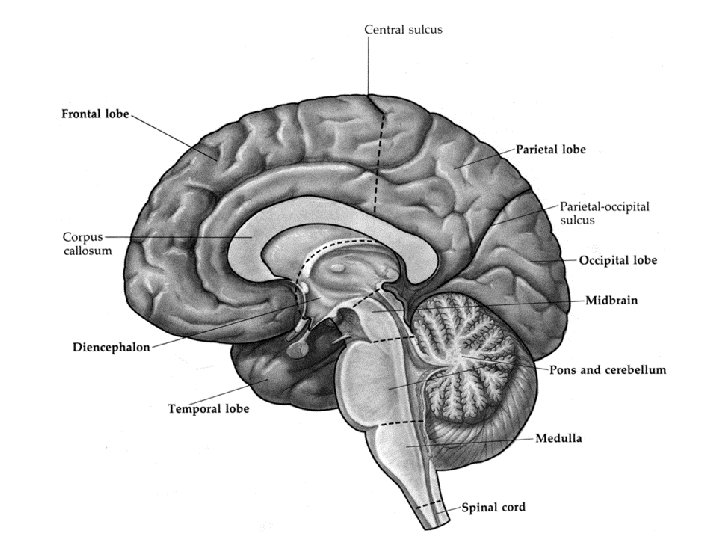
1 2 3 4 5 Parasagittal View of

 gray matter (cell bodies)")

 caudate cingulate")
 Cut through anterior commissure caudate cingulate gyrus corpus")
 caudate cingulate gyrus putamen")


 Superior colliculus Tectum Cerebral aqueduct")


- Slides: 12
1 2 3 4 5 Parasagittal View of the Human Brain Showing the Location of Your 6 Coronal Cuts (red lines) 6
Cut #1 Cut anterior to corpus callosum white matter (axons) gray matter (cell bodies)
Cut #2 Cut through genu of corpus callosum cingulate gyrus corpus callosum lateral ventricle temporal lobe insular lobe
Cut #3 Cut through body of corpus callosum internal capsule (white band) caudate cingulate gyrus corpus callosum lateral ventricle putamen
Cut #4 internal capsule (white band) Cut through anterior commissure caudate cingulate gyrus corpus callosum putamen anterior commissure optic tract Pencil is pointing to anterior commissure amygdala
Cut #5 Cut through mammillary body internal capsule (white band) caudate cingulate gyrus putamen corpus callosum thalamus globus pallidus hypothalamus mammillary body hippocampus
Cut #6 Cut through splenium of corpus callosum cingulate gyrus spenium of the corpus callosum thalamus posterior horn of the lateral ventricle
1 2 3 Parasagittal View of the Human Brain Showing the Location of Your 3 Brainstem Cuts (red lines)
Brainstem Cut #1 Cut through superior colliculus (rostral midbrain) Superior colliculus Tectum Cerebral aqueduct 1 Tegmentum Red nucleus Substantia nigra Crus cerebri Superior colliculus Crus cerebri (cerebral peduncle) Oculomotor nerve (III)
Brainstem Cut #2 Cut through mid-pons 2 Vermis Fourth ventricle Pontine tegmentum Middle cerebellar peduncle Basilar pons Superior cerebellar peduncle
Brainstem Cut #3 Cut through caudal-medulla Fasciculus graciclis Nucleus gracilis 3 Nucleus cuneatus Spinal nucleus V Pyramidal tract Fasciculus cuneatus