05 The Trigeminal System Somatic Sensation of the


 1.")


 trigeminal nucleus")
 trigeminal nucleus • Mediates fine touch, joint position,")
 trigeminal nucleus • Mediates pain and temperature. •")


 Spinal trigeminal tract & spinal trig nucleus Med lem ALS")


- Slides: 18
05. The Trigeminal System Somatic Sensation of the Face and Head David A. Morton, Ph. D. Thursday February 7 th, 2013
Objectives: 1. Outline the two pathways for facial sensation from the head. 2. Contrast facial sensation from the head and somatic sensation from the body. In what ways are they similar? Different? Try drawing this on the Haines atlas diagram at the end of the lecture. 3. Diagram the corneal reflex: the afferent and efferent limbs as well as nuclei involved in the brainstem. 4. If a person does not blink, how would you determine if the problem were in the sensory (afferent) limb, motor (efferent) limb, or brainstem interconnections for the corneal reflex? 5. Explain how a single, small medullary vascular lesion could abolish pain and temperature from the face on the right side and pain and temperature from the body on the left side. What vessel is most likely occluded?
Peripheral Receptors and Sensation • Structures served by trigeminal system CN V-1 CN V-2 ? Great auricular nerve (C 2 -C 3) CN V-3
How many neurons in sensory pathway? DCML/ALS Trigeminal system 1. Peripheral ganglion (DRG) 1. Peripheral ganglion (Trig. gang. ) 2. Spinal cord, medulla 2. 2 nuclei in brainstem (pons, medulla, sp cord) 3. VPL nucleus of the thalamus 3. VPM nucleus of the thalamus
II. Primary Sensory Neurons: Trigeminal ganglion CN V 1 CN V-3 CN V-2 Trigeminal ganglion
Primary Sensory Neurons: Trigeminal ganglion CN V 1 CN V-3 CN V-2 Primary sensory neuron
Second-Order Trigeminal Neurons Originate in two brain stem nuclei: • Principal (chief) trigeminal nucleus • Spinal (descending) trigeminal nucleus Young, Young and Tolbert, 2008 Fig 11 -10
Second-Order Trigeminal Neurons A. Principal (chief) trigeminal nucleus • Mediates fine touch, joint position, vibration. • Located in pons lateral to motor nucleus of CN V. This would be equivalent to what nucleus of the brainstem? _______ Young, Young and Tolbert, 2008 Fig 11 -10
Second-Order Trigeminal Neurons B. Spinal (descending) trigeminal nucleus • Mediates pain and temperature. • Spinal (descending) trigeminal tract • Descends caudally as far as C 2 -C 3 and is continuous with dorsal horn. This would be equivalent to what region in the spinal cord? _______ Young, Young and Tolbert, 2008 Fig 11 -10
Second-Order Trigeminal Neurons C. Ascending Trigeminal Pathways • Axons of second-order neurons form the trigeminothalamic pathway to the Secondary sensory neuron VPM nucleus of the thalamus. Secondary sensory neuron a. Axons from neurons in the chief sensory nucleus carry sensations such as: _____ b. Axons from neurons in the spinal trigeminal nucleus carry sensations such as: ____ Young, Young and Tolbert, 2008 Fig 11 -10
Second-Order Trigeminal Neurons Secondary sensory neuron Young, Young and Tolbert, 2008 Fig 11 -10
Third-Order Trigeminal Neurons Project from VPM in the thalamus to the Postcentral gyrus (primary sensory cortex) • Axons course through the internal capsule
Pons Chief trig nucleus ALS Med lem
Medulla oblongata (rostral) Spinal trigeminal tract & spinal trig nucleus Med lem ALS
The right side of the pons is lesioned. What sensory loss would you expect?
The right side of the pons is lesioned. What sensory loss would you expect? Below the lesion: • Loss of facial sensation on right • Loss of pain and temp from left side • Loss of proprioception/vibration from left side L
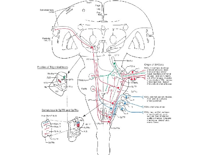
Self-assessment: When you think you have mastered the pathways, select 2 colors in both a dark and light shade. Use the dark color for the body and the lighter color for the face pathways. e. g. Light blue-Trigeminothalamic (ventral) Dark blue – ALS Pink – trigeminothalamic (dorsal) from chief Red – DCML