Introduction of the osteology bone 206 Classification of
。成人有206块骨 一、骨的分类 Classification of bone • 依据骨的位置可分为")
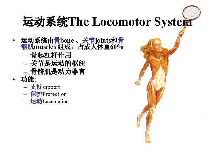
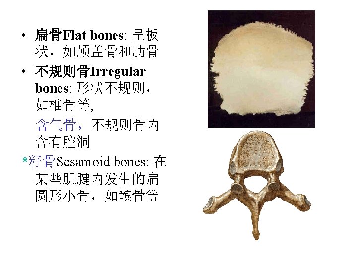
第一节 骨学总论Introduction of the osteology • 骨bone主要由骨组织构成,骨组织(骨细 胞、胶原纤维和基质)。成人有206块骨 一、骨的分类 Classification of bone • 依据骨的位置可分为 according to their positions – 颅骨skull – 躯干骨bones of trunk – 四肢骨appendicular skeleton • 依据骨的形态可分为according to their shape – 长骨long bone – 短骨short bone – 扁骨 flat bone
: – 体又称骨干Diaphysis or shaft, 髓腔medullary cavity, 容纳骨")
• 长骨Long bones (found in limbs): – 体又称骨干Diaphysis or shaft, 髓腔medullary cavity, 容纳骨 髓 – 两端Two ends-骺epiphysis 关 节面articular surface, 干骺端metaphysis, 骺软骨epiphysial cartilage, 骺线 epiphysial line • 短骨Short bones: 呈立方体形, 如腕骨和跗骨

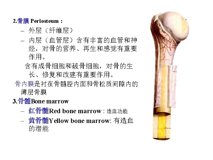
二、骨的构造General structures of bone 1. 骨质Bone substance – 骨密质compact bone – 骨松质spongy bone 呈海绵状,由骨小梁构成 ※颅盖骨,外板和内板outer plate and inner plate, 板障


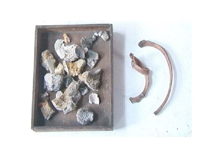
三、骨的化学成分和物理性质 Chemical composition and physical properties • 有机质Organic material: 主要是骨胶原纤维束和粘多糖蛋白。构成支架,赋 予骨的弹性和韧性。 • 无机质Inorganic salts: 主要是碱性磷酸钙。赋予骨硬度和脆性 Organic material Inorganic salts Children 1 1 Adult 3 7 Old 2 8

躯干骨 Bones of trunk 组成: ■ 24块椎骨 vertebrae ■ 1块骶骨 sacrum ■ 1块尾骨coccyx ■ 1块胸骨sternum ■ 12对肋 ribs

椎骨Vertebrae • There are 33 vertebrae in children, arranged as follows: • 颈椎 Cervical vertebrae C. 7 • 胸椎Thoracic vertebrae T. 12 • 腰椎Lumbar vertebrae L. 5 • 骶椎 Sacral vertebrae S. 5 骶骨sacrum • 尾椎 Coccygeal vertebrae Co. 3~4 尾骨 coccyx

1. 椎骨的一般特征General features of vertebrae • 椎体Vertebral body • 椎弓Vertebral arch – 椎弓根pedicle of vertebral arch : 椎上、下切迹 sup. and inf. Vertebral notch – 椎弓板lamina of vertebral arch • process (7): – 棘突spinous process – 横突transverse process – 上、下关节突sup. and inf. articular processes • Vertebral foramen 椎孔 • Vertebral canal 椎管 • Intervertebral foramen 椎间孔

2. 各部椎骨的主要特征 main characteristics of regional vertebrae 颈椎Cervical vertebrae • Vertebral Body: small • Vertebral foramen: larger and triangular • Spinous processes: short and bifid in C 2 to C 6, however longer in C 7 • Transverse processes: short and bifid, 横突孔transverse foramen • Articular processes: horizontal

椎体钩uncus of vertebral body和 钩椎关节Luschka joint
 – Body and spinous process absent,")
非典型颈椎Atypical cervical vertebrae • 寰椎 Atlas (C 1) – Body and spinous process absent, consists of anterior and posterior arches, and two lateral masses – 椎动脉沟Groove for vertebral artery 齿突凹
: distinguished by齿突 dens which articulates with dental fovea")
• 枢椎 Axis (C 2): distinguished by齿突 dens which articulates with dental fovea of anterior arch of atlas

• 颈动脉结节Carotid tubercle: anterior tubercle of transverse process of C 6 • 隆椎 prominent Vertebra(C 7): contains long and non-bifid spinous process, it is visible with neck flexed, used as clinical landmark in counting cervical and thoracic spinous processes

胸椎Thoracic vertebrae • Vertebral Body : heart-shape, 上、下肋凹superior and inferior costal fovea • Vertebral foramen: smaller, rounder • Spinous processes: long, point obliquely downward • Transverse processes: 横突肋凹 transverse costal fovea • Articular processes: coronal position

腰椎 Lumbar vertebrae • Vertebral Body: larger, kidney-shape • Vertebral foramen: larger and triangular • Spinous processes: projects horizontally • Transverse processes: long • Articular processes: sagittal position
 • Posterior surface: 骶正中嵴median")
骶骨Sacrum • Anterior surface: 岬promontory, 骶前孔anterior sacral foramina (four pairs) • Posterior surface: 骶正中嵴median sacral crest, 骶后孔posterior sacral foramina (four pairs), 骶管裂孔sacral hiatus, 骶角sacral cornu • Lateral part: 耳状面auricular surface, 骶粗隆 sacral tuberosity
 anaesthesia")
Cornu Sacral hiatus palpation 骶管麻醉Transsacral (epidural) anaesthesia

二、胸骨Sternum • 胸骨柄Manubrium sterni : 颈静脉切迹jugular notch, 锁切迹clavicular notch • 胸骨体 Body of sternum • 剑突Xiphoid process ★胸骨角Sternal angle : the junction of manubrium and body, which connects 2 nd costal cartilage laterally, and lies opposite lower border of T 4 posteriorly

★胸骨角 Sternal angle which connects 2 nd costal cartilage laterally, and lies opposite lower border of T 4 posteriorly

三、肋Ribs-12 pair • 一般特征General features 肋rib包括肋骨costal bone和 肋软骨costal cartilage – 真肋Ribs 1~7 called true ribs – 假肋Ribs 8~12 called false ribs – 浮肋Ribs 11~12 called floating ribs

典型肋骨的特征Characteristics of “typical” costal bone • Posterior end: 肋头costal head, 肋颈costal neck, 肋结节 costal tubercle • Shaft: 肋角costal angle, 肋沟costal groove • Anterior end

非典型肋骨的特征Atypical costal bone • First rib: 前斜角肌结节tubercle for scalenus anterior, 锁骨下动、静脉沟 sulcus for subclavian artery and vein § 11 th and 12 th ribs lack costal necks, tubercles and angles

Bones of upper limbs Composition: • Should girdle 上肢带骨 clavicle锁骨,scapula肩胛骨 • Bones of free upper limb 自由上肢骨 – Humerus肱骨 in arm – Radius桡骨 and ulna尺骨 in forearm – Carpal腕骨, metacarpals掌骨 and phalanges指骨 in hand

Clavicle 锁骨 • “S” shaped, medial 2/3 convex forward and lateral 1/3 convex backward • Sternal end胸骨端 medially and acromial end肩峰端 laterally

Scapula 肩胛骨 • Three borders – Superior: coracoid process 喙突 , scapular notch 肩胛切迹 – Lateral (axillary) border腋缘 – Medial (vertebral) border脊柱缘 • Three angles – Superior: opposite to the 2 nd rib – Inferior: opposite to the 7 th rib or 7 th intercostals space – Lateral: glenoid cavity关节盂, supra- and infraglenoid tubercles 盂上、下结节 • Two surfaces – Anterior surface concave: subscapular fossa肩胛下窝 – Posterior surface: supra- and infraspinous fossae冈上、 下窝, spine of scapula 肩胛冈, acromion 肩峰

Humerus 肱骨 • Upper end: head of humerus肱骨头, anatomical neck解剖颈, greater and lesser tubercles大、小结节, crests of greater and lesser tubercle, 大、小结节嵴, intertubercular groove结节间沟, surgical neck外科颈 • Shaft: deltoid tuberosity三角肌粗隆on lateral surface, and a groove for radial nerve桡神经沟 on posterior surface • Lower end: lateral and medial epicondyles内、外上髁, capitulum 肱骨小 头, trochlea 肱骨滑车, coranoid fossa 冠突 窝and radial fossa 桡窝 (anteriorly) and olecranon fossa鹰嘴窝 (posteriorly), and sulcus for ulnar nerve 尺神经沟

Radius • Upper end: head of radius桡骨头, neck of radius桡骨颈, radial tuberosity 桡骨粗隆, and articular circumference环状关节面 • Shaft:interosseous border骨间缘 • Lower end: styloid process 茎突 laterally, ulnar notch尺切迹 medially, and carpal articular surface腕关节面 inferiorly

Fracture of the distal end pf the radius

Ulnar • Upper end: olecranon鹰嘴 coronoid process 冠突 trochlear notch滑车切迹 radial notch 桡切迹 ulnar tubersity尺骨粗隆 • Lower end styloid process 尺骨茎突 head of ulna 尺骨头
 scaphoid 手舟骨, lunate月骨, triquetral三角骨")
Carpal bones 腕骨 • Proximal row ― (lateral to medial) scaphoid 手舟骨, lunate月骨, triquetral三角骨 and pisiform豌豆骨 • Distal row ― (lateral to medial) trapezium 大多角骨, trapezoid小多角骨, capitate头状骨 and hamate钩骨 Metacarpal bones掌骨 • Numbered one to five from thumb to little finger • Structure of each―base (proximally), shaft, and head (distally) Phalanges of fingers 指骨 • Consist of 14 ―two for first digit (thumb) and three for each of other four digits • Structure of each ―base (proximally), shaft, and trochlea of phalanx (distally), tuberosity of distal phalanx远节指骨粗隆

Bones of Lower Limb Composition: • Pelvic girdle: hip bone髋骨 • Bones of free lower limb: – Femur股骨 in thigh ( patella 髌骨) – Tibia胫骨 and fibula腓骨 in leg – Tarsals跗骨, metatarsals跖骨, phalanges of toes趾骨 in foot

Hip bone Consisting of three fused bones, ilium, ischium, pubis • Ilium髂骨 – Body of ilium 髂骨体 to form superior 2/5 of acetabulum髋臼 – Ala of ilium 髂骨翼: iliac crest 髂嵴, anterior superior iliac spine 髂前上棘, anterior inferior iliac spine 髂前下棘, tubercle of iliac crest髂结节, posterior superior iliac spine 髂后上棘,posterior inferior iliac spine 髂后下棘, greater sciatic notch 坐骨大切迹, gluteal surface 臀面,iliac fossa 髂窝, arcuate line弓状线, auricular surface 耳状面, iliac tuberosity 髂粗隆,

• Ischium 坐骨 – Body of ischium 坐骨体―to form posterior and inferior 2/5 of acetabulum, ischial spine坐骨棘, lesser sciatic notch 坐骨小切迹 – Ramus of ischium 坐骨支, ischial tuberosity 坐 骨结节 • Pubis 耻骨 – Body of pubis 耻骨体―to form anterior and inferior 1/5 of acetabulum, iliopubic eminence 髂耻隆起 – Superior ramus of pubis 耻骨上支: pecten of pubis 耻骨梳, pubic tubercle 耻骨结节, pubic crest 耻骨嵴, symphysial surface耻骨联合面 – Inferior ramus of pubis耻骨下支: obturator foramen 闭孔 • Acetabulum 髋臼―formed by bodies of ischium, ilium and pubis Lunate surface月状面, acetabular fossa 髋臼窝, acetabular notch髋臼切迹

Femur 股骨 • Upper end: femoral head股骨头, fovea of femoral head 股骨头凹, neck of femur 股 骨颈, greater trochanter 大转子,lesser trochanter 小转子, intertrochanteric line 转子间线, intertrochanteric crest 转子间 嵴 • Shaft: linea aspera 粗线, gluteal tuberosity 臀肌粗隆, pectineal line耻骨肌线 , popliteal surface 腘面 • Lower end: medial and lateral condyles内、 外侧髁, medial and lateral epicondyles内、 外上髁, adductor tubercle 收肌结节, intercondylar fossa髁间窝, patellar surface 髌面

Fracture of the femoral neck

Tibia 胫骨 • Upper end: medial and lateral condyles 内、外侧髁, intercondylar eminence 髁间 隆起, fibular articular surface 腓关节面, tuberosity of tibia 胫骨粗隆 • Shaft: interosseous border 骨 间缘, soleal line 比目鱼肌线 • Lower end: fibular notch腓切 迹, medial malleolus 内踝

Fibula 腓骨 • Upper end: fibular head 腓骨头, neck of fibula 腓骨颈 • Shaft: interosseous border骨间缘 • Lower end: lateral malleolus 外踝 Patella 髌骨 • triangular sesamoid bone

Tarsal bones 跗骨 Consist of seven short bones arranged in three row • Posterior row ―talus 距骨, calcaneus 跟骨 • Intermediate row ―navicular bone 足舟骨 • Anterior row ―medial, intermediate, and lateral cuneiforms 内侧、中间和外侧楔 骨, cuboid bone 骰骨

Metatarsal bones 跖骨 • Consist of five elongated bones, numbered one to five medial to lateral • Structure of each ―base (proximally), shaft, and head (distally) in each Phalanges of toes 趾骨 • Consist of 14―two for first toe (hallux) and three for each of other four toes ※Surface landmarks: trochlea of talus距骨滑车, calcaneal tuberosity 跟骨结节, tuberosity of navicular bone 舟骨粗隆, and tuberosity of fifth metatarsal bone 第五跖骨粗隆
- Slides: 44